Introduction
Breast, lung and bronchial and colorectal cancer are
the three most commonly diagnosed types of cancer among women and
represent 50% of all cases. However, breast cancer alone is
expected to account for 30% of all new cancer cases diagnosed among
women (1). Breast cancer is a
collection of conditions that have different biological properties
and can arise from various genetic abnormalities. Treatment for
breast cancer is based on clinical classification, such as hormone
receptor [estrogen receptor (ER)/progesterone receptor
(PgR)]-positive breast cancer, human epidermal growth factor
receptor 2 (HER2)-positive breast cancer and triple-negative breast
cancer (TNBC). Luminal type breast cancer is ER/PgR-positive and
HER2-negative and accounts for 70-80% of breast cancer cases.
HER2-type breast cancer is characterized by HER2 protein expression
and accounts for 15-20% of breast cancer cases. TNBC is
characterized by the lack of ER, PgR and HER2 expression and
accounts for 15-20% of breast cancer cases (2). Hormone therapy, including aromatase
inhibitors, selective ER modulators and ER downregulators, has
considerably improved the clinical outcomes of patients with
ER-positive breast cancer. Tamoxifen treatment can reduce the
5-year risk of recurrence and death by 41 and 33%, respectively
(3). Luminal A-type breast cancer
is characterized by the expression of hormone receptors and has a
good overall prognosis under hormone therapy. Chemotherapy is
currently considered for luminal B-type breast cancer, which has a
high growth potential. Moreover, HER2-targeted drugs, such as
trastuzumab (Herceptin®), pertuzumab
(Perjeta®) and lapatinib (Tykerb®) have
improved the outcomes and survival rates of women with
HER2-positive breast cancer (4).
Although differences between intrinsic breast cancer
subtypes are well known, one of the issues with breast cancer
treatment is that a practical 'targeted therapy' for TNBC is not
widely available and the effectiveness of chemotherapy for patients
with TNBC remains limited (5).
Olaparib, an oral poly (adenosine diphosphate-ribose) polymerase
inhibitor, is available for chemotherapy-resistant, BRCA
mutation-positive, HER2-negative inoperable or recurrent breast
cancer. It provides a significant benefit over standard therapy for
patients with metastatic breast cancer having a germline
BRCA mutation (6). Breast
cancer has historically been viewed as an immunologically silent
disease. Previous clinical trials have reported that nivolumab, an
anti-PD-1 antibody, has demonstrated an objective response rate of
only 5-10% in patients with metastatic TNBC (7). Preclinical and clinical trials have
demonstrated that low-dose induction chemotherapy or radiation
followed by immune checkpoint inhibitor treatment, such as with
nivolumab, may be useful for the stimulation of anticancer immune
responses (8).
Prognostic biomarker tests for breast cancer are
available, including Oncotype DX, Mama Print and Prosigma. However,
these biomarkers test different sets of breast cancer-related genes
(9). Although these diagnostic
tests are typically used as prognostic markers in early-stage
breast cancer, they are costly and require multiple markers
(10). Therefore, simpler and
easily measurable biomarkers with highly accurate assays need to be
developed for the treatment of patients with breast cancer.
To identify target molecules for the diagnosis and
treatment of cancer, gene expression analysis and subsequent tissue
microarray analysis of solid tumor tissues have been conducted.
Dozens of oncoantigens that play essential roles in the progression
of various solid tumors have been isolated (11-31),
including members of the kinesin superfamily, which are involved in
essential mechanisms during intracellular transport and cell
division in cancers. The present study focused on the genes that
encode kinesin proteins that are overexpressed in the majority of
breast cancers, whereas are scarcely expressed in normal tissues.
During this screening process, kinesin family member 20A (KIF20A),
a member of the kinesin super-family‑6, was identified as a
candidate molecular target that was frequently and highly expressed
in breast cancer. KIF20A possesses a conserved motor domain that
binds to microtubules and couples adenosine triphosphate (ATP)
hydrolysis to generate mechanical forces (32). In addition, KIF20A has a
microtubule plus end-directed motility and is involved in different
cellular processes, such as mitotic spindle formation, chromosome
partitioning and cytokinesis (33). Previous studies have indicated that
KIF20A is highly expressed in several types of human cancers,
including pancreatic (34), breast
(35), glioma (36), prostate (37) and bladder cancers (38). FOXM1 has been reported to regulate
KIF20A expression, resulting in the promotion of cell growth.
Conversely, the depletion of KIF20A has been reported to enhance
the antitumor effects of paclitaxel in luminal type breast cancer
cells (35). To date, it has not
been completely elucidated whether KIF20A expression is related to
clinical breast cancer subtypes. Furthermore, the mechanistic
potential of KIF20A inhibition as a novel molecular therapy for
various subtypes of breast cancer has not yet been fully
investigated. Therefore, in the present study, KIF20A was
characterized as a potential diagnostic and therapeutic target for
different types of breast cancer.
Materials and methods
Cells lines and clinical samples
In total, 11 breast cancer cell lines (T-47D,
ZR-75-1, MCF-7, AU565, SK-BR-3, HCC1599, HCC1143, HCC1937,
MDA-MB-231, BT-20 and MDA-MB-468) and normal adult breast
epithelial cell line (184A1) were used in the present study.
Table I details the derivation of
these cells and the suppliers. All cells were grown in monolayers
in the appropriate medium supplemented with 10% fetal bovine serum
(FBS; Gibco; Thermo Fisher Scientific, Inc.). The cells were
incubated at 37°C in a humidified air atmosphere with 5%
CO2, apart from the MDA-MB-468 and MDA-MB-231 cells,
which were incubated with 0% CO2. The 184A1 cells were
grown in medium supplemented with EpiLife Defined Growth Supplement
(Thermo Fisher Scientific, Inc.). Furthermore, 10 breast cancer or
normal breast tissue samples obtained from patients who had
undergone surgery at Kanagawa Cancer Center Hospital during March,
2008 to December, 2009 (Table II)
were used for reverse transcription-quantitative PCR (RT-qPCR)
experiments. A total of 257 formalin‑fixed primary breast cancer
tissues (obtained from female patients; median patient age, 57
years; age range, 28-89 years) and adjacent healthy tissues were
obtained from patients at the Kanagawa Cancer Center Hospital,
Japan (Table III). The clinical
stage of the samples was determined according to the Union for
International Cancer Control TNM classification. The present study
and the use of all clinical materials were approved by individual
institutional ethics committees. The project to establish tumor
tissue micro-arrays from archival formalin‑fixed and
paraffin‑embedded surgically resected tissues and to use the tissue
microarrays for later unspecified research works was approved by
the Kanagawa Cancer Center Ethics Committee with the approval no.
Rin-177, 27 (September, 2010). The patients involved in the breast
cancer tissue microarrays received mastectomy operations for breast
cancer at Kanagawa Cancer Center Hospital from 2004 to 2006.
Written comprehensive informed consent was obtained from the
patients for the use of their clinical information and for
specimens remaining after clinically required examinations, such as
archival formalin‑fixed and paraffin‑embedded specimens following
diagnosis.
 | Table IThe human breast cancer cell lines
and normal breast cells used in the present study. |
Table I
The human breast cancer cell lines
and normal breast cells used in the present study.
| Cell line | Histology | Subtype |
|---|
| T-47D | Ductal
carcinoma | Luminal |
| ZR-75-1 | Ductal
carcinoma | Luminal |
| MCF-7 | Ductal
carcinoma | Luminal |
| AU565 | Adenocarcinoma | HER2/neu
positive |
| SK-BR-3 | Adenocarcinoma | HER2/neu
positive |
| HCC1599 | Ductal
carcinoma | TNBC |
| HCC1143 | Ductal
carcinoma | TNBC |
| HCC1937 | Primary ductal
carcinoma | TNBC |
| MDA-MB-231 | Adenocarcinoma | TNBC |
| BT-20 | Epithelial
carcinoma | TNBC |
| MDA-MB-468 | Adenocarcinoma | TNBC |
| 184A1 | Human mammary
epithelial cells | |
| Table IICharacteristics of breast cancer
tissues used for RT-qPCR. |
Table II
Characteristics of breast cancer
tissues used for RT-qPCR.
|
Characteristics | No. of
patients |
|---|
| Sex | |
| Male | 0 |
| Female | 10 |
| Age (years) | |
| <65 | 9 |
| ≥65 | 1 |
| Stage | |
| I | 0 |
| II | 7 |
| III | 3 |
| Histology | |
| Papillotubular
carcinoma | 2 |
| Scirrhous
carcinoma | 3 |
| Solid tubular
carcinoma | 5 |
| Table IIIAssociation between KIF20A-positivity
in breast cancer tissues and patient characteristics. |
Table III
Association between KIF20A-positivity
in breast cancer tissues and patient characteristics.
|
Characteristics | Total no. of
patients (n=257) | KIF20A expression
| P-value positive
vs. absent expression |
|---|
| Strong positive
(n=106) | Weak positive
(n=89) | Absent (n=62) |
|---|
| Age (years) | | | | | 0.8639 |
| <65 | 193 | 85 | 62 | 46 | |
| ≥65 | 64 | 21 | 27 | 16 | |
| ER status | | | | | |
| Negative | 71 | 35 | 30 | 6 | 0.0002a |
| Positive | 186 | 71 | 59 | 56 | |
| PgR status | | | | | |
| Negative | 98 | 44 | 38 | 16 | 0.0245a |
| Positive | 159 | 62 | 51 | 46 | |
| HER2/neu
status | | | | | |
| Negative | 212 | 84 | 71 | 57 | 0.0333a |
| Positive | 45 | 22 | 18 | 5 | |
| Triple-negative
breast | | | | | |
| cancer (TNBC) | | | | | |
| TNBC | 39 | 20 | 16 | 3 | |
| Others | 218 | 86 | 73 | 59 | |
| Histology | | | | | |
| Invasive lobular
carcinoma | 6 | 2 | 1 | 3 | |
| Scirrhous
carcinoma | 87 | 30 | 30 | 27 | 0.0348a,b |
| Papillotubular
carcinoma | 65 | 28 | 26 | 11 | |
| Solid tubular
carcinoma | 64 | 32 | 24 | 8 | |
| Invasive ductal
carcinoma | 5 | 1 | 2 | 2 | |
| Mucinous
carcinoma | 10 | 6 | 1 | 3 | |
| DCIS | 13 | 4 | 2 | 7 | |
| Apocrine
carcinoma | 3 | 1 | 2 | 0 | |
| Others | 4 | 2 | 1 | 1 | |
| pT factor | | | | | |
| T1 | 91 | 25 | 34 | 32 | 0.0035a |
| T2-T3 | 166 | 81 | 55 | 30 | |
| pN factor | | | | | |
| N0 | 148 | 50 | 56 | 42 | 0.0767 |
| N1-N2 | 109 | 56 | 33 | 20 | |
RT‑qPCR
Total ribonucleic acid (RNA) was extracted from the
cultured cells and clinical tissues using a Maxwell® 16
LEV simplyRNA Cells kit and Tissue kit (Promega Corp.), according
to the manufacturer's protocol. Complementary DNA was synthesized
using a ReverTra Ace® RT-qPCR lit (Toyobo). The mRNAs
were quantified by RT‑qPCR analysis using TaqMan®
Universal Master Mix II and TaqMan® Gene Expression
Assays on a StepOne Plus (Thermo Fisher Scientific, Inc.),
according to the manufacturer's instructions. The reaction
conditions were as follows: Initial denaturation for 2 min at 50°C
and 10 min at 95°C followed by 40 cycles of denaturation (15 sec at
95°C and 60 sec at 60°C). Each experiment was performed in
triplicate. For the assays, KIF20A (Hs00993573_m1) primer
was used and actin beta (ACTB; Hs01060665_g1) was run as an
internal control (Thermo Fisher Scientific, Inc.).
Western blot analysis
Cells were lysed in Pierce RIPA buffer (Thermo
Fisher Scientific, Inc.) that included a 1% protease inhibitor
cocktail (Thermo Fisher Scientific, Inc.). Following
homogenization, the cell lysates were incubated on ice for 30 min
and centrifuged at 14,000 × g for 15 min at 4°C to separate the
supernatant from cellular debris. Total protein was measured using
a Qubit Protein assay kit (Thermo Fisher Scientific, Inc.). The
proteins were then mixed with sodium dodecyl sulfate sample buffer,
boiled at 100°C for 5 min, and incubated at room temperature for 5
min. Following electrophoresis on 7.5 or 12%
Mini-Protean® TGX gels (Bio-Rad Laboratories, Inc.), the
proteins were transferred onto Trans-Blot® Turbo
0.2-µm polyvinylidene difluoride membranes (Bio-Rad
Laboratories, Inc.). The membranes were blocked with Block
Ace® (Dainippon Pharmaceutical) for 1 h, following which
they were incubated overnight with rabbit polyclonal anti-KIF20A
antibody (1:500, cat. no. A300‑879A; Bethyl Laboratories) at 4°C.
Immunoreactive proteins were incubated with anti-rabbit horseradish
peroxidase (HRP)-conjugated secondary antibodies (1:5,000, GE
Healthcare Life Sciences) for 1 h at room temperature. Protein
bands were visualized by chemiluminescence western blotting
detection reagents using an ImageQuant LAS 4000 mini (GE Healthcare
Life Sciences).
Immunocytochemistry
Cultured cells were washed twice with Dulbecco's
phosphate-buffered saline [PBS(-); cat. no. C14190500; Life
Technologies; Thermo Fisher Scientific, Inc.] and fixed in 4%
paraformaldehyde solution for 15 min at room temperature. Cells
were rendered permeable using PBS(-) containing 0.2% Triton X-100
for 2 min at room temperature. Thereafter, the cells were covered
with blocking solution containing 3% bovine serum albumin in PBS(-)
for 30 min to block non‑specific binding prior to the primary
antibody reaction. Subsequently, the cells were incubated with the
rabbit polyclonal anti-KIF20A antibody (1:200, cat. no. A300-879A;
Bethyl Laboratories) in a wet chamber for 1 h at room temperature.
The immune complexes were stained with Alexa Fluor®
488-conjugated goat anti-rabbit IgG secondary antibody (1:1,000,
cat. no. A11008; Life Technologies; Thermo Fisher Scientific, Inc.)
in a wet chamber for 1 h at room temperature. Mounting medium with
DAPI (Vector Laboratories, Inc.) was used to coverslip the slides.
KIF20A staining was visualized under a microscope (BZ-X710, Keyence
Corp.).
Immunohistochemistry and tissue
microarray analysis
Tumor tissue microarrays were constructed according
to previously published procedures (39). Formalin-fixed breast cancers were
obtained at the Kanagawa Cancer Center. The tissue areas selected
for sampling were determined by visual alignment with the
corresponding hematoxylin and eosin-stained sections on slides.
Three, four or five tissue cores (diameter, 0.6 mm; height, 3-4 mm)
obtained from donor tumor blocks were placed into recipient
paraffin blocks using a tissue micro-arrayer (Beecher Instruments).
The tissue microarray slides were de‑paraffinized and heat‑induced
antigen retrieval was conducted in an autoclave using Target
Retrieval Solution (pH 9). Endogenous peroxidase was blocked (Dako;
Agilent Technologies, Inc.) and the anti-KIF20A antibody (1:100,
cat. no. A300-879A; Bethyl Laboratories) was added. The sections
were incubated at room temperature for 1 h prior to incubation with
EnVision+System-HRP labeled polymer anti-rabbit secondary antibody
(cat. no. K4003; Dako; Agilent Technologies, Inc.) for 30 min at
room temperature. 3,3'-Diaminobenzidine (DAB) chromogen and DAB
substrate buffer were added, and the specimens were counterstained
with hematoxylin and eosin for 1 min room temperature. The staining
intensity within each tumor tissue core was mostly homogenous,
facilitating the semi-quantitative evaluation of the KIF20A
staining intensity by three independent investigators. KIF20A
expression patterns in the tissue arrays were classified ranging
from absent/weak to strong. Cases were defined as strongly positive
if all of the three reviewers independently classified them as
such.
RNA interference assay
siRNA oligonucleotides (Sigma-Aldrich; Merck KGaA)
were used to evaluate the biological functions of KIF20A in breast
cancer cells. The sequences targeting each gene were as follows:
Control 1 si‑Luciferase (LUC), 5′-CGUACGCGGAAUACUUCGATT-3′;
Control 2 si‑EGFP, 5′-GAAGCAGCACGACUUCUUCTT-3′;
KIF20A-suppressing siRNA (si‑KIF20A) #1, 5′-GUUCUC
AGCCAUUGCUAGCTT-3′; and si‑KIF20A #2, 5′-CCCUUA
UGCCCGGAUCCUATT-3′. Breast cancer cell lines were incubated with
medium containing siRNAs (50 µM) and Lipofectamine 2000
reagent (Thermo Fisher Scientific, Inc.), for 4 h at 37°C in a
CO2 incubator. Following incubation, the transfection
mixture was removed and replace with 1X normal growth medium
Cell viability assay
The transfected cells were seeded on a 6-well plate
at a density of 4×10⁴ cells/well. Cell viability was
evaluated 7 days following transfection using a Cell Counting Kit-8
(Dojindo Laboratories, Inc.).
Colony formation assay
The colony formation assay was performed using
Giemsa solution (WAKO) staining. Images were captured by
MP990-PIXUS (Canon). The transfected cells were seeded on a 10 cm
dish at a density of 2×10⁵ cells/well. Following
incubation for 7 days, the treated cells were fixed with 100%
methanol for 30 min at room temperature and stained with Giemsa
solution (Wako Pure Chemical Industries, Ltd.) for 20 min at room
temperature.
KIF20A inhibitor assay
First, dose-response experiments with paprotrain
using the ZR-75-1 cells at concentrations of 50, 100 or 200
µM were performed; 200 µM of paprotrain (Merck KGaA)
treatment significantly inhibited the cell growth and was thus
adopted to compare the effect of paprotrain on various cell lines
under the same conditions. The ZR-75-1, SK-BR-3 and HCC1937 cells
were treated with 200 µM of paprotrain for 24 h (Merck KGaA)
to examine the growth suppressive effects of a selective
cell-permeable KIF20A inhibitor in breast cancer cells using MTT
assay, flow cytometry and live‑cell imaging.
Flow cytometric analysis
Flow cytometric analysis was performed using the
CycletestPlus DNA Reagent kit (cat. no. 340242; BD Biosciences) and
the FACSVerse system (BD Biosciences). The breast cancer cells
lines were transfected with siRNA oligonucleotides or treated with
200 µM of paprotrain for 24 h. Following treatment,
1.0×10⁶ cells were harvested for DNA ploidy staining.
The samples were filtered through a 70-µm nylon mesh and
stored on ice in the dark. The cell cycle was analyzed within 3 h
using the FACSVerse flow cytometer. The DNA content was analyzed in
20,000 ungated cells.
Live‑cell imaging
Live-cell imaging was performed using the Evos FL
Auto Imaging System (Thermo Fisher Scientific, Inc.) to monitor
cytokinetics. Breast cancer cells were seeded into 35-mm glass
dishes in medium containing 10% FBS. Images were captured every 15
min following si-KIF20A transfection or paprotrain treatment using
a fully-automated imaging system of Evos FL Auto Imaging
System.
Matrigel invasion assay
The ZR-75-1, SK-BR-3 and HCC1937 cells transfected
either with siRNA against KIF20A or with si-control (LUC)
were grown to near confluence in culture medium containing 10% FBS.
The cells were harvested by trypsinization, washed in medium
without the addition of serum or protease inhibitor, and suspended
in medium at a concentration of 2×10⁵ per milliliter.
Before preparing the cell suspension, the dried layer of Matrigel
matrix (BD Biosciences) was rehydrated with medium for 2 h at room
temperature. Medium (0.75 ml) containing 10% FBS was added to each
lower chamber in 24-well Matrigel invasion chambers, and 0.5 ml
(1×10⁵ cells) of cell suspension in growth medium with
0.1% FBS was added to each insert of the upper chamber. The plates
of inserts were incubated for 22 h at 37°C, in which condition, no
growth-promoting effect by si-KIF20A expression was also confirmed
by MTT assay. The chambers were processed, and cells invading
through the Matrigel were fixed and stained with Giemsa for 1 h at
room temperature as directed by the supplier (BD Biosciences).
Statistical analysis
Statistical analyses were conducted using StatView
(JMP) and SPSS 25 software (IBM, Inc.). The significance test
analyzing the difference between 2 groups of cell-based assays was
performed using the Student's t-test. One-way ANOVA followed by
Tukey's post hoc test were performed to compare the means of each
group with the means of every other group when performing multiple
comparisons. Fisher's exact test was used to assess the association
between KIF20A expression and the clinicopathological variables of
the patients, including age, histology type, ER status, PgR status,
HER2 status and pathological T (pT) and N (pN) factors. An overall
survival (OS) curve was calculated from the date of surgery to the
time of breast cancer-related mortality or to the final follow‑up
observation. Kaplan‑Meier curves were calculated for each relevant
variable and for KIF20A expression. Differences in survival
duration among patient subgroups were analyzed using the log-rank
test. Univariate and multivariate analyses were conducted using the
Cox proportional hazard regression model to determine associations
between clinico-pathological variables and cancer-related
mortality. Individual associations were first analyzed between
death and possible prognostic factors, including age, ER status,
PgR status, HER2 status, pT classification and pN classification.
Subsequently, a multivariate analysis was conducted using backward
(stepwise) procedures that forced KIF20A expression into the model,
along with each variable that satisfied an entry level of
P<0.05. As factors were continually added to the model,
independent factors did not exceed an exit level of P<0.05.
Database analysis
The association between KIF20A gene expression and
the survival of breast cancer patients was evaluated by PrognoScan
(http://www.prognoscan.org/). Signaling
pathways related to KIF20A were screened by ONCOMINE database
(https://www.oncomine.org/resource/login.html) and the
GSEA database (software.broadinstitute.org/gsea/msigdb/).
Results
KIF20A expression in breast cancer cell
lines and tissues
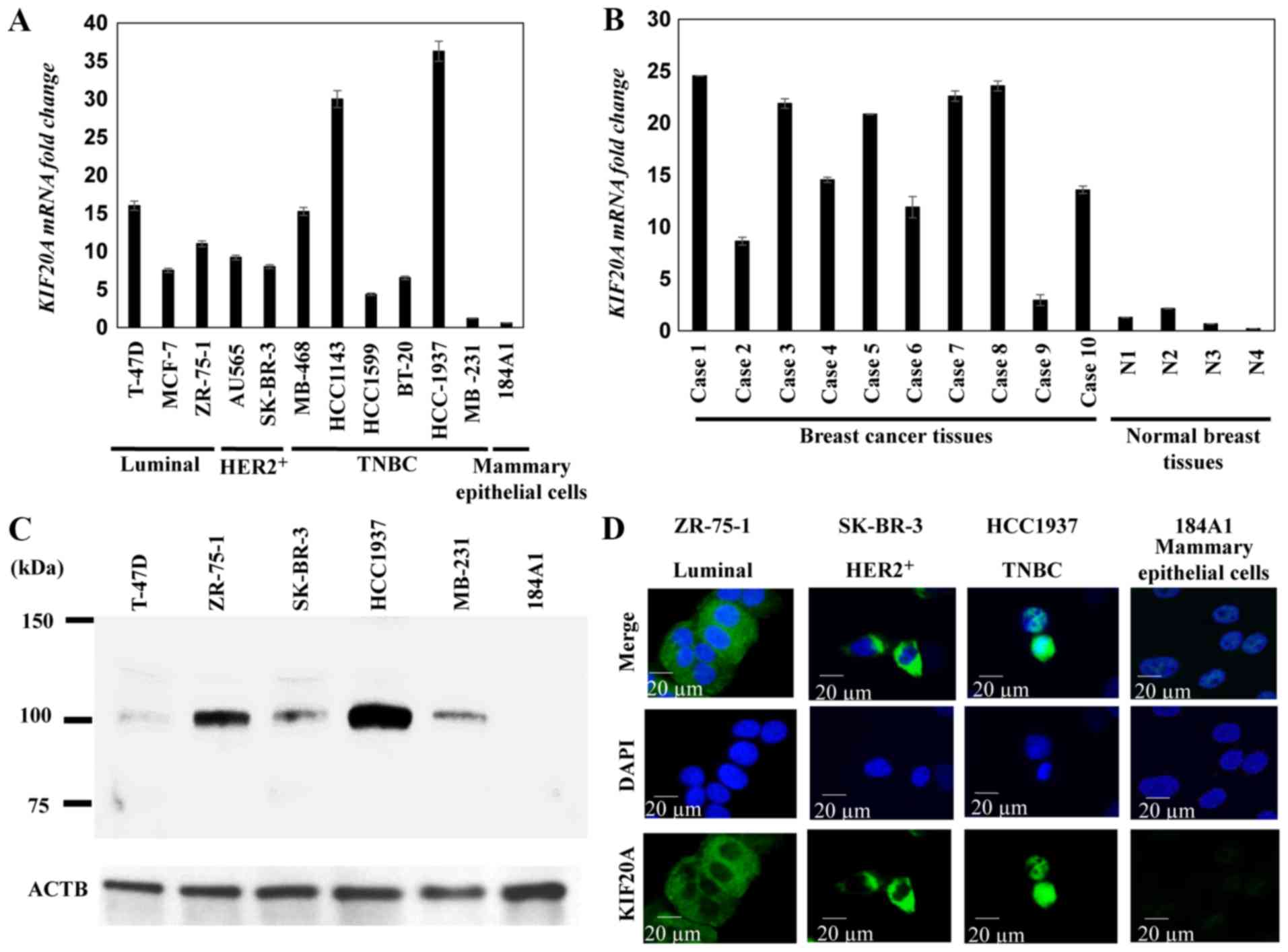
RT-qPCR revealed higher KIF20A mRNA
expression levels in the majority of the 11 breast cancer cell
lines compared with the 184A1 breast epithelial cells (Fig. 1A). Furthermore, KIF20A was
expressed in the majority of the clinical breast cancer tissues,
whereas it was barely detectable in the adjacent breast tissues
(Fig. 1B). Western blot analysis
revealed that KIF20A protein was expressed in the majority of the
breast cancer cells, whereas it was barely detectable in normal
breast cells (Fig. 1C).
Immunocytochemical analysis revealed that KIF20A protein was
detected in the cytoplasm and/or nucleus of ZR-75-1 (Luminal A),
SK-BR-3 (HER2/neu-positive) and HCC1937 (TNBC with BRCA1
mutation) cell lines (Fig. 1D).
KIF20A was localized in the cytoplasm and the nucleus of breast
cancer cells.
KIF20A expression is associated with the
poor prognosis of patients with breast cancer
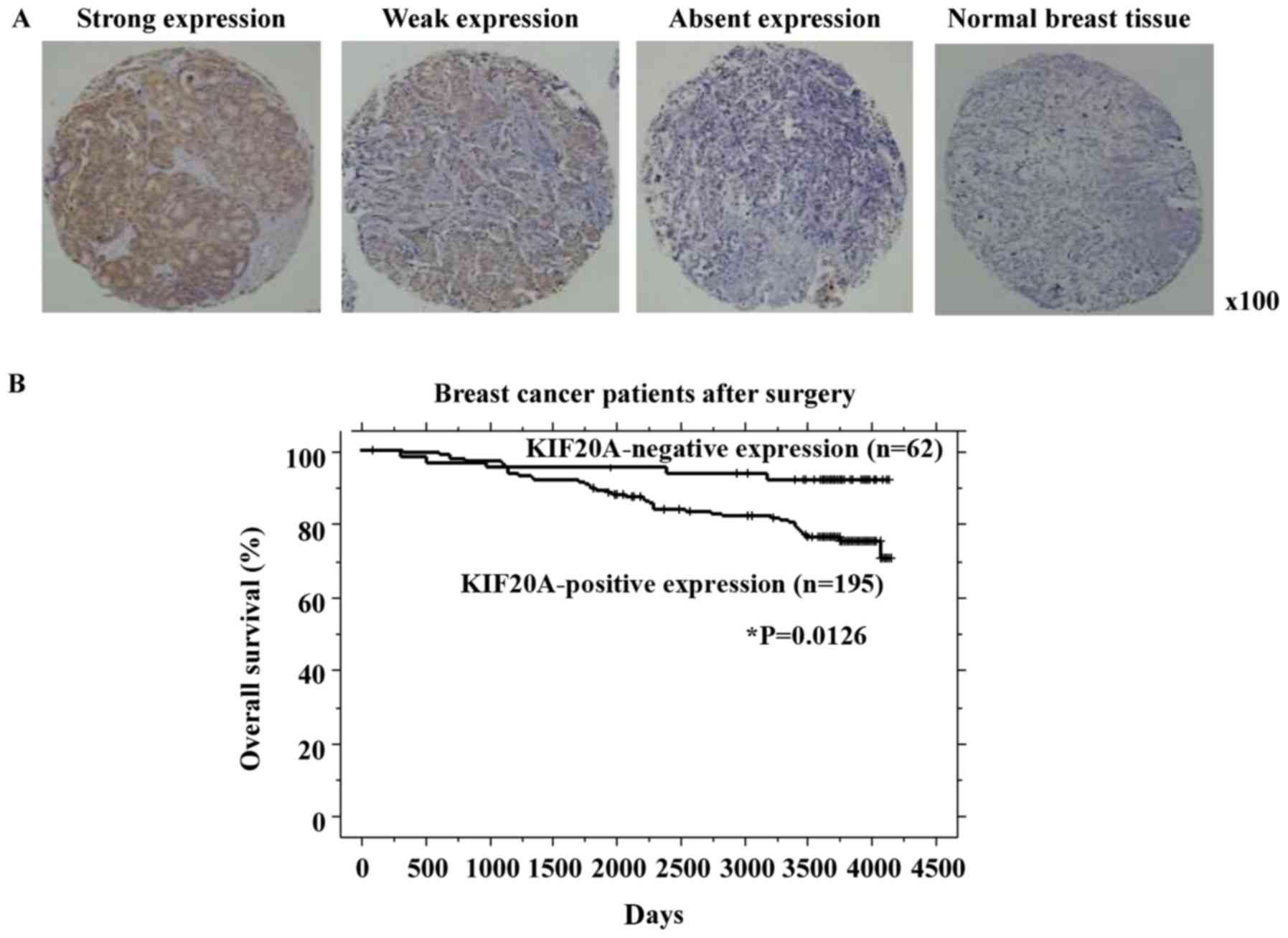
Immunohistochemistry revealed that KIF20A was
expressed in 195 of the 257 (75.9%) breast cancer cases
investigated. A strong expression was present in 106 cases (41.3%),
a weak expression was present in 89 cases (34.6%), and the
expression was absent in 62 cases (24.1%) (Fig. 2A and Table III). On classification based on
the breast cancer subtype, KIF20A was expressed in 130 of 186
(69.9%) hormone receptor-positive breast cancers, in 40 of 45
(88.9%) HER2-positive breast cancers, and in 36 of 39 (92%) TNBCs.
KIF20A was more frequently expressed in HER2-positive breast cancer
and TNBC compared to the other types.
Furthermore, the associations between KIF20A protein
expression and the patient clinicopathological parameters were
assessed. The ER status factor (higher expression in ER-negative;
P=0.0002, Fisher's exact test), PgR status factor (higher
expression in PgR-negative; P=0.0245, Fisher's exact test), HER2
status factor (higher expression in HER2-positive; P=0.0333,
Fisher's exact test) and pT factor (higher expression in T2‑3;
P=0.0035, Fisher's exact test) were significantly associated with
KIF20A expression (Table III).
Kaplan-Meier analysis revealed that a positive KIF20A expression
was significantly associated with a shorter OS compared with no
KIF20A expression (P=0.0126, log-rank test, Fig. 2B). Univariate analysis was also
performed to investigate the association of patient prognosis with
factors, including KIF20A expression status (positive vs.
negative), age (≥65 vs. <65 years), ER status (negative vs.
positive), PgR status (negative vs. positive), HER2 status
(positive vs. negative), pT classification (T2‑3 vs. T1) and pN
classification (N1‑2 vs. N0). Univariate analysis revealed that a
positive KIF20A expression (P=0.0147), an advanced pT stage
(P=0.0223) and an advanced pN stage (P<0.0001) were
significantly associated with a worse prognosis. Furthermore,
multivariate analysis revealed that a positive KIF20A expression
and an advanced pN stage were independent prognostic factors
(P=0.0357 and P=0.0001, respectively, Table IV).
| Table IVCox proportional hazards model
analysis of prognostic factors in patients with breast cancer. |
Table IV
Cox proportional hazards model
analysis of prognostic factors in patients with breast cancer.
| Variables | Hazards ratio | 95% CI |
Unfavorable/favorable | P-value |
|---|
| Univariate
analysis | | | | |
| KIF20A
expression | 3.152 | 1.253-7.929 |
Positive/absent | 0.0147a |
| Age (years) | 1.045 | 0.558‑1.958 | ≥65/<65 | 0.8908 |
| ER status | 1.687 | 0.959-2.967 |
Negative/positive | 0.0695 |
| PgR status | 1.65 | 0.957-2.845 |
Negative/positive | 0.0713 |
| HER2/neu
status | 1.142 | 0.573-2.278 |
Positive/negative | 0.7051 |
| T-factor | 2.173 | 1.117-4.230 | T2-3/T1 | 0.0223a |
| N-factor | 3.744 | 2.054-6.823 | N1-2/N0 | <0.0001a |
| Multivariate
analysis | | | | |
| KIF20A
expression | 2.698 | 1.068-6.813 |
Positive/absent | 0.0357a |
| T-factor | 1.46 | 0.739-2.886 | T2-3/T1 | 0.2762 |
| N-factor | 3.311 | 1.795-6.108 | N1-2/N0 | 0.0001a |
To validate the potential of KIF20A as a prognostic
biomarker, the prognostic value of KIF20A gene expression
was also investigated using the PrognoScan database. KIF20A
expression was significantly associated with the poor prognosis of
breast cancer patients (dataset no. GSE1456-GPL96; P=0.001501). The
data independently support the immunohistochemical data of the
present study.
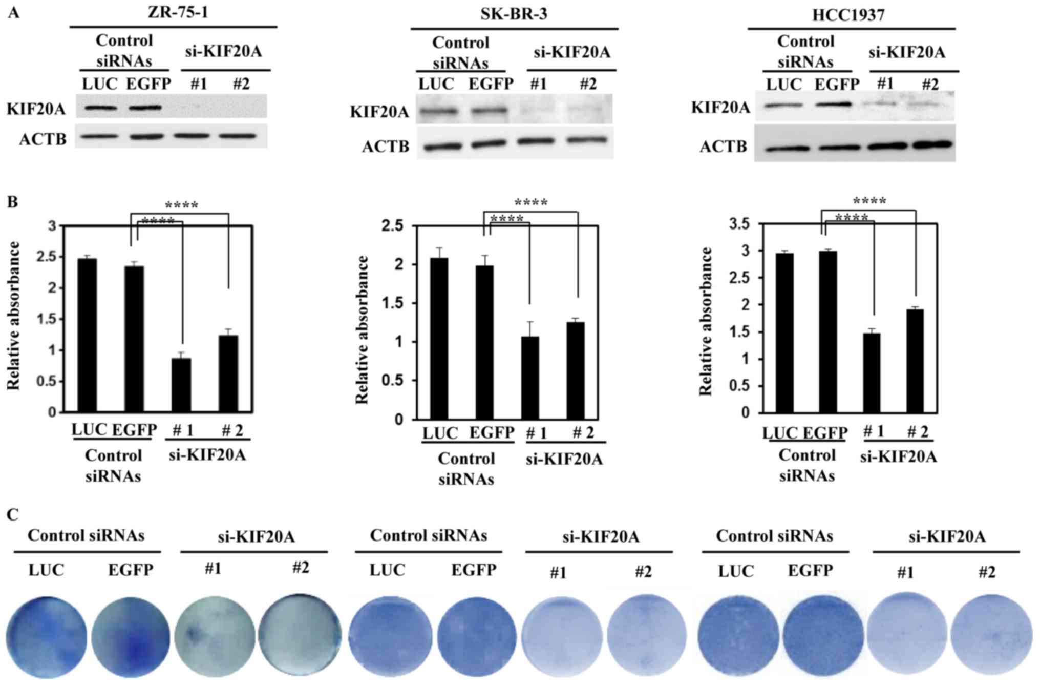
si‑KIF20A inhibits breast cancer cell
growth
To elucidate whether KIF20A upregulation plays a
significant role in breast cancer cell growth, ZR-75-1 (Luminal A),
SK-BR-3 (HER2/neu-positive) and HCC1937 (TNBC with BRCA1
mutation) cell lines were transfected with si-KIF20As to suppress
KIF20A expression. Western blot analysis revealed that si-KIF20A
decreased the KIF20A protein levels in the cancer cells compared
with the control siRNA (Fig. 3A).
In addition, si‑KIF20A significantly inhibited breast cancer cell
viability (Fig. 3B). Moreover,
colony formation assays revealed that si-KIF20A decreased the
number of breast cancer cells (Fig.
3C).
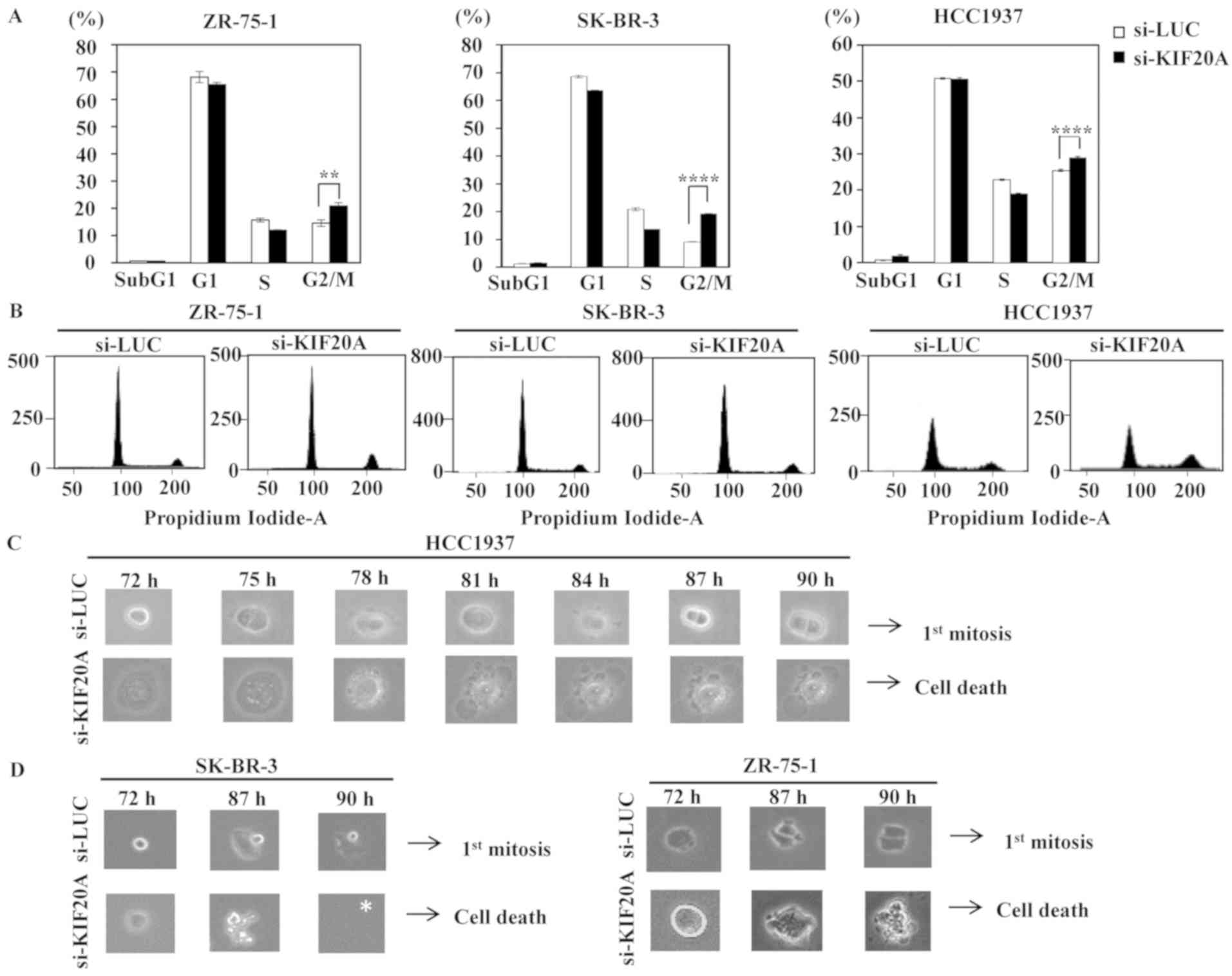
si‑KIF20A inhibits the cell cycle
progression of breast cancer cells
To further investigate the mechanisms of tumor
growth regulated by KIF20A, flow cytometric analysis of the cell
cycle was performed following siRNA transfection. Flow cytometric
analysis revealed that compared with the control siRNA, si‑KIF20A
significantly increased the population of cells in the G2/M phase
at 72 h post-transfection (ZR-75-1 cells, P=0.0019; SK-BR-3 cells,
P<0.0001; and HCC1937 cells, P<0.0001; population of cells at
each cell cycle is shown as a percentage and in flow cytometric
images in Fig. 4A and B).
Morphological changes were monitored using live-cell imaging of the
ZR-75-1, SK-BR-3 and HCC1937 cells transfected with si-KIF20A
(Fig. 4C and D). Time-lapse
imaging detected regular cell division in cells transfected with
control siRNA, whereas very few cell divisions, as well as
subsequent death were observed in all cells transfected with
si-KIF20A.
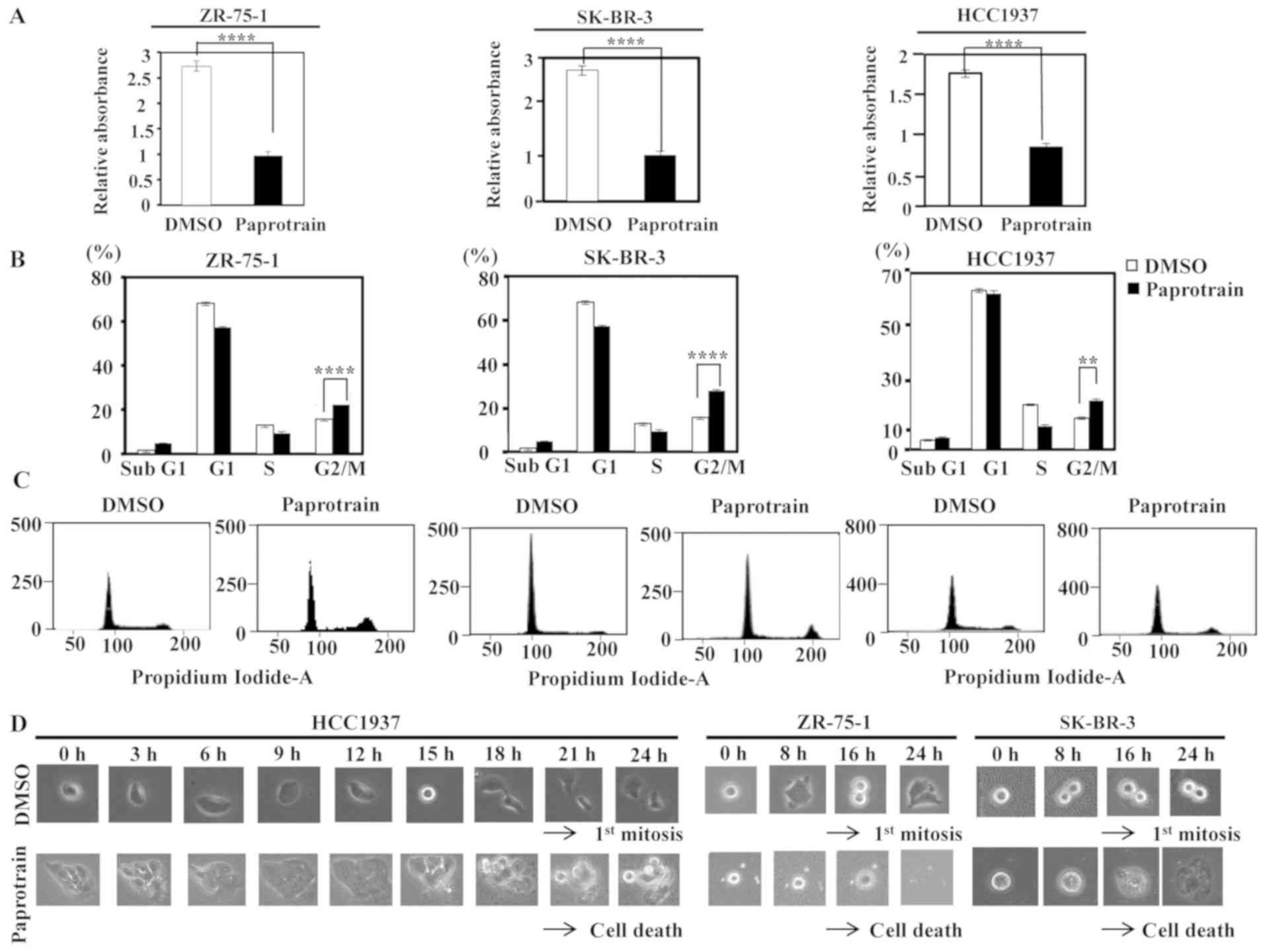
Paprotrain inhibits breast cancer cell
growth by blocking the kinesin motor ATPase activity of KIF20A
The breast cancer cells, ZR-75-1 (Luminal A),
SK-BR-3 (HER2/neu-positive) and HCC1937 (TNBC with BRCA1
mutation) were incubated in medium with or without paprotrain to
assess two lines of research: The first was to observe the
functional role of the kinesin motor ATPase activity of KIF20A via
pharmacological inhibition and the second was to assess the
potential of selective KIF20A inhibitor for clinical use. MTT assay
revealed that incubation with paprotrain for 24 h significantly
decreased breast cancer cell viability (Fig. 5A). Furthermore, flow cytometric
analysis performed 24 h following paprotrain treatment revealed
that the number of cells in the G2/M phase was significantly higher
than the number of cells without paprotrain treatment (population
of cells in each cell cycle is shown as a percentage and in flow
cytometric images in Fig. 5B and
C). Live-cell imaging revealed that the cancer cells without
paprotrain treatment divided regularly, whereas paprotrain
treatment resulted in cell cycle arrest in the G2/M phase and
subsequent cell death (Fig.
5D).
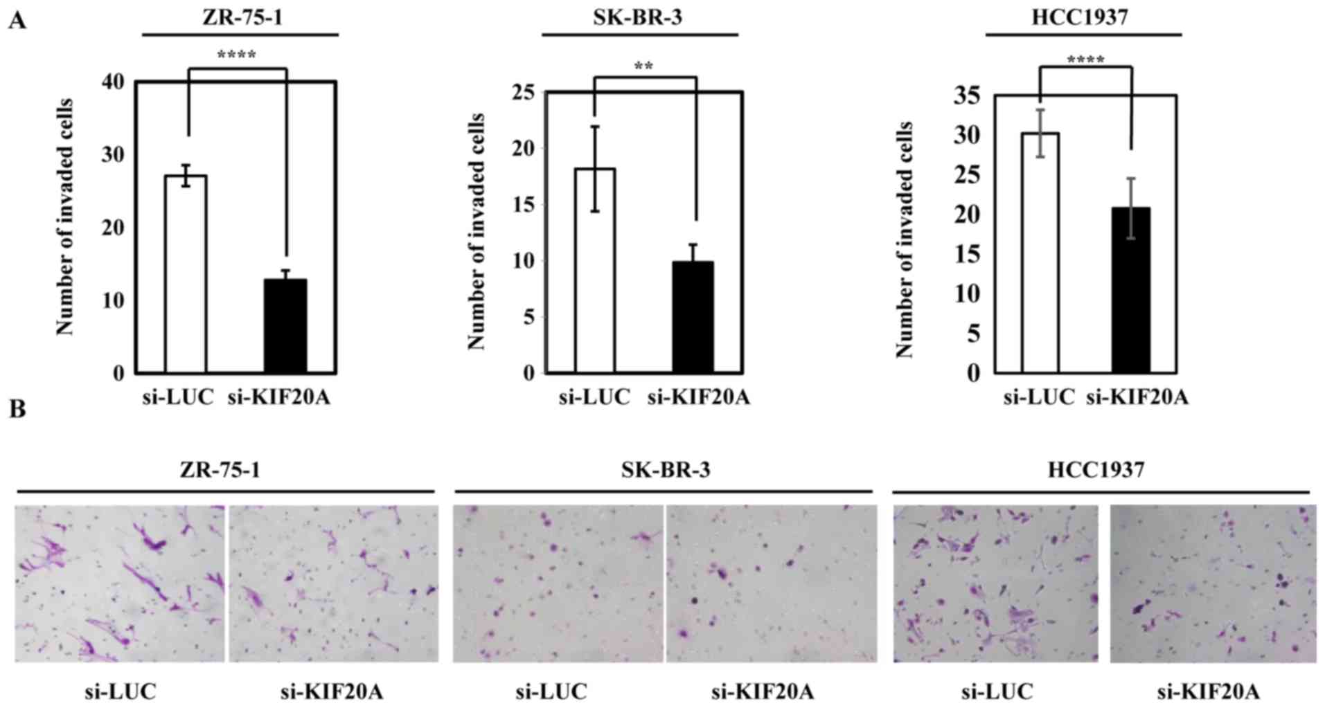
si‑KIF20A inhibits invasive phenotype of
breast cancer cells
The possible role of KIF20A in cellular invasion was
examined by Matrigel assays using the ZR-75-1, SK-BR-3 and HCC1937
cells. Transfection with si-KIF20A or control siRNA into either of
the cells significantly inhibited their invasive activity (Fig. 6). The results also indicated that
KIF20A contributed to the more malignant phenotype of breast cancer
cells.
In silico analysis of KIF20A‑related
pathways
Since KIF20A expression in breast cancer appears to
play a role in mitosis, as reported in other types of cells
(33,34), the co-expression and
co-localization of KIF20A protein with Aurora kinase B and PLK1 was
initially confirmed in mitotic breast cancer cells by western blot
analysis, the ONCOMINE database and immunocytochemistry (data not
shown). To further screen novel KIF20A-related pathways, the GSEA
database was used and it was found that KIF20A expression was
likely to be related to various pathways, including genes for cell
cycle and cell invasion, as well as Aurora B and PLK1 signaling
(data not shown). The information functionally supports the data of
the cell cycle analysis in the present study and live-cell imaging,
as well as cell invasion assay using siRNAs for KIF20A.
Discussion
The current understanding of the molecular
mechanisms and basic signaling pathways in breast cancer
pathogenesis has led to the development of certain novel molecular
targeted therapies, such as therapeutic antibodies and small
compounds. Molecular targeted agents are expected to exhibit good
clinical effectiveness against cancer cells due to their specific
anticancer mechanisms of action. However, no agent can completely
regulate the disease for prolonged periods of time due to the
genetic and biological heterogeneity of breast cancer cells, as
well as their drug resistance. Therefore, the development of novel,
cost-effective therapeutic agents and intensive therapies, as well
as the identification of precision medicine biomarkers are required
to overcome highly malignant breast tumors. Effective potential
molecular targets for cancer should exhibit a restricted expression
in normal adult tissues. This reduces the cytotoxic off-target
effects of the therapy and is indicative of specific cancer
biomarkers (40–42). In the present study, KIF20A was
expressed in the majority of breast cancer cells and tissues,
whereas it was seldom detected in normal breast epithelial tissue.
With the exception of the testes, KIF20A is expressed at
minimal levels in normal tissues and organs (BioGPS database;
http://biogps.org/#goto=welcome),
suggesting that it is an ideal diagnostic and therapeutic target.
To investigate the mechanisms of KIF20A activation in breast
cancer, comparative genome hybridization and the genome sequencing
database for KIF20A (https://cancer.sanger.ac.uk/cosmic) were used.
Missense mutations in KIF20A were detected in 0.29% of
breast cancer tissues (4/1,397 cases); however, no KIF20A
gene amplification or translocation were reported. According to
cBioportal for Cancer Genomics (http://www.cbioportal.org/), among 10,967 cases of
breast cancer, missense mutations, deletions, and genetic
amplification of KIF20A were detected in only 106 cases
(0.9%). It has been demonstrated that KIF20A expression is
regulated by glioma-associated oncogene 2 via the FOXM1-MMB complex
in hepatocellular cells and pancreatic cancer (43). Another study reported that
KIF20A transcriptional activity was regulated by E2F/DP
binding to the promoter region of KIF20A (44). Therefore, KIF20A overexpression may
be involved in several epigenetic mechanisms.
In the present study, the original tissue microarray
analysis revealed that KIF20A overexpression was significantly
associated with the poor prognosis of patients with breast cancer.
In addition, PrognoScan revealed a significant association between
a high KIF20A expression and a reduced OS of patients with
breast cancer (dataset no. GSE1456-GPL96; P=0.001501), thereby
independently supporting the current data that KIF20A may be a
prognostic biomarker for these patients.
The present study demonstrated the potential
clinical application of KIF20A inhibition for breast cancer
treatment by selectively inhibiting the ATPase activity of KIF20A
and suppressing KIF20A expression using siRNAs. Breast cancer cells
were treated with the KIF20A inhibitor, paprotrain, which
reversibly blocks KIF20A function by being uncompetitive with ATP
and noncompetitive with microtubules (45-47).
Similar to si-KIF20A, paprotrain decreased the viability of the
ZR-75-1 (Luminal A), SK-BR-3 (HER2/neu-positive) and HCC1937 (TNBC
with BRCA1 mutation) breast cancer cell lines via G2/M
arrest and subsequent mitotic cell death as monitored by live-cell
imaging. Targeting KIF20A with more selective and potent small
molecule inhibitors may be an effective therapeutic strategy for a
wide variety of breast cancers. The newly developed, potent KIF20A
inhibitor, BKS0349, was recently reported to suppress KIF20A ATPase
activity at levels 2-10-fold greater than paprotrain in various
cancer cell lines and in a xenograft mouse model without noticeable
variation (48,49). Further preclinical studies
investigating KIF20A inhibitors are crucial for the development of
novel molecular targeted drugs that can be used to treat highly
malignant breast cancers, such as TNBC. As the information obtained
by gene expression and inhibition assays using small number of
breast cancer cell lines is limited, further detailed cell line
assays considering KIF20A expression levels and cell phenotypes
that can be categorized by breast cancer subtypes, as well as
genome-wide genetic aberrations detected using whole genome
sequencing are eagerly warranted.
To examine a mechanistic insight of KIF20A
expression in breast cancer, the co-expression and co-localization
of KIF20A protein with Aurora kinase B and PLK1 in mitotic breast
cancer cells was initially confirmed by western blot analysis, the
ONCOMINE database and immunocytochemistry; this was consistent with
the findings described in other type of cells (33,34,44,45).
To further identify novel KIF20A-related pathways, the GSEA
database was screened and it was found that KIF20A expression was
likely to be related to various pathways, including cell cycle and
cell invasion, as well as Aurora B and PLK1 signaling pathways.
In conclusion, these findings suggest that KIF20A is
a common oncoprotein in breast cancer. KIF20A plays an essential
role in breast cancer proliferation and invasion, and is a
candidate prognostic marker. Therefore, targeting KIF20A may be
useful for the development of novel treatments, such as
immunotherapies and molecular targeted therapies, which can exert
potent biological effects on cancer with minimal adverse
effects.
Funding
Not applicable.
Availability of data and materials
All data generated or analyzed during this study are
included in this published article.
Authors' contributions
MN, AT and YD were involved in the conception and
design of the study. MN, AT, PMT and YD were involved in the
development of the study methodology. TYo, TYa, YM and YD were
involved in the acquisition of data (such as acquiring and managing
patients and providing facilities). MN, AT, PMT, BT, MZ and YD were
involved in the analysis and interpretation of data (e.g.,
statistical analysis, biostatistics and computational analysis).
MN, AT and YD were involved in the writing, reviewing and/or
revision of the manuscript. MN, AT, PMT and YD were involved in
administrative, technical or material support (i.e., reporting or
organizing data, constructing databases). YD supervised the study.
All authors read and approved the final manuscript.
Ethics approval and consent to
participate
The present study and the use of all clinical
materials were approved by individual institutional ethics
committees. The project to establish tumor tissue microarrays from
archival formalin-fixed and paraffin-embedded surgically resected
tissues and to use the tissue microarrays for later unspecified
research works was approved by the Kanagawa Cancer Center Ethics
Committee with the approval no. Rin-177, 27 (September, 2010).
Written comprehensive informed consent was obtained from the
patients for the use of their clinical information and for
specimens remaining after clinically required examinations, such as
archival formalin‑fixed and paraffin‑embedded specimens following
diagnosis.
Patient consent for publication
Not applicable.
Competing interests
The authors declare that they have no competing
interests.
Acknowledgments
Not applicable.
Abbreviations:
|
ATP
|
adenosine triphosphate
|
|
FBS
|
fetal bovine serum
|
|
DAB
|
3,3'-diaminobenzidine
|
|
MTT
|
3-(4,5-dimethylthiazol-2-yl)-2,5-diphenyltetrazolium bromide
|
|
OS
|
overall survival
|
|
RNA
|
ribonucleic acid
|
|
TNBC
|
triple-negative breast cancer
|
References
|
1
|
Siegel RL, Miller KD and Jemal A: Cancer
statistics, 2019. CA Cancer J Clin. 69:7–34. 2019. View Article : Google Scholar : PubMed/NCBI
|
|
2
|
Dai X, Xiang L, Li T and Bai Z: Cancer
hallmarks, biomarkers and breast cancer molecular subtypes. J
Cancer. 7:1281–1294. 2016. View Article : Google Scholar : PubMed/NCBI
|
|
3
|
Loi S, Haibe-Kains B, Desmedt C, Lallemand
F, Tutt AM, Gillet C, Ellis P, Harris A, Bergh J, Foekens JA, et
al: Definition of clinically distinct molecular subtypes in
estrogen receptor-positive breast carcinomas through genomic grade.
J Clin Oncol. 25:1239–1246. 2007. View Article : Google Scholar : PubMed/NCBI
|
|
4
|
Schroeder RL, Stevens CL and Sridhar J:
Small molecule tyrosine kinase inhibitors of ErbB2/HER2/Neu in the
treatment of aggressive breast cancer. Molecules. 19:15196–15212.
2014. View Article : Google Scholar : PubMed/NCBI
|
|
5
|
Hudis CA and Gianni L: Triple-negative
breast cancer: An unmet medical need. Oncologist. 16(Suppl 1):
1–11. 2011. View Article : Google Scholar : PubMed/NCBI
|
|
6
|
Robson M, Im SA, Senkus E, Xu B, Domchek
SM, Masuda N, Delaloge S, Li W, Tung N, Armstrong A, et al:
Olaparib for metastatic breast cancer in patients with a germline
BRCA mutation. N Engl J Med. 377:523–533. 2017. View Article : Google Scholar : PubMed/NCBI
|
|
7
|
Adams S, Schmid P, Rugo HS, Winer EP,
Loirat D, Awada A, Cescon DW, Iwata H, Campone M, Nanda R, et al:
Pembrolizumab monotherapy for previously treated metastatic
triple-negative breast cancer: Cohort A of the phase II KEYNOTE-086
study. Ann Oncol. 30:397–404. 2019. View Article : Google Scholar
|
|
8
|
Voorwerk L, Slagter M, Horlings HM,
Sikorska K, van de Vijver KK, de Maaker M, Nederlof I, Kluin RJC,
Warren S, Ong S, et al: Immune induction strategies in metastatic
triple-negative breast cancer to enhance the sensitivity to PD-1
blockade: The TONIC trial. Nat Med. 25:920–928. 2019. View Article : Google Scholar : PubMed/NCBI
|
|
9
|
Ovcaricek T, Takac I and Matos E:
Multigene expression signatures in early hormone receptor positive
HER 2 negative breast cancer. Radiol Oncol. 53:285–292. 2019.
View Article : Google Scholar : PubMed/NCBI
|
|
10
|
Yamauchi H, Nakagawa C, Yamashige S, Takei
H, Yagata H, Yoshida A, Hayashi N, Hornberger J, Yu T, Chao C, et
al: Societal cost-effectiveness analysis of the 21-gene assay in
estrogen-receptor-positive, lymph-node-negative early-stage breast
cancer in Japan. BMC Health Serv Res. 14:3722014. View Article : Google Scholar : PubMed/NCBI
|
|
11
|
Daigo Y and Nakamura Y: From cancer
genomics to thoracic oncology: Discovery of new biomarkers and
therapeutic targets for lung and esophageal carcinoma. Gen Thorac
Cardiovasc Surg. 56:43–53. 2008. View Article : Google Scholar : PubMed/NCBI
|
|
12
|
Daigo Y, Takano A, Teramoto K, Chung S and
Nakamura Y: A systematic approach to the development of novel
therapeutics for lung cancer using genomic analyses. Clin Pharmacol
Ther. 94:218–223. 2013. View Article : Google Scholar : PubMed/NCBI
|
|
13
|
Ishikawa N, Daigo Y, Takano A, Taniwaki M,
Kato T, Hayama S, Murakami H, Takeshima Y, Inai K, Nishimura H, et
al: Increases of amphiregulin and transforming growth factor-alpha
in serum as predictors of poor response to gefitinib among patients
with advanced non-small cell lung cancers. Cancer Res.
65:9176–9184. 2005. View Article : Google Scholar : PubMed/NCBI
|
|
14
|
Ishikawa N, Daigo Y, Yasui W, Inai K,
Nishimura H, Tsuchiya E, Kohno N and Nakamura Y: ADAM8 as a novel
serological and histochemical marker for lung cancer. Clin Cancer
Res. 10:8363–8370. 2004. View Article : Google Scholar : PubMed/NCBI
|
|
15
|
Kakiuchi S, Daigo Y, Ishikawa N, Furukawa
C, Tsunoda T, Yano S, Nakagawa K, Tsuruo T, Kohno N, Fukuoka M, et
al: Prediction of sensitivity of advanced non-small cell lung
cancers to gefitinib (Iressa, ZD1839). Hum Mol Genet. 13:pp.
3029–3043. 2004, View Article : Google Scholar : PubMed/NCBI
|
|
16
|
Kato T, Daigo Y, Hayama S, Ishikawa N,
Yamabuki T, Ito T, Miyamoto M, Kondo S and Nakamura Y: A novel
human tRNA-dihydrouridine synthase involved in pulmonary
carcinogenesis. Cancer Res. 65:5638–5646. 2005. View Article : Google Scholar : PubMed/NCBI
|
|
17
|
Kikuchi T, Daigo Y, Katagiri T, Tsunoda T,
Okada K, Kakiuchi S, Zembutsu H, Furukawa Y, Kawamura M, Kobayashi
K, et al: Expression profiles of non-small cell lung cancers on
cDNA microarrays: Identification of genes for prediction of
lymph‑node metastasis and sensitivity to anti-cancer drugs.
Oncogene. 22:2192–2205. 2003. View Article : Google Scholar : PubMed/NCBI
|
|
18
|
Suzuki C, Daigo Y, Ishikawa N, Kato T,
Hayama S, Ito T, Tsuchiya E and Nakamura Y: ANLN plays a critical
role in human lung carcinogenesis through the activation of RHOA
and by involvement in the phosphoinositide 3-kinase/AKT pathway.
Cancer Res. 65:11314–11325. 2005. View Article : Google Scholar : PubMed/NCBI
|
|
19
|
Kakiuchi S, Daigo Y, Tsunoda T, Yano S,
Sone S and Nakamura Y: Genome-wide analysis of organ-preferential
metastasis of human small cell lung cancer in mice. Mol Cancer Res.
1:485–499. 2003.PubMed/NCBI
|
|
20
|
Taniwaki M, Daigo Y, Ishikawa N, Takano A,
Tsunoda T, Yasui W, Inai K, Kohno N and Nakamura Y: Gene expression
profiles of small‑cell lung cancers: Molecular signatures of lung
cancer. Int J Oncol. 29:567–575. 2006.PubMed/NCBI
|
|
21
|
Oshita H, Nishino R, Takano A, Fujitomo T,
Aragaki M, Kato T, Akiyama H, Tsuchiya E, Kohno N, Nakamura Y, et
al: RASEF is a novel diagnostic biomarker and a therapeutic target
for lung cancer. Mol Cancer Res. 11:937–951. 2013. View Article : Google Scholar : PubMed/NCBI
|
|
22
|
Hayama S, Daigo Y, Yamabuki T, Hirata D,
Kato T, Miyamoto M, Ito T, Tsuchiya E, Kondo S and Nakamura Y:
Phosphorylation and activation of cell division cycle associated 8
by aurora kinase B plays a significant role in human lung
carcinogenesis. Cancer Res. 67:4113–4122. 2007. View Article : Google Scholar : PubMed/NCBI
|
|
23
|
Ishikawa N, Daigo Y, Takano A, Taniwaki M,
Kato T, Tanaka S, Yasui W, Takeshima Y, Inai K, Nishimura H, et al:
Characterization of SEZ6L2 cell-surface protein as a novel
prognostic marker for lung cancer. Cancer Sci. 97:737–745. 2006.
View Article : Google Scholar : PubMed/NCBI
|
|
24
|
Kato T, Sato N, Hayama S, Yamabuki T, Ito
T, Miyamoto M, Kondo S, Nakamura Y and Daigo Y: Activation of
Holliday junction recognizing protein involved in the chromosomal
stability and immortality of cancer cells. Cancer Res.
67:8544–8553. 2007. View Article : Google Scholar : PubMed/NCBI
|
|
25
|
Suzuki C, Takahashi K, Hayama S, Ishikawa
N, Kato T, Ito T, Tsuchiya E, Nakamura Y and Daigo Y:
Identification of Myc-associated protein with JmjC domain as a
novel therapeutic target oncogene for lung cancer. Mol Cancer Ther.
6:542–551. 2007. View Article : Google Scholar : PubMed/NCBI
|
|
26
|
Takahashi K, Furukawa C, Takano A,
Ishikawa N, Kato T, Hayama S, Suzuki C, Yasui W, Inai K, Sone S, et
al: The neuromedin U-growth hormone secretagogue receptor
1b/neurotensin receptor 1 oncogenic signaling pathway as a
therapeutic target for lung cancer. Cancer Res. 66:9408–9419. 2006.
View Article : Google Scholar : PubMed/NCBI
|
|
27
|
Taniwaki M, Takano A, Ishikawa N, Yasui W,
Inai K, Nishimura H, Tsuchiya E, Kohno N, Nakamura Y and Daigo Y:
Activation of KIF4A as a prognostic biomarker and therapeutic
target for lung cancer. Clin Cancer Res. 13:6624–6631. 2007.
View Article : Google Scholar : PubMed/NCBI
|
|
28
|
Yamabuki T, Takano A, Hayama S, Ishikawa
N, Kato T, Miyamoto M, Ito T, Ito H, Miyagi Y, Nakayama H, et al:
Dikkopf-1 as a novel serologic and prognostic biomarker for lung
and esophageal carcinomas. Cancer Res. 67:2517–2525. 2007.
View Article : Google Scholar : PubMed/NCBI
|
|
29
|
Fujitomo T, Daigo Y, Matsuda K, Ueda K and
Nakamura Y: Identification of a nuclear protein, LRRC42, involved
in lung carcinogenesis. Int J Oncol. 45:147–156. 2014. View Article : Google Scholar : PubMed/NCBI
|
|
30
|
Nguyen MH, Koinuma J, Ueda K, Ito T,
Tsuchiya E, Nakamura Y and Daigo Y: Phosphorylation and activation
of cell division cycle associated 5 by mitogen-activated protein
kinase play a crucial role in human lung carcinogenesis. Cancer
Res. 70:5337–5347. 2010. View Article : Google Scholar : PubMed/NCBI
|
|
31
|
Hayama S, Daigo Y, Kato T, Ishikawa N,
Yamabuki T, Miyamoto M, Ito T, Tsuchiya E, Kondo S and Nakamura Y:
Activation of CDCA1-KNTC2, members of centromere protein complex,
involved in pulmonary carcinogenesis. Cancer Res. 66:10339–10348.
2006. View Article : Google Scholar : PubMed/NCBI
|
|
32
|
Hirokawa N, Noda Y and Okada Y: Kinesin
and dynein super-family proteins in organelle transport and cell
division. Curr Opin Cell Biol. 10:60–73. 1998. View Article : Google Scholar : PubMed/NCBI
|
|
33
|
Hill E, Clarke M and Barr FA: The
Rab6-binding kinesin, Rab6-KIFL, is required for cytokinesis. EMBO
J. 19:5711–5719. 2000. View Article : Google Scholar : PubMed/NCBI
|
|
34
|
Taniuchi K, Nakagawa H, Nakamura T, Eguchi
H, Ohigashi H, Ishikawa O, Katagiri T and Nakamura Y:
Down-regulation of RAB6KIFL/KIF20A, a kinesin involved with
membrane trafficking of discs large homologue 5, can attenuate
growth of pancreatic cancer cell. Cancer Res. 65:105–112.
2005.PubMed/NCBI
|
|
35
|
Khongkow P, Gomes AR, Gong C, Man EP,
Tsang JW, Zhao F, Monteiro LJ, Coombes RC, Medema RH, Khoo US, et
al: Paclitaxel targets FOXM1 to regulate KIF20A in mitotic
catastrophe and breast cancer paclitaxel resistance. Oncogene.
35:990–1002. 2016. View Article : Google Scholar
|
|
36
|
Saito K, Ohta S, Kawakami Y, Yoshida K and
Toda M: Functional analysis of KIF20A, a potential
immunotherapeutic target for glioma. J Neurooncol. 132:63–74. 2017.
View Article : Google Scholar : PubMed/NCBI
|
|
37
|
Zhang Z, Chai C, Shen T, Li X, Ji J, Li C,
Shang Z and Niu Y: Aberrant KIF20A Expression is associated with
adverse clinical outcome and promotes tumor progression in prostate
cancer. Dis Markers. 2019:47827302019. View Article : Google Scholar : PubMed/NCBI
|
|
38
|
Shen T, Yang L, Zhang Z, Yu J, Dai L, Gao
M, Shang Z and Niu Y: KIF20A affects the prognosis of bladder
cancer by promoting the proliferation and metastasis of bladder
cancer cells. Dis Markers. 2019:48631822019. View Article : Google Scholar : PubMed/NCBI
|
|
39
|
Takano A, Ishikawa N, Nishino R, Masuda K,
Yasui W, Inai K, Nishimura H, Ito H, Nakayama H, Miyagi Y, et al:
Identification of nectin-4 oncoprotein as a diagnostic and
therapeutic target for lung cancer. Cancer Res. 69:6694–6703. 2009.
View Article : Google Scholar : PubMed/NCBI
|
|
40
|
Kobayashi Y, Takano A, Miyagi Y, Tsuchiya
E, Sonoda H, Shimizu T, Okabe H, Tani T, Fujiyama Y and Daigo Y:
Cell division cycle-associated protein 1 overexpression is
essential for the malignant potential of colorectal cancers. Int J
Oncol. 44:69–77. 2014. View Article : Google Scholar
|
|
41
|
Thang PM, Takano A, Yoshitake Y, Shinohara
M, Murakami Y and Daigo Y: Cell division cycle associated 1 as a
novel prognostic biomarker and therapeutic target for oral cancer.
Int J Oncol. 49:1385–1393. 2016. View Article : Google Scholar : PubMed/NCBI
|
|
42
|
Daigo K, Takano A, Thang PM, Yoshitake Y,
Shinohara M, Tohnai I, Murakami Y, Maegawa J and Daigo Y:
Characterization of KIF11 as a novel prognostic biomarker and
therapeutic target for oral cancer. Int J Oncol. 52:155–165.
2018.
|
|
43
|
Shi C, Huang D, Lu N, Chen D, Zhang M, Yan
Y, Deng L, Lu Q, Lu H and Luo S: Aberrantly activated Gli2-KIF20A
axis is crucial for growth of hepatocellular carcinoma and predicts
poor prognosis. Oncotarget. 7:26206–26219. 2016. View Article : Google Scholar : PubMed/NCBI
|
|
44
|
Fontijn RD, Goud B, Echard A, Jollivet F,
van Marle J, Pannekoek H and Horrevoets AJ: The human kinesin-like
protein RB6K is under tight cell cycle control and is essential for
cytokinesis. Mol Cell Biol. 21:2944–2955. 2001. View Article : Google Scholar : PubMed/NCBI
|
|
45
|
Tcherniuk S, Skoufias DA, Labriere C, Rath
O, Gueritte F, Guillou C and Kozielski F: Relocation of Aurora B
and survivin from centromeres to the central spindle impaired by a
kinesin‑specific MKLP‑2 inhibitor. Angew Chem Int Ed Engl.
49:8228–8231. 2010. View Article : Google Scholar : PubMed/NCBI
|
|
46
|
Labrière C, Talapatra SK, Thoret S,
Bougeret C, Kozielski F and Guillou C: New MKLP-2 inhibitors in the
paprotrain series: Design, synthesis and biological evaluations.
Bioorg Med Chem. 24:721–734. 2016. View Article : Google Scholar : PubMed/NCBI
|
|
47
|
Sakai R, Morikawa Y, Kondo C, Oka H,
Miyajima H, Kubo K and Uehara T: Combinatorial measurement of
CDKN1A/p21 and KIF20A expression for discrimination of DNA
damage-induced clastogenicity. Int J Mol Sci. 15:17256–17269. 2014.
View Article : Google Scholar : PubMed/NCBI
|
|
48
|
Düzgün ŞA, Yerlikaya A, Zeren S, Bayhan Z,
Okur E and Boyacı İ: Differential effects of p38 MAP kinase
inhibitors SB203580 and SB202190 on growth and migration of human
MDA-MB-231 cancer cell line. Cytotechnology. 69:711–724. 2017.
View Article : Google Scholar : PubMed/NCBI
|
|
49
|
Ferrero H, Corachán A, Quiñonero A,
Bougeret C, Pouletty P, Pellicer A and Domínguez F: Inhibition of
KIF20A by BKS0349 reduces endometriotic lesions in a xenograft
mouse model. Mol Hum Reprod. 25:562–571. 2019. View Article : Google Scholar : PubMed/NCBI
|