Introduction
The incidence of gastric cancer has been on the
decline globally over the last few years, although it remains
rampant in several countries and is currently the fourth most
common type of cancer worldwide (1,2). In
China, in particular, more new cases of gastric cancer are
diagnosed annually compared to any other country. Gastric cancer is
difficult to diagnose in an early stage and the long-term prognosis
remains disappointing (3,4). Over the last few decades, there have
been no notable advances in conventional therapy and the median
survival of patients with advanced gastric cancer ranges between 9
and 11 months. Targeted therapy is a novel promising strategy for
the treatment of malignancies. In gastric cancer, trastuzumab was
shown to reduce the risk of death by 26% in the ToGA trial, when
combined with chemotherapy in patients exhibiting high human
epidermal growth factor receptor 2 (HER2) expression (5). However, the median overall survival
in that study was only 13.8 months and the results were
unsatisfactory. Moreover, a series of clinical trials indicated
that there is currently no molecular-targeted agent superior to
trastuzumab (6). Therefore,
further investigation is required to elucidate the molecular
mechanisms underlying the development of gastric cancer, identify
novel treatment targets or design therapeutic strategies.
Lemur tyrosine kinase (LMTK)3, which is a member of
the LMTK family, is a serine/threonine-protein kinase that may be
involved in the β-catenin pathway and leukemic cell survival
(7,8); however, the biological function of
the gene remains unclear. Giamas et al (9) recently demonstrated that LMTK3 is an
estrogen receptor-α (ERα) regulator, with a central role in
endocrine resistance. Moreover, the expression of LMTK3 was
associated with breast cancer phenotype and patient prognosis
(10).
It was reported that estrogens may play a role in
gastric carcinogenesis (11) and
tamoxifen may prevent gastric cancer in Helicobacter
pylori-infected INS-GAS mice (12). Furthermore, estrogen receptor (ER)
has been detected in gastric cancer tissues and proven to be
correlated with prognosis (13–15).
Therefore, ER and related genes may represent potential targets for
the treatment of gastric cancer (16). In view of the role of LMTK3 in
breast cancer, we herein investigated the expression of LMTK3 and
its correlation with the clinicopathological parameters and
prognosis of gastric cancer patients.
Materials and methods
Materials
The gastric cancer tissue microarray (TMA) was
purchased form Outdo Biotech Co., Shanghai, China. The tumor
samples were obtained from 83 patients with primary operable
gastric cancer who had undergone surgical resection between 2006
and 2007. All the patients in this series were Chinese, including
55 men and 28 women, with a mean age of 64.3 years (range, 37–84
years). All the cases were evaluable after array construction. All
the patients underwent potentially curative tumor resection and
none had received chemotherapy or radiotherapy prior to surgery.
The tumor histological types and grading were reviewed and
classified according to the WHO classification criteria; the
disease stage was determined according to the TNM staging
system.
Immunohistochemistry
Anti-LMTK3 mouse monoclonal antibody (Santa Cruz
Biotechnology, Inc., Heidelberg, Germany) was optimized to a
working concentration of 2 μg/ml on full-face excisional tissue
sections. Subsequently, gastric cancer TMA was conducted,
comprising 4-μm formalin-fixed paraffin-embedded tissue cores
immunostained with the optimized anti-LMTK3 monoclonal antibody on
the Leica BOND-MAX automated system (Leica Microsystems Inc.,
Buffalo Grove, IL, USA) according to the manufacturer’s
instructions. Heat-induced epitope retrieval was performed in
citrate buffer (ER1) for 5 min. Detection was achieved using the
Polymer Detection kit (Leica Microsystems Inc., Newcastle Upon
Tyne, UK). These detection systems contain peroxidase block,
protein block, post primary block, Novolink polymer,
3,3′-diaminobenzidine (DAB) chromogen, Novolink DAB substrate
buffer (polymer) and hematoxylin for subsequent counterstaining of
the TMAs. Negative controls were performed by omission of the
primary antibody.
LMTK3 immunoreactivity was detected in the nucleus
and cytoplasm of gastric cancer cells to a variable degree. The
stained specimens were then categorized into 6-degree classes
according to the quantitative score. Initially, 4 degrees of the
proportional score (PS) for the positively-stained cells were
assigned as follows, according to the frequency of positive tumor
cells: 0, none; 1, 1/100–1/4; 2, 1/4–1/2; and 3, >1/2.
Thereafter, 4 degrees of the intensity score (IS) were assigned as
follows, according to the intensity of the staining: 0, none; 1,
weak; 2, intermediate; and 3, strong. The PS and the IS were then
added to obtain a total score (TS), which ranged between 0 and 6.
According to the TS, the LMTK3 expression of the tumor was
classified as negative when the score was 0–3 and as positive when
the score was 4–6. All the cases were independently scored by two
of the investigators who were blinded to the clinicopathological or
outcome data. In case of discrepancies between the two
investigators, a consensus was reached via simultaneous examination
using a double-headed microscope.
Statistics
The statistical analysis for TMAs was performed
using SPSS version 16.0 statistical software (SPSS Inc., Chicago,
IL, USA). The statistical significance was evaluated using the
Chi-square test or the Fisher’s exact test. A multivariate COX
regression analysis was used to evaluate independent associations.
The survival curves were analyzed using the Kaplan-Meier method and
the significance was determined by the log-rank test. The odds
ratio and 95% confidence interval (95% CI) was calculated for each
variable. P<0.05 was considered to indicate statistically
significant differences.
Results
Detection of the LMTK3 expression in
gastric cancer
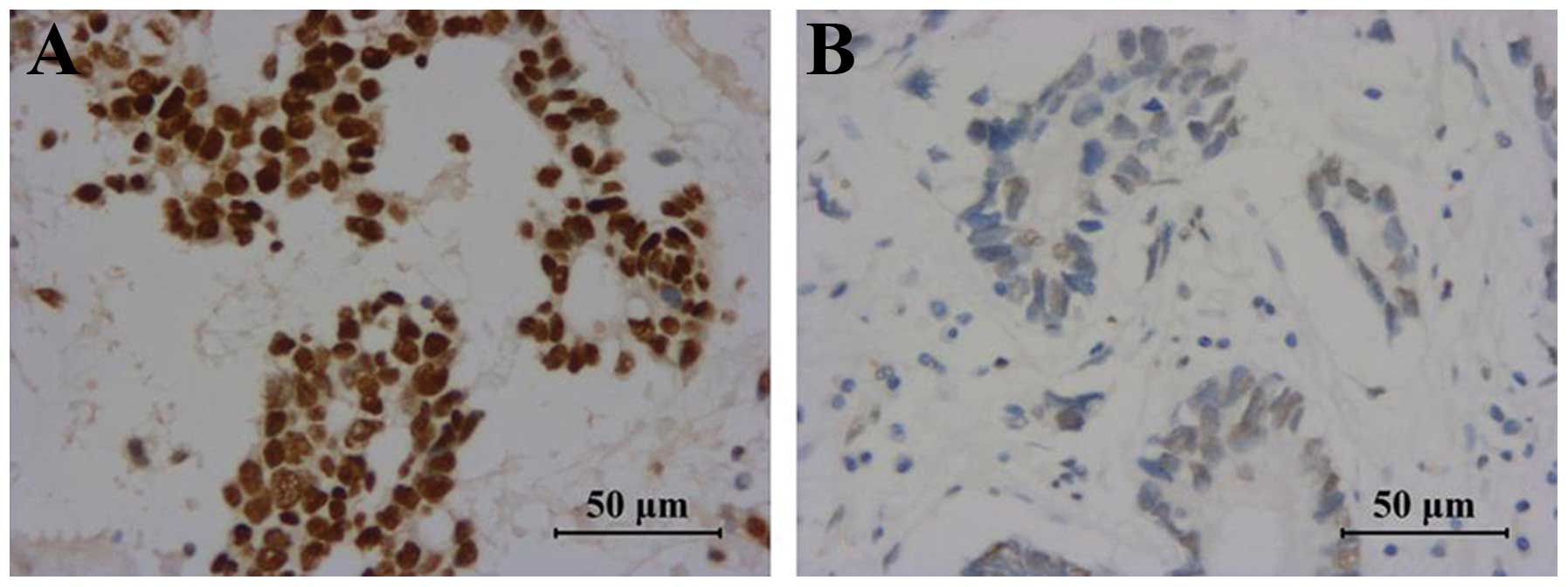
Immunostaining analysis revealed that LMTK3 was
detected predominantly in the nucleus of gastric cancer cells, with
variable cytoplasmic staining (Fig.
1). LMTK3 was also expressed in the adjacent non-cancerous
mucosa; however, the positive rate in gastric cancer tissue was
significantly higher compared to that in non-cancerous mucosa (79.5
vs. 45.8%, respectively; P=0.000; Table I).
 | Table IAssociation between LMTK3 expression
and clinicopathological characteristics in gastric cancer. |
Table I
Association between LMTK3 expression
and clinicopathological characteristics in gastric cancer.
| | LMTK3 | | |
|---|
| |
| | |
|---|
| Characteristics | Cases | Negative | Positive (%) | χ2 | P-value |
|---|
| Tissue | | | | 25.739 | 0.000 |
| Gastric cancer | 83 | 17 | 66 (79.5) | | |
| Non-cancerous
mucosa | 83 | 45 | 38 (45.8) | | |
| Age (years) | | | | 0.004 | 0.948 |
| ≤50 | 7 | 2 | 5 (6.0) | | |
| >50 | 76 | 15 | 61 (73.5) | | |
| Gender | | | | 0.996 | 0.318 |
| Male | 55 | 13 | 42 (50.6) | | |
| Female | 28 | 4 | 24 (28.9) | | |
| Stage | | | | 4.445 | 0.035 |
| I+II | | 11 | 20 (24.1) | | |
| III+IV | | 8 | 44 (53.0) | | |
| Tumor size (cm) | | | | 0.284 | 0.594 |
| ≤5 | 49 | 11 | 38 (45.8) | | |
| >5 | 34 | 6 | 28 (33.7) | | |
| Depth of
invasion | | | | 9.272 | 0.002 |
| T1+T2 | 17 | 8 | 9 (10.8) | | |
| T3+T4 | 66 | 9 | 57 (68.7) | | |
| Lymph node
metastasis | | | | 0.328 | 0.567 |
| N0+N1 | | 8 | 26 (31.3) | | |
| N2+N3 | | 9 | 40 (48.2) | | |
| Distant
metastasis | | | | 1.381 | 0.240 |
| M0 | 74 | 17 | 57 (68.7) | | |
| M1 | 9 | 0 | 9 (10.8) | | |
| Lymphatic and
vascular invasion | | | | 0.987 | 0.321 |
| Negative | 69 | 16 | 53 (63.9) | | |
| Positive | 14 | 1 | 13 (15.7) | | |
| Location | | | | 0.125 | 0.940 |
| Upper | 8 | 2 | 6 (7.2) | | |
| Middle | 29 | 6 | 23 (27.7) | | |
| Lower | 46 | 9 | 37 (44.6) | | |
| Grade | | | | 1.99 | 0.158 |
| I+II | 37 | 5 | 32 (38.6) | | |
| III+IV | 46 | 12 | 34 (41.0) | | |
Correlation of LMTK3 expression with
clinicopathological parameters and postoperative survival in
gastric cancer
In the present study, we investigated the
correlation of LMTK3 expression with clinicopathological parameters
and postoperative survival. Our results suggested that LMTK3
expression was significantly correlated with the depth of invasion
(P=0.002) and disease stage (P=0.035), which were obviously
associated with the prognosis of gastric cancer patients. There was
no significant correlation with the other clinicopathological
parameters (Table I).
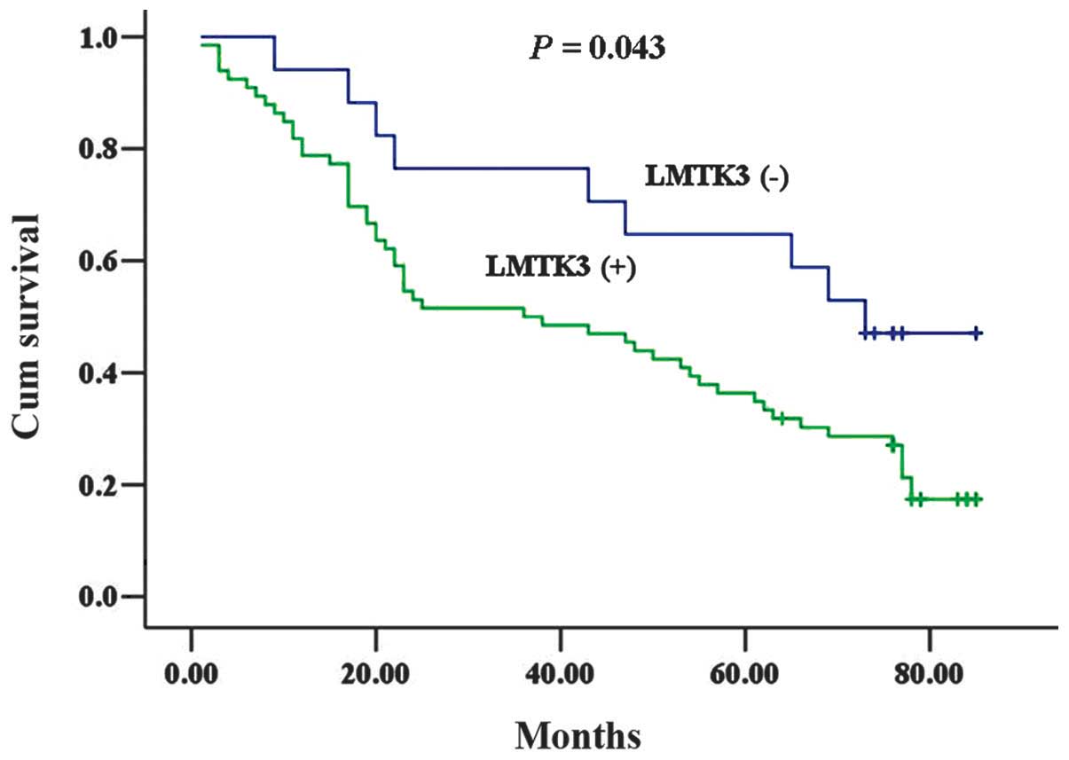
The Kaplan-Meier analysis revealed that the
postoperative survival of the LMTK3-negative group tended to be
superior to that of the LMTK3-positive group, with a statistically
significant difference (P=0.043; Fig.
2). Furthermore, as shown in Table II, the multivariate analysis
confirmed that the hazard risk (HR) of death was significantly
higher in the group with positive LMTK3 staining compared to the
negative group (HR=3.071, 95% CI: 1.375–9.283). Therefore, it was
suggested that positive LMTK3 staining may be associated with a
poorer prognosis in patients with gastric cancer.
| Table IIMultivariate survival analysis of
prognostic factors by Cox regression analysis. |
Table II
Multivariate survival analysis of
prognostic factors by Cox regression analysis.
| Clinicopathological
parameters | HR (95% CI) | P-value |
|---|
| LMTK3 |
| Higher/lower | 3.071
(1.375–9.283) | 0.019 |
| Gender |
| Female/male | 0.684
(0.375–1.247) | 0.215 |
| Age (years) |
| ≤50/>50 | 0.896
(0.350–2.295) | 0.819 |
| Grade |
| III+IV/I+II | 1.373
(0.881–2.139) | 0.161 |
| Size (cm) |
| >5/≤5 | 1.279
(0.720–2.727) | 0.401 |
| TNM stage |
| III+IV/I+II | 3.642
(2.934–10.887) | 0.015 |
Discussion
Gastric cancer is the second leading cause of
cancer-related mortality worldwide; however, over the last few
decades, there have been no significant advances in the treatment
of gastric cancer and the conventionally used methods have been
proven unsatisfactory. Molecular-targeted therapy has become
indispensable in the treatment of malignancies, such as trastuzumab
for breast cancer and gefitinib or erlotinib for non-small-cell
lung cancer. However, to date, only a limited number of targeted
agents were shown to be of clinical benefit for gastric cancer,
including trastuzumab and ramucirumab (17). Due to the significant heterogeneity
of gastric cancer, HER2 is only overexpressed in only a few
patients and the majority of the patients cannot benefit from
trastuzumab (18,19). To overcome the difficulties in
gastric cancer treatment, more efficient biomarkers, molecular
targets and novel therapeutic strategies are urgently required.
Endocrine therapy is important for the treatment of
hormone-dependent malignancies, such as breast and prostate cancer.
Estrogen and ER also play and important role in gastric cancer.
According to epidemiological evidence and animal studies, estrogen
plays a protective role in gastric tumorigenesis (20–22).
Although not a direct target organ of sex hormones, ERα and ERβ
were both found to be expressed in gastric cancer tissue. Further
investigation demonstrated that the expression of ER was correlated
with the prognosis of gastric cancer patients, with the two
subtypes playing different roles (13,14,23–26).
As regards ER, certain studies on anti-estrogen endocrine therapy
have been conducted on gastric cancer; however, the results were
not satisfactory and did not significantly affect the treatment
outcome (27–30). Despite the unsatisfactory attempts,
it was suggested that endocrine therapy may be a useful strategy
for the treatment of gastric cancer, provided another efficient
related target was utilized to improve the effect, rather than
directly blocking ER (31).
The first studies on LMTK3 were mainly focused on
breast cancer. However, we recently demonstrated the presence of
LMTK3 protein in the blood of patients with colorectal cancer and
serum LMTK3 may be a valuable biomarker for predicting disease
progression and prognosis in such patients (32). Wakatsuki et al (33) reported that LMTK3 polymorphisms are
correlated with the prognosis of gastric cancer, but they did not
extensively investigate LMTK3 protein expression. In the present
study, we demonstrated that the positive rate of LMTK3 protein
expression in gastric cancer was significantly higher compared to
that in the adjacent non-cancerous mucosa. Furthermore, the
expression of LMTK3 was correlated with depth of invasion and
cancer stage. In addition, LMTK3 appeared to be strongly positive
in all 9 specimens with distant metastasis (Table I), strongly indicating that the
expression of LMTK3 is correlated with metastasis of gastric
cancer, if the sample is of adequate size. Furthermore, the
survival analysis demonstrated that the expression of LMTK3 was
negatively associated with postoperative survival in gastric cancer
patients. The multivariate analysis identified the expression of
LMTK3 as an independent negative prognostic factor in gastric
cancer.
In breast cancer, LMTK3 was shown to induce the
estrogen pathway via ERα phosphorylation and high baseline LMTK3
expression was found to be associated with a poorer overall and
disease-free survival (9,10). Of note, LMTK3 silencing through RNA
interference significantly enhanced the growth inhibitory effect of
tamoxifen in tamoxifen-resistant breast cancer cell lines (9). According to the function of LMTK3 in
breast cancer, targeted inhibition of LMTK3 aimed at enhancing the
effect of endocrine therapy may be an efficient treatment strategy
for gastric cancer, particularly in patients expressing ERα.
In conclusion, our results demonstrated that the
expression of LMTK3 may be a useful biomarker as a negative
prognostic factor in gastric cancer and a potential novel target
for the treatment of gastric cancer. Given the differences in the
ER subtype distribution between breast and gastric cancer, LMTK3
may be more than a modulator of ERα in gastric cancer (33). The association between LMTK3 and
ERβ requires further investigation and the role of LMTK3 in the
development of gastric cancer should be elucidated by further
studies.
Acknowledgements
This study was supported by grants from the
Technology Project of Changzhou Social Development (no. CS20102016)
and the Natural Science Funds for Young Teacher of Soochow
University (no. Q3124943).
References
|
1
|
Siegel R, Naishadham D and Jemal A: Cancer
statistics, 2012. CA Cancer J Clin. 62:10–29. 2012. View Article : Google Scholar
|
|
2
|
Kamangar F, Dores GM and Anderson WF:
Patterns of cancer incidence, mortality, and prevalence across five
continents: defining priorities to reduce cancer disparities in
different geographic regions of the world. J Clin Oncol.
24:2137–2150. 2006. View Article : Google Scholar
|
|
3
|
Huang JY, Xu YY, Sun Z, et al: Comparison
different methods of intraoperative and intraperitoneal
chemotherapy for patients with gastric cancer: a meta-analysis.
Asian Pac J Cancer Prev. 13:4379–4385. 2012. View Article : Google Scholar
|
|
4
|
Meyer HJ and Wilke H: Treatment strategies
in gastric cancer. Dtsch Arztebl Int. 108:698–705. 2011.
|
|
5
|
Bang YJ, Van Cutsem E, Feyereislova A, et
al: Trastuzumab in combination with chemotherapy versus
chemotherapy alone for treatment of HER2-positive advanced gastric
or gastro-oesophageal junction cancer (ToGA): a phase 3,
open-label, randomised controlled trial. Lancet. 376:687–697. 2010.
View Article : Google Scholar
|
|
6
|
De Vita F, Giuliani F, Silvestris N, et
al: Current status of targeted therapies in advanced gastric
cancer. Expert Opin Ther Targets. 16(Suppl 2): S29–S34.
2012.PubMed/NCBI
|
|
7
|
Naik S, Dothager RS, Marasa J, Lewis CL
and Piwnica-Worms D: Vascular endothelial growth factor receptor-1
is synthetic lethal to aberrant beta-catenin activation in colon
cancer. Clin Cancer Res. 15:7529–7537. 2009. View Article : Google Scholar : PubMed/NCBI
|
|
8
|
Tyner JW, Deininger MW, Loriaux MM, et al:
RNAi screen for rapid therapeutic target identification in leukemia
patients. Proc Natl Acad Sci USA. 106:8695–8700. 2009. View Article : Google Scholar : PubMed/NCBI
|
|
9
|
Giamas G, Filipovic A, Jacob J, et al:
Kinome screening for regulators of the estrogen receptor identifies
LMTK3 as a new therapeutic target in breast cancer. Nat Med.
17:715–719. 2011. View
Article : Google Scholar : PubMed/NCBI
|
|
10
|
Stebbing J, Filipovic A, Ellis IO, et al:
LMTK3 expression in breast cancer: association with tumor phenotype
and clinical outcome. Breast Cancer Res Treat. 132:537–544. 2011.
View Article : Google Scholar : PubMed/NCBI
|
|
11
|
Camargo MC, Goto Y, Zabaleta J, Morgan DR,
Correa P and Rabkin CS: Sex hormones, hormonal interventions, and
gastric cancer risk: a meta-analysis. Cancer Epidemiol Biomarkers
Prev. 21:20–38. 2012. View Article : Google Scholar : PubMed/NCBI
|
|
12
|
Sheh A, Ge Z, Parry NM, et al:
17β-estradiol and tamoxifen prevent gastric cancer by modulating
leukocyte recruitment and oncogenic pathways in Helicobacter
pylori-infected INS-GAS male mice. Cancer Prev Res (Phila).
4:1426–1435. 2011.
|
|
13
|
Ryu WS, Kim JH, Jang YJ, et al: Expression
of estrogen receptors in gastric cancer and their clinical
significance. J Surg Oncol. 106:456–461. 2012. View Article : Google Scholar : PubMed/NCBI
|
|
14
|
Xu CY, Guo JL, Jiang ZN, et al: Prognostic
role of estrogen receptor alpha and estrogen receptor beta in
gastric cancer. Ann Surg Oncol. 17:2503–2509. 2010. View Article : Google Scholar : PubMed/NCBI
|
|
15
|
Deng H, Huang X, Fan J, et al: A variant
of estrogen receptor-α, ER-α36 is expressed in human gastric cancer
and is highly correlated with lymph node metastasis. Oncol Rep.
24:171–176. 2010.
|
|
16
|
Zhou J, Teng R, Xu C, et al:
Overexpression of ERα inhibits proliferation and invasion of MKN28
gastric cancer cells by suppressing β-catenin. Oncol Rep.
30:1622–1630. 2013.
|
|
17
|
Cho JY: Molecular diagnosis for
personalized target therapy in gastric cancer. J Gastric Cancer.
13:129–135. 2013. View Article : Google Scholar : PubMed/NCBI
|
|
18
|
Gravalos C and Jimeno A: HER2 in gastric
cancer: a new prognostic factor and a novel therapeutic target. Ann
Oncol. 19:1523–1529. 2008. View Article : Google Scholar : PubMed/NCBI
|
|
19
|
Jorgensen JT and Hersom M: HER2 as a
prognostic marker in gastric cancer - a systematic analysis of data
from the literature. J Cancer. 3:137–144. 2012. View Article : Google Scholar : PubMed/NCBI
|
|
20
|
Furukawa H, Iwanaga T, Koyama H and
Taniguchi H: Effect of sex hormones on the experimental induction
of cancer in rat stomach - a preliminary study. Digestion.
23:151–155. 1982. View Article : Google Scholar : PubMed/NCBI
|
|
21
|
Lindblad M, Ye W, Rubio C and Lagergren J:
Estrogen and risk of gastric cancer: a protective effect in a
nationwide cohort study of patients with prostate cancer in Sweden.
Cancer Epidemiol Biomarkers Prev. 13:2203–2207. 2004.PubMed/NCBI
|
|
22
|
Chandanos E and Lagergren J: Oestrogen and
the enigmatic male predominance of gastric cancer. Eur J Cancer.
44:2397–2403. 2008. View Article : Google Scholar : PubMed/NCBI
|
|
23
|
Gan L, He J, Zhang X, et al: Expression
profile and prognostic role of sex hormone receptors in gastric
cancer. BMC Cancer. 12:5662012. View Article : Google Scholar : PubMed/NCBI
|
|
24
|
Matsuyama S, Ohkura Y, Eguchi H, et al:
Estrogen receptor beta is expressed in human stomach
adenocarcinoma. J Cancer Res Clin Oncol. 128:319–324. 2002.
View Article : Google Scholar : PubMed/NCBI
|
|
25
|
Wang M, Pan JY, Song GR, Chen HB, An LJ
and Qu SX: Altered expression of estrogen receptor alpha and beta
in advanced gastric adenocarcinoma: correlation with prothymosin
alpha and clinicopathological parameters. Eur J Surg Oncol.
33:195–201. 2007. View Article : Google Scholar
|
|
26
|
Chandanos E, Rubio CA, Lindblad M, et al:
Endogenous estrogen exposure in relation to distribution of
histological type and estrogen receptors in gastric adenocarcinoma.
Gastric Cancer. 11:168–174. 2008. View Article : Google Scholar : PubMed/NCBI
|
|
27
|
Kitaoka H: Chemo-endocrine therapy of
diffuse carcinoma of the stomach and its clinical evaluation. Jpn J
Cancer Clinics. 31(Suppl 9): 1189–1194. 1985.(In Japanese).
|
|
28
|
Harrison JD, Morris DL, Ellis IO, Jones JA
and Jackson I: The effect of tamoxifen and estrogen receptor status
on survival in gastric carcinoma. Cancer. 64:1007–1010. 1989.
View Article : Google Scholar : PubMed/NCBI
|
|
29
|
Kojima O and Takahashi T: Endocrine
therapy of scirrhous carcinoma of the stomach. Jpn J Cancer
Chemother. 13:2526–2531. 1986.(In Japanese).
|
|
30
|
Kitaoka H: Sex hormone dependency in
diffuse carcinoma of the stomach and results of chemo-endocrine
therapy. Jpn J Cancer Clinics. 30(Suppl 6): 741–748. 1984.(In
Japanese).
|
|
31
|
Kim MJ, Cho SI, Lee KO, Han HJ, Song TJ
and Park SH: Effects of 17β-estradiol and estrogen receptor
antagonists on the proliferation of gastric cancer cell lines. J
Gastric Cancer. 13:172–178. 2013.
|
|
32
|
Shi H, Wu J, Ji M, et al: Serum lemur
tyrosine kinase 3 expression in colorectal cancer patients predicts
cancer progression and prognosis. Med Oncol. 30:7542013. View Article : Google Scholar : PubMed/NCBI
|
|
33
|
Wakatsuki T, LaBonte MJ, Bohanes PO, et
al: Prognostic role of lemur tyrosine kinase-3 germline
polymorphisms in adjuvant gastric cancer in Japan and the United
States. Mol Cancer Ther. 12:2261–2272. 2013. View Article : Google Scholar : PubMed/NCBI
|