Introduction
Axillary surgery in breast cancer has transitioned
from level III to level II dissection. Axillary lymph node
dissection (ALND) is commonly associated with complications, such
as sensory and motor nervous system disorders and edema of the arm.
To avoid such complications, sentinel LN biopsy (SLNB) is accepted
as a standard technique for clinically node-negative breast cancer
patients. SLNB alone (without ALND) is currently the standard
course of action for axillary management in patients with negative
SLNs (1).
The American College of Surgeons Oncology Group
(ACOSOG) Z0011 randomized controlled trial compared overall
survival between patients with positive SLNs undergoing SLNB alone
and those undergoing SLNB with ALND (2). The trial demonstrated that, for patients
with stage T1–2 and ≤2 metastatic SLNs who were treated with
breast-conserving surgery followed by whole-breast irradiation and
adjuvant systemic therapy, SLNB alone resulted in a similar
disease-free and overall survival compared with ALND (2). Furthermore, the International Breast
Cancer Study Group (IBCSG) 23-01 trial randomized patients with
micrometastases to SLNB alone or ALND groups, and reported a
similar disease-free survival for the two patient groups (3). Additionally, the European Organisation
for Research and Treatment of Cancer 10981–22023 AMAROS trial
compared the axillary recurrence rate between the ALND and axillary
irradiation patient groups; both groups consisted of patients with
positive SLNs, and the results of the trial demonstrated a similar
disease-free and overall survival between the two, similar to the
findings of the ACOSOG and IBCSG trials (4). Based on these results, the clinical
significance of ALND has been reduced and the selection of patients
with positive SLNs in whom ALND may be omitted is attracting
increasing attention.
The presence of LN metastases is the most
significant prognostic indicator for breast cancer and a major
factor in determining adjuvant therapy. Previously, if the presence
of LN metastasis was confirmed, postoperative chemotherapy was
deemed essential. However, following the St. Gallen Consensus
Conference in 2011, the intrinsic subtype of breast cancer has
become a more important determinant of adjuvant therapy, rather
than the presence of LN metastasis, although the presence of ≥4 LN
metastases remains an important indicator, as additional
chemotherapy is recommended for such patients, regardless of the
cancer subtype (5). Furthermore, when
≥4 LN metastases are identified, irradiation of the supra- and
subclavian regions, in addition to the preserved breast, has been
reported to improve patient survival as well as local disease
control (6,7). Therefore, it is important to determine
whether ≥4 LN metastases are present, in order to optimize
treatment. Several previous studies have reported a number of
methods to predict ≥4 LN metastases (8–11).
However, the majority of these relied on factors that were
determined postoperatively, and are therefore not widely applied in
clinical practice.
The one-step nucleic acid amplification (OSNA) assay
is a method for diagnosing LN metastasis through solubilization of
LNs and amplification and detection of cytokeratin (CK)19 mRNA. The
OSNA assay is able to assess the entire LN, while histopathological
examination usually evaluates only the maximum cut surface. The
OSNA assay is completed in 30–40 min and is thus suitable for
intraoperative diagnosis of SLN metastasis. Several previous
studies have reported the usefulness of the OSNA assay for
predicting the extent of LN metastasis based on its ability to
semi-quantitatively measure CK19 mRNA copy number (12–15,16).
The aim of the present study was to evaluate whether
it is possible to intraoperatively diagnose the presence of ≥4 LN
metastases in breast cancer patients using the OSNA assay.
Patients and methods
Patients
A total of 621 patients with invasive breast cancer
underwent SLNB evaluated by the OSNA assay between May, 2010 and
December, 2014 at Kinki University Hospital, Osaka, Japan. Of the
621 patients, 134 who subsequently underwent ALND of the positive
SLNs were analyzed. Patients who received neoadjuvant drug therapy,
axillary LN sampling alone, and patients with non-invasive cancer,
were excluded from this study. Staging was based on the 7th edition
of the Union for International Cancer Control TNM classification of
malignant tumors (17).
SLN detection
Detection of SLNs was performed using a radioisotope
tracer (technetium-99m phytate) and dye (indocyanine green). The
day prior to surgery, 85 MBq (0.5 ml) of tracer was injected into
the subdermal space in the outer border of the areola.
Lymphoscintigraphy was performed 2 h following the injection.
During surgery, 5 mg (1 ml) of indocyanine green was injected into
the subdermal space in the outer border of the areola and the SLNs
were identified using a hand-held gamma probe and dye mapping. SLN
metastases were evaluated intraoperatively using the OSNA assay,
and ALND was performed when at least one SLN was found to be
OSNA-positive (+ or ++, as defined below).
SLN assessment
Whole SLNs were evaluated using the OSNA assay, as
previously described (18). SLNs were
assessed as OSNA-negative (CK19 mRNA <2.5×10²
copies/µl), OSNA+ (2.5×10² to <5.0×10³
copies/µl), and OSNA++ (≥5.0×10³ copies/µl). When an SLN
was assessed as OSNA + inhibition (+I; ≥2.5×10²
copies/µl in the diluted samples), the patients were excluded from
the study, as the reading was not accurate. Total tumor load (TTL)
was defined as the total number of CK19 mRNA copies in all positive
SLNs. The SLN ratio was defined as the ratio of positive:removed
SLNs. Non-SLN tissue sections were evaluated by hematoxylin and
eosin staining.
Examination of clinical tumor size and
axillary nodal status
Clinical tumor size was defined as the largest tumor
size measured by magnetic resonance imaging, ultrasonography (US)
or computed tomography (CT). Clinical axillary LN metastasis
diagnosis was established using contrast-enhanced CT and US. If an
LN was diagnosed as metastatic using fine-needle aspiration biopsy,
SLNB was not performed.
Statistical analysis
JMP software version 11 (SAS Institute, Cary, NC,
USA) was used for statistical analysis. Univariate and multivariate
analyses (logistic regression model) were performed to assess the
association of the variables with ≥4 LN metastases. A P-value of
<0.05 was considered to indicate statistically significant
differences. The accuracy of the TTL was estimated by constructing
a receiver operator characteristic (ROC) curve and measuring the
area under the curve (AUC).
Results
Clinicopathological
characteristics
Of the 621 patients who had SLNs evaluated using the
OSNA assay, 170 (27.3%) were OSNA-positive. Of these 170 patients,
61 (35.9%) were found to be OSNA+, 102 (60.0%) were OSNA++, and 7
(4.1%) were OSNA + I. Of the OSNA-positive patients, 134 (35 OSNA+
and 99 OSNA++) who underwent ALND were eligible for inclusion in
our study (Table I). A total of 31
patients had one OSNA+ SLN, 4 patients had two OSNA+ SLNs, 62
patients had one OSNA++ SLN, 14 patients had two OSNA++ SLNs, 3
patients had three OSNA++ SLNs, 17 patients had one OSNA+ and one
OSNA++ SLNs, and 3 patients had one OSNA+ and two OSNA++ SLNs. The
median number of dissected LNs was 18.3 (range, 5–43). Of the 134
patients, 31 (23.1%) had ≥4 LN metastases.
 | Table I.Patient characteristics (n=134). |
Table I.
Patient characteristics (n=134).
| Characteristics | No. (%) |
|---|
| Age, years [median
(range)] | 55.5 (27–81) |
| Menopausal
status |
|
|
Premenopausal | 51
(38.1) |
|
Postmenopausal | 83
(61.9) |
| Estrogen receptor
status |
|
| + | 109 (81.3) |
| − | 25
(18.7) |
| Progesterone receptor
status |
|
| + | 94
(70.1) |
| − | 40
(29.9) |
| HER2 status |
|
| + | 29
(21.6) |
| − | 105 (78.4) |
| Breast cancer
subtype |
|
|
Luminal/HER2− | 95
(70.9) |
|
Luminal/HER2+ | 20
(14.9) |
|
HER2+ |
9 (6.7) |
|
Triple-negative | 10
(7.5) |
| Type of breast
surgery |
|
| Partial
mastectomy | 70
(52.2) |
| Total
mastectomy | 64
(47.8) |
| Clinical T
classification |
|
| cT1 | 66
(49.3) |
| cT2 | 63
(47.0) |
| cT3 |
5 (3.7) |
| Clinical N
classification |
|
| cN0 | 59
(44.0) |
| cN1
suspected | 75
(56.0) |
| OSNA diagnosis |
|
| + | 35
(26.1) |
| ++ | 99
(73.9) |
| Histological
grade |
|
| 1 | 22
(16.5) |
| 2 | 44
(32.8) |
| 3 | 24
(17.9) |
|
Unknown | 44
(32.8) |
| Ki-67 |
|
|
<20% | 46
(34.3) |
| ≥20% | 35
(26.1) |
|
Unknown | 53
(39.6) |
| Lymphovascular
invasion |
|
| No | 53
(39.5) |
| Yes | 79
(59.0) |
|
Unknown |
2 (1.5) |
| Histological
type |
|
|
Ductal | 116 (86.6) |
|
Lobular | 11
(8.2) |
|
Other |
7 (5.2) |
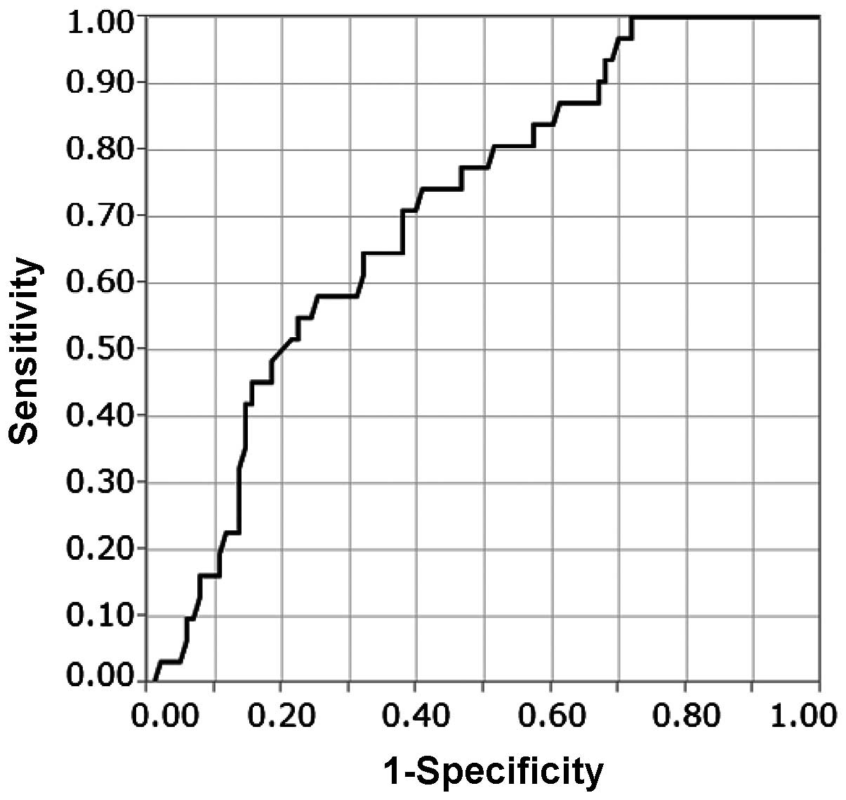
Association between TTL and ≥4 LN
metastases
The association between TTL and ≥4 LN metastases was
evaluated using ROC curve analysis (Fig.
1). The median of TTL was 4.5×10⁴ copies/µl
(2.5×10²-6.0×10⁶). The AUC of the ROC curve
was 0.708 and the TTL cut-off was 5.4×10⁴ copies/µl. Of
the patients with TTL <5.4×10⁴ copies/µl, 8 (11.6%)
had ≥4 LN metastases and, of the patients with TTL
≥5.4×10⁴ copies/µl, 23 (35.4%) had ≥4 LN metastases
(Table II). With a TTL cut-off of
5.4×10⁴ copies/µl, the sensitivity, specificity,
positive and negative predictive values were 74, 59, 35 and 88%,
respectively.
| Table II.Univariate analysis of variables
correlated with ≥4 lymph node metastases. |
Table II.
Univariate analysis of variables
correlated with ≥4 lymph node metastases.
|
| LN metastases, no.
(%) |
|
|---|
|
|
|
|
|---|
| Variables | <4 (n=103) | ≥4 (n=31) | P-value |
|---|
| Menopausal
status |
|
| 0.177 |
|
Premenopausal | 36 (70.6) | 15 (29.4) |
|
|
Postmenopausal | 67 (80.7) | 16 (19.3) |
|
| Clinical T
classification |
|
| 0.010 |
|
cT1 | 57 (86.4) | 9
(13.6) |
|
|
≥cT2 | 46 (67.6) | 22 (32.4) |
|
| Clinical N
classification |
|
| 0.006 |
|
cN0 | 52 (88.1) | 7
(11.9) |
|
| cN1
suspected | 51 (68.0) | 24 (32.0) |
|
| ER status |
|
| 0.909 |
| + | 84 (77.1) | 25 (22.9) |
|
| − | 19 (76.0) | 6
(24.0) |
|
| PR status |
|
| 0.910 |
| + | 72 (76.6) | 22 (23.4) |
|
| − | 31 (77.5) | 9
(22.5) |
|
| HER2 status |
|
| 0.102 |
| + | 19 (65.5) | 10 (34.5) |
|
| − | 84 (80.0) | 21 (20.0) |
|
| TTL
(copies/µl) |
|
| 0.001 |
|
<5.4×10⁴ | 61 (88.4) | 8
(11.6) |
|
|
≥5.4×10⁴ | 42 (64.6) | 23 (35.4) |
|
| Ki-67 |
|
| 0.144 |
|
<20% | 39 (84.8) | 7
(15.2) |
|
|
≥20% | 25 (71.4) | 10 (28.6) |
|
| Histological
grade |
|
| 0.339 |
| ≤2 | 53 (80.3) | 13 (19.7) |
|
| 3 | 17 (70.8) | 7
(29.2) |
|
| SLN ratio |
|
| 0.103 |
|
<0.67 | 34 (91.9) | 3
(8.1) |
|
|
≥0.67 | 69 (71.1) | 28 (28.9) |
|
| Lymphovascular
invasion |
|
| 0.063 |
| No | 45 (84.9) | 8
(15.1) |
|
|
Yes | 56 (70.9) | 23 (29.1) |
|
Variables correlated with ≥4 LN
metastases
The association between clinicopathological
variables and ≥4 LN metastases was evaluated. In the univariate
analysis, clinical T classification (T1 vs. ≥ T2, P=0.01), clinical
N classification (P=0.006), and TTL ≥5.4×10⁴ copies/µl
(P=0.001) were correlated with ≥4 LN metastases (Table II). In the multivariate analysis, TTL
≥5.4×10⁴ copies/µl was the only factor significantly
correlated with ≥4 LN metastases (OR=2.95, 95% CI: 1.17–7.97,
P=0.022; Table III).
| Table III.Multivariate analysis of variables
correlated with ≥4 lymph node metastases. |
Table III.
Multivariate analysis of variables
correlated with ≥4 lymph node metastases.
| Variables | Odds ratio | 95% CI | P-value |
|---|
| Clinical T
classification | 2.39 | 0.98–6.15 | 0.088 |
|
cT1 |
|
|
|
|
≥cT2 |
|
|
|
| Clinical N
classification | 2.31 | 0.88–6.57 | 0.055 |
|
cN0 |
|
|
|
| cN1
suspected |
|
|
|
| TTL
(copies/µl) | 2.95 | 1.17–7.97 | 0.022 |
|
<5.4×10⁴ |
|
|
|
|
≥5.4×10⁴ |
|
|
|
Discussion
A number of previous studies have reported on the
prediction of non-SLN metastasis in SLN-positive patients. Osako
et al demonstrated that SLN copy number, number of
macrometastatic SLNs and lymphovascular invasion were significant
factors for the identification of non-SLN metastasis (15). According to a meta-analysis of studies
investigating non-SLN metastasis prediction, size of SLN metastasis
>2 mm, extracapsular extension in SLNs, ≥2 positive SLNs, ≤1
negative SLN, tumor size >2 cm, ratio of positive SLNs >50%
and lymphovascular invasion in the primary tumor, have been
reported to be strongly correlated with non-SLN metastasis
(19).
It was previously reported that ~50% of SLN-positive
patients have non-SLN metastases (20,21).
However, even in patients with positive SLNs who did not undergo
ALND, but instead received appropriate adjuvant therapy, the
locoregional and distant recurrence rates were reported to be 1–2
and 5%, respectively (3), which are
significantly lower compared with those in patients with non-SLN
metastases. Therefore, even in patients with non-SLN metastases, it
is not always necessary to perform ALND. Thus, we hypothesized that
it may be more important to predict ≥4 LN metastases rather than
non-SLN metastases, and conducted this study to evaluate the
factors that predict the presence of ≥4 LN metastases.
Several previous studies have focused on factors
associated with ≥4 LN metastases. Maretoja et al (8) reported an international multicenter
predictive tool for the risk of ≥4 LN metastases in patients with
SLN macrometastases and demonstrated that the prevalence of ≥4 LN
metastases in each center, the number of positive SLNs, the number
of negative SLNs, the histological size of the primary tumor and
the presence of extracapsular extension of SLN metastases were
strongly correlated with the presence of ≥4 LN metastases.
Furthermore, Katz et al (9)
reported that the number of involved SLNs, extranodal extension,
lymphovascular invasion, number of uninvolved SLNs, size of largest
SLN metastasis, histology (lobular vs. other), and pathological
tumor size were significant factors for identifying ≥4 LN
metastases.
In the present study, only TTL was significantly
correlated with ≥4 LN metastases. Our analysis demonstrated that
pathological factors reported in previous studies (tumor size,
histological grade, lymphovascular invasion and histology) were not
correlated with ≥4 LN metastases. Although extranodal extension of
SLNs was shown to be correlated with ≥4 LN metastases in a number
of previous studies, SLNs were solubilized for the OSNA assay in
the present study and, for this reason, it was not possible to
evaluate extranodal extension. The SLN ratio, which has also been
identified as an important factor in previous studies, was not
found to be correlated with ≥4 LN metastases in our analysis.
A number of the aforementioned factors are
clinically confounding. For example, as the clinical tumor size and
size of SLN metastases differed according to the imaging modality
used, there is a limit to their accurate evaluation. Moreover, the
majority of these factors, such as extranodal SLN extension,
lymphovascular invasion and histological grade, are difficult to
assess accurately, pre- or intra-operatively. Therefore, these
factors cannot be used during surgery to determine the need for
ALND.
In the present study, OSNA diagnosis was
significantly correlated with ≥4 LN metastases. Additionally, OSNA
diagnosis was one of the factors correlated with ≥4 LN metastases
in the univariate analysis (data not shown). As the OSNA assay is
an objective method that may be rapidly evaluated during surgery,
it is useful for intraoperatively determining the necessity of
ALND. An OSNA+ result corresponds to micrometastasis on
histopathological examination (18)
and, as a previous randomized controlled trial demonstrated, ALND
is of no clinical significance with respect to disease-free
survival and the survival rate of patients with micrometastases
(3,22,23). By
contrast, for patients with SLN macrometastases, it is more
important to identify those in whom ALND may be safely omitted.
Therefore, we consider omission of ALND based on OSNA diagnosis
alone to be inadequate.
The usefulness of TTL, assessed by the OSNA assay,
was investigated as an alternative to OSNA diagnosis. TTL is
considered to reflect the tumor burden in LNs more accurately
compared with the number of copies of one SLN. Furthermore, TTL has
been previously reported to be useful for predicting the extent of
LN metastases. Peg et al (16)
reported that TTL is an independent predictor of non-SLN metastasis
and, if patients have TTL ≥1.5×10⁴ copies/µl, non-SLN
metastasis occurs at a higher frequency. Similar studies have
reported a correlation between TTL and non-SLN metastasis (13,15). In
the present study, we evaluated the association between TTL and ≥4
LN metastases, and found it to be significant when TTL
≥5.4×10⁴ copies/µl. Ohi et al (14) investigated the correlation between ≥4
LN metastases and the maximum copy number of SLNs, and reported
that 1.0×10⁵ copies/µl were correlated with ≥4 LN
metastases; however, they only evaluated one SLN, namely the one
with the maximum number of copies. We consider that the total copy
number of all the SLNs is more significantly correlated with the
extent of LN metastases compared with the maximum copies of one
SLN. To the best of our knowledge, the present study was the first
to investigate the correlation between TTL and ≥4 LN metastases. We
demonstrated that, of the patients with TTL >5.4×10⁴ copies/µl,
23 (35.4%) had ≥4 LN metastases, which suggests that ALND cannot be
omitted in these cases.
There major limitation of our study was the AUC of
TTL, which was 0.708, and is of moderate accuracy. The development
of novel molecular markers associated with LN metastases may
improve the accuracy of the TTL.
In conclusion, TTL ≥5.4×10⁴ copies/µl
significantly correlated with ≥4 LN metastases. Therefore, TTL is
likely to become an objective tool for intraoperatively deciding
the omission of ALND in SLN-positive breast cancer patients.
Further studies are required to improve the accuracy of this
assessment.
Acknowledgements
The present study was supported in part by
grants-in-aid for scientific research from the Japanese Breast
Cancer Society.
References
|
1
|
Lyman GH, Giuliano AE, Somerfield MR,
Benson AB III, Bodurka DC, Burstein HJ, Cochran AJ, Cody HS III,
Edge SB, Galper S, et al: American Society of Clinical Oncology:
American Society of Clinical Oncology guideline recommendations for
sentinel lymph node biopsy in early-stage breast cancer. J Clin
Oncol. 23:7703–7720. 2005. View Article : Google Scholar : PubMed/NCBI
|
|
2
|
Giuliano AE, Hunt KK, Ballman KV, Beitsch
PD, Whitworth PW, Blumencranz PW, Leitch AM, Saha S, McCall LM and
Morrow M: Axillary dissection vs. no axillary dissection in women
with invasive breast cancer and sentinel node metastasis, A
randomized clinical trial. JAMA. 305:569–579. 2011. View Article : Google Scholar : PubMed/NCBI
|
|
3
|
Galimberti V, Cole BF, Zurrida S, Viale G,
Luini A, Veronesi P, Baratella P, Chifu C, Sargenti M, Intra M, et
al: Axillary dissection versus no axillary dissection in patients
with sentinel-node micrometastases (IBCSG 23-01): A phase 3
randomised controlled trial. Lancet Oncol. 14:297–305. 2013.
View Article : Google Scholar : PubMed/NCBI
|
|
4
|
Donker M, van Tienhoven G, Straver ME,
Meijnen P, van de Velde CJ, Mansel RE, Cataliotti L, Westenberg AH,
Klinkenbijl JH, Orzalesi L, et al: Radiotherapy or surgery of the
axilla after a positive sentinel node in breast cancer (EORTC
10981-22023 AMAROS): A randomised, multicentre, open-label, phase 3
non-inferiority trial. Lancet Oncol. 15:1303–1310. 2014. View Article : Google Scholar : PubMed/NCBI
|
|
5
|
Goldhirsch A, Wood WC, Coates AS, Gelber
RD, Thürlimann B and Senn HJ: Panel members: Strategies for
subtypes - dealing with the diversity of breast, cancer. Highlights
of the St. Histopathology. Ann Oncol. 22:1736–1747. 2011.
View Article : Google Scholar : PubMed/NCBI
|
|
6
|
Overgaard M, Nielsen HM and Overgaard J:
Is the benefit of postmastectomy irradiation limited to patients
with four or more positive nodes, as recommended in international
consensus reports? A subgroup analysis of the DBCG 82 b&c
randomized trials. Radiother Oncol. 82:247–253. 2007. View Article : Google Scholar : PubMed/NCBI
|
|
7
|
Recht A, Edge SB, Solin LJ, Robinson DS,
Estabrook A, Fine RE, Fleming GF, Formenti S, Hudis C, Kirshner JJ,
et al: Postmastectomy radiotherapy: Clinical practice guidelines of
the American Society of Clinical Oncology. J Clin Oncol.
19:1539–1569. 2001.PubMed/NCBI
|
|
8
|
Meretoja TJ, Audisio RA, Heikkilä PS, Bori
R, Sejben I, Regitnig P, Luschin-Ebengreuth G, Zgajnar J, Perhavec
A, Gazic B, et al: International multicenter tool to predict the
risk of four or more tumor-positive axillary lymph nodes in breast
cancer patients with sentinel node macrometastases. Breast Cancer
Res Treat. 138:817–827. 2013. View Article : Google Scholar : PubMed/NCBI
|
|
9
|
Katz A, Smith BL, Golshan M, Niemierko A,
Kobayashi W, Raad RA, Kelada A, Rizk L, Wong JS, Bellon JR, et al:
Nomogram for the prediction of having four or more involved nodes
for sentinel lymph node-positive breast cancer. J Clin Oncol.
26:2093–2098. 2008. View Article : Google Scholar : PubMed/NCBI
|
|
10
|
Chagpar AB, Scoggins CR, Martin RC II,
Cook EF, McCurry T, Mizuguchi N, Paris KJ, Carlson DJ, Laidley AL,
El-Eid SE, et al: Predicting patients at low probability of
requiring postmastectomy radiation therapy. Ann Surg Oncol.
14:670–677. 2007. View Article : Google Scholar : PubMed/NCBI
|
|
11
|
Rivers AK, Griffith KA, Hunt KK, Degnim
AC, Sabel MS, Diehl KM, Cimmino VM, Chang AE, Lucas PC and Newman
LA: Clinicopathologic features associated with having four or more
metastatic axillary nodes in breast cancer patients with a positive
sentinel lymph node. Ann Surg Oncol. 13:36–44. 2006. View Article : Google Scholar : PubMed/NCBI
|
|
12
|
Sagara Y, Ohi Y, Matsukata A, Yotsumoto D,
Baba S, Tamada S and Sagara Y, Matsuyama Y, Ando M, Rai Y and
Sagara Y: Clinical application of the one-step nucleic acid
amplification method to detect sentinel lymph node metastasis in
breast cancer. Breast Cancer. 20:181–186. 2013. View Article : Google Scholar : PubMed/NCBI
|
|
13
|
Espinosa-Bravo M, Sansano I, Pérez-Hoyos
S, Ramos M, Sancho M, Xercavins J, Rubio IT and Peg V: Prediction
of non-sentinel lymph node metastasis in early breast cancer by
assessing total tumoral load in the sentinel lymph node by
molecular assay. Eur J Surg Oncol. 39:766–773. 2013. View Article : Google Scholar : PubMed/NCBI
|
|
14
|
Ohi Y, Umekita Y, Sagara Y, Rai Y,
Yotsumoto D, Matsukata A, Baba S, Tamada S, Matsuyama Y, Ando M, et
al: Whole sentinel lymph node analysis by a molecular assay
predicts axillary node status in breast cancer. Br J Cancer.
107:1239–1243. 2012. View Article : Google Scholar : PubMed/NCBI
|
|
15
|
Osako T, Iwase T, Kimura K, Horii R and
Akiyama F: Sentinel node tumour burden quantified based on
cytokeratin 19 mRNA copy number predicts non-sentinel node
metastases in breast cancer, Molecular whole-node analysis of all
removed nodes. Eur J Cancer. 49:1187–1195. 2013. View Article : Google Scholar : PubMed/NCBI
|
|
16
|
Peg V, Espinosa-Bravo M, Vieites B,
Vilardell F, Antúnez JR, de Salas MS, Delgado-Sánchez JJ, Pinto W,
Gozalbo F, Petit A, et al: Intraoperative molecular analysis of
total tumor load in sentinel lymph node: A new predictor of
axillary status in early breast cancer patients. Breast Cancer Res
Treat. 139:87–93. 2013. View Article : Google Scholar : PubMed/NCBI
|
|
17
|
Sobin LH, Gospodarowicz MK and Wittekind
C: UICC International Union Against Cancer TMN Classification of
Malignant Tumours (7th). Hoboken, NJ: Wiley-Blackwell. 181–193.
2009.
|
|
18
|
Tsujimoto M, Nakabayashi K, Yoshidome K,
Kaneko T, Iwase T, Akiyama F, Kato Y, Tsuda H, Ueda S, Sato K, et
al: One-step nucleic acid amplification for intraoperative
detection of lymph node metastasis in breast cancer patients. Clin
Cancer Res. 13:4807–4816. 2007. View Article : Google Scholar : PubMed/NCBI
|
|
19
|
van la Parra RF, Peer PG, Ernst MF and
Bosscha K: Meta-analysis of predictive factors for non-sentinel
lymph node metastases in breast cancer patients with a positive
SLN. Eur J Surg Oncol. 37:290–299. 2011. View Article : Google Scholar : PubMed/NCBI
|
|
20
|
Chu KU, Turner RR, Hansen NM, Brennan MB,
Bilchik A and Giuliano AE: Do all patients with sentinel node
metastasis from breast carcinoma need complete axillary node
dissection? Ann Surg. 229:536–541. 1999. View Article : Google Scholar : PubMed/NCBI
|
|
21
|
Reynolds C, Mick R, Donohue JH, Grant CS,
Farley DR, Callans LS, Orel SG, Keeney GL, Lawton TJ and Czerniecki
BJ: Sentinel lymph node biopsy with metastasis, Can axillary
dissection be avoided in some patients with breast cancer? J Clin
Oncol. 17:1720–1726. 1999.PubMed/NCBI
|
|
22
|
Yi M, Giordano SH, Meric-Bernstam F,
Mittendorf EA, Kuerer HM, Hwang RF, Bedrosian I, Rourke L and Hunt
KK: Trends in and outcomes from sentinel lymph node biopsy (SLNB)
alone vs. SLNB with axillary lymph node dissection for
node-positive breast cancer patients, Experience from the SEER
database. Ann Surg Oncol. 17((Suppl 3)): S343–S351. 2010.
View Article : Google Scholar
|
|
23
|
Bilimoria KY, Bentrem DJ, Hansen NM,
Bethke KP, Rademaker AW, Ko CY, Winchester DP and Winchester DJ:
Comparison of sentinel lymph node biopsy alone and completion
axillary lymph node dissection for node-positive breast cancer. J
Clin Oncol. 27:2946–2953. 2009. View Article : Google Scholar : PubMed/NCBI
|