Introduction
Gastrointestinal stromal tumors (GISTs) are common
mesenchymal tumors of the gastrointestinal tract, which arise from
the interstitial cells of Cajal (ICCs). GISTs are rare tumors,
comprising 0.1–0.3% of all GI malignancies (1).
The majority of GISTs are located in the stomach
(60–70%) and the small intestine (20–25%). GISTs are difficult to
diagnose and early diagnosis may be challenging. A definitive
diagnosis requires immunohistochemical confirmation. These tumors
have a known potential for malignancy and their prognosis is poor
when the mitotic index is high.
GI bleeding is the most common presentation (50%) of
GISTs and is usually associated with ulceration of the tumor into
the intestinal lumen (2).
This is the report of an unusual case of an
exophytic GIST originating from the small bowel, which was
asymptomatic, apart from a weakly positive occult blood test in a
young adult woman. At first, the occult blood was attributed to
hemorrhoids. However, the patient had a family history of colon
cancer; therefore, she required more thorough investigation,
despite the lack of obvious symptoms. GISTs in the small intestine
may be identified by capsule endoscopy following negative upper
endoscopy and colonoscopy findings and are confirmed through
histological examination of the surgical specimen.
Case report
A 35-year-old woman presented with a weakly positive
occult blood test, following a regular health screening. The
patient had no abdominal discomfort, weight loss or any other
clinical manifestations; however, she had a family history of colon
cancer. The patient's blood pressure was 117/86 mmHg, her heart
rate was 80 beats/min, and her temperature 36.9°C. There was no
anemia on examination.
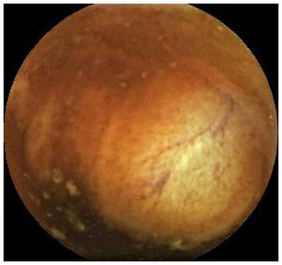
An endoscopic assessment of the upper and lower GI
tract (gastroscopy and colonoscopy) was performed, but did not
reveal any abnormal findings. Subsequently, an exophytic tumor was
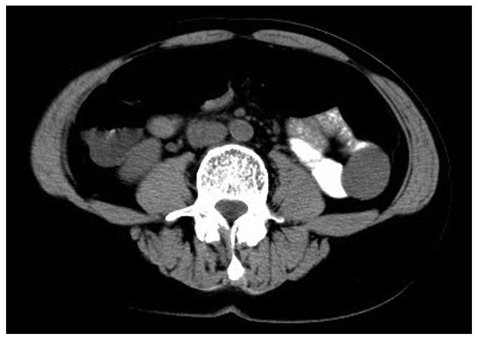
identified in the small intestine by capsule endoscopy (Fig. 1). A computed tomography scan revealed
a mass sized 1.5×2.0×2.5 cm in the lower left quadrant (Fig. 2). An enhanced computed tomography scan
revealed a smoothly outlined hypervascular solid mass. The computed
tomography scan was suggestive of a soft tissue tumor arising from
the small intestine. Based on the capsule endoscopic and
radiological findings, a diagnosis of a small intestinal mass was
established. The patient was subsequently referred for surgery and
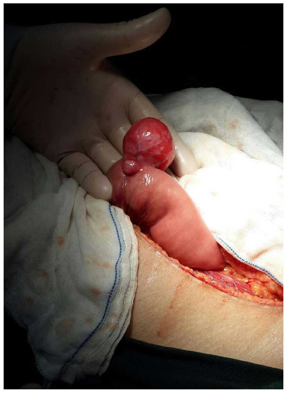
underwent laparotomy. The intraoperative findings included an
isolated exophytic pedunculated mass, sized 1.5×2.0×2.5 cm, on the
wall of the mid-ileum (Fig. 3). There
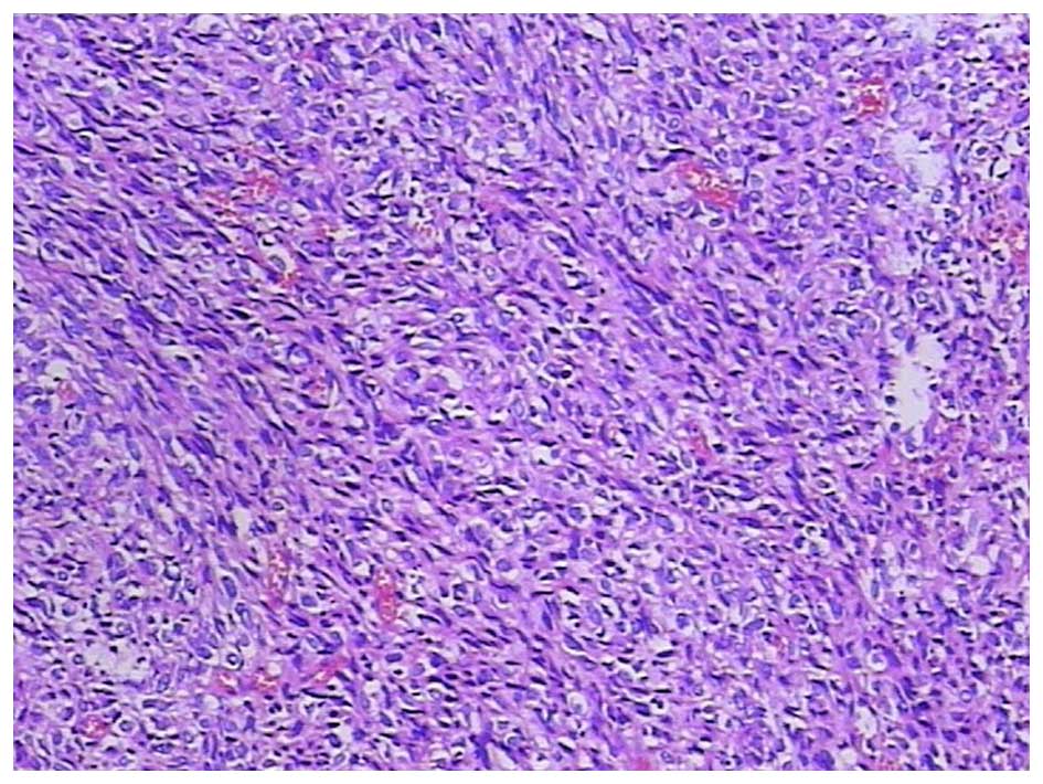
was no macroscopic evidence of distant spread. The
histopathological report was that of a GIST arising from the small
intestine (Fig. 4). On pathological
examination of the surgical specimen, the tumor was established as
low-risk according to the Fletcher's criteria, with a mitotic index
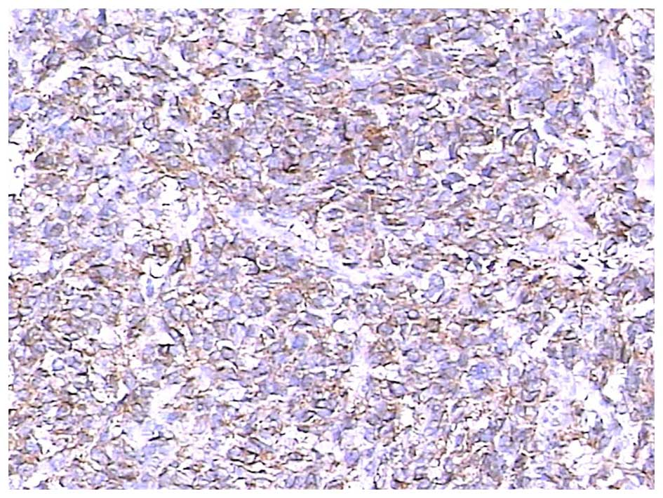
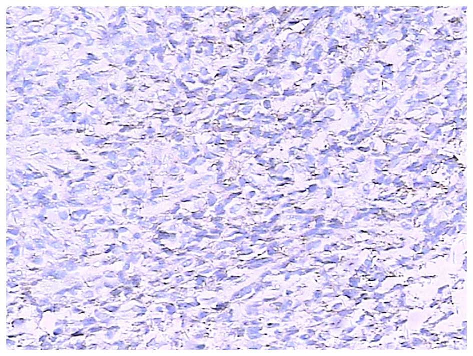
of <5 mitoses/50 high-power fields (HPFs) (3). The immunohistochemistry results revealed
a high expression of CD117 (Fig. 5)
and discovered on GIST-1 (DOG-1) (Fig.
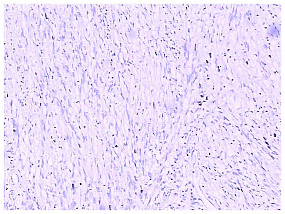
6); however, desmin, S100 and pancytokeratin were negative. The
Ki-67 expression revealed a low proliferative index (2%) (Fig. 7).
Discussion
GISTs are mesenchymal tumors of the GI tract that
express the tyrosine kinase receptor and originate from the ICCs.
GISTs arise from the muscularis mucosa or muscularis propria layers
and mainly exhibit an endophytic growth pattern, which may present
with evidence of obstruction or bleeding. Exophytic tumors present
as a large mass lesion or, if perforation has occurred, with
symptoms of peritonitis (4).
The diagnosis is confirmed by histological
examination. Immunohistochemistry is crucial for differentiating
GISTs from leiomyomas and neurogenic tumors (5). Specific abnormalities of c-Kit (CD117)
and platelet-derived growth factor receptor α (PDGFRA) are the main
oncogenic event in GISTs. Approximately 75–80% of GISTs harbor
gain-of-function c-Kit mutations, 7–10% harbor gain-of-function
PDGFRA mutations, and the remaining are defined as wild-type, as
they have no c-Kit or PDGFRA mutations (6,7).
In this study, immunohistochemistry was conclusive
in determining the histology of the tumor, based on CD117 and DOG-1
positivity, resulting in the final diagnosis of small intestinal
GIST.
For GISTs, the main symptoms are bleeding and
obstruction, which is caused by the growing tumor. Pressure
necrosis and ulceration of the overlying mucosa may cause GI
bleeding. In the present case, a weakly positive occult blood test
was the most important finding beyond atypical abdominal
discomfort. The GI bleeding was only obscure, as there was no
obvious necrosis or ulceration of the overlying mucosa on capsule
endoscopy. This case was found incidentally during a colorectal
cancer screening. As the patient had a family history of colon
cancer, endoscopic examination was suggested. Subsequently, an
exophytic GIST arising from the small intestine was identified.
GISTs may be classified as low- or high-risk tumors
by taking into consideration the possibility of metastasis or
recurrence based on the macroscopic/histological findings. GISTs
have a known potential for malignancy and their prognosis is poor
when the mitotic index is high. The poor prognostic predictive
factors include tumor size >5 cm, mitotic index >5/50 HPFs,
tumor necrosis, and a Ki-67 (MIB-1) index of >10%, and are
associated with high mortality (2,8).
Taking into consideration the prognostic predictive
factors found in our case, such as the small size of the tumor (2.5
cm), low mitotic index (﹤5/50 HPFs), absence of necrosis and a
Ki-67 (MIB-1) index of ﹤10%, there is little chance of recurrence
of the tumor. Surgical resection is the only effective therapy and
recommended for nearly all adult patients.
GISTs are mesenchymal tumors of the GI tract that
typically occur in adults aged >40 years, whereas they are rare
in in younger (<40 years) patients (4). Studies on GISTs in young adult patients
are limited due to their rarity. However, as our patient was a
34-year-old woman, GI stromal tumors should be included in the
differential diagnosis of intestinal mesenchymal tumors presenting
as a single mass in young female adults, despite diagnostic
physical findings not being readily apparent.
Wireless capsule endoscopy is considered to be the
first-line non-invasive diagnostic tool of the small intestine for
patients with obscure GI bleeding, enabling early diagnosis and
application of a definitive therapy, thereby increasing the chance
of survival.
Although GISTs of the small intestine presenting
with obscure GI bleeding in young adult women are a rare finding,
clinicians should consider capsule endoscopy even following
negative findings on upper endoscopy and colonoscopy.
References
|
1
|
Badshah MB, Riaz H, Korsten MA, Dhala A,
Park YH, Abadi M and Badshah MB: Gastro-intestinal stromal tumor
(GIST) complicating a colonic interposition, A novel case report.
BMC Res Notes. 7:6042014. View Article : Google Scholar : PubMed/NCBI
|
|
2
|
Fletcher CD, Berman JJ, Corless C,
Gorstein F, Lasota J, Longley BJ, Miettinen M, O’Leary TJ, Remotti
H, Rubin BP, Shmookler B, Sobin LH and Weiss SW: Diagnosis of
gastrointestinal stromal tumors, a consensus approach. Hum Pathol.
33:459–465. 2002. View Article : Google Scholar : PubMed/NCBI
|
|
3
|
Misawa S, Takeda M, Sakamoto H, Kirii Y,
Ota H and Takagi H: Spontaneous rupture of a giant gastrointestinal
stromal tumor of the jejunum, A case report and literature review.
World J Surg Oncol. 12:1532014. View Article : Google Scholar : PubMed/NCBI
|
|
4
|
Sezer A, Yagci MA, Hatipoglu AR, Coskun I,
Cicin I, Usta U and Temizoz O: A rare cause of intestinal
obstruction due to an exophytic gastrointestinal stromal tumor of
the small bowel. Signa Vitae. 4:32–34. 2009.
|
|
5
|
Manxhuka-Kerliu S, Sahatciu-Meka V, Kerliu
I, Juniku-Shkololli A, Kerliu L, Kastrati M and Kotorri V: Small
intestinal gastrointestinal stromal tumor in a young adult woman, A
case report and review of the literature. J Med Case Rep.
8:3212014. View Article : Google Scholar : PubMed/NCBI
|
|
6
|
Hirota S, Isozaki K, Moriyama Y, Hashimoto
K, Nishida T, Ishiguro S, Kawano K, Hanada M, Kurata A, Takeda M,
et al: Gain-of-function mutations of c-kit in human
gastrointestinal stromal tumors. Science. 279:577–580. 1998.
View Article : Google Scholar : PubMed/NCBI
|
|
7
|
Nannini M, Biasco G, Di Scioscio V, Di
Battista M, Zompatori M, Catena F, Castellucci P, Paterini P, Dei
Tos AP, Stella F, et al: Clinical, radiological and biological
features of lung metastases in gastrointestinal stromal tumors
(case reports). Oncol Rep. 25:113–120. 2011.PubMed/NCBI
|
|
8
|
Emory TS, Sobin LH, Lukes L, Lee DH and
O'Leary TJ: Prognosis of gastrointestinal smooth-muscle (stromal)
tumors Dependence on anatomic site. Am J Surg Pathol. 23:82–87.
1999. View Article : Google Scholar : PubMed/NCBI
|