Introduction
Mediastinal lymph node (LN) metastasis is one of the
most significant prognostic factors in patients with non-small-cell
lung cancer (NSCLC) and accurate N staging is a crucial step prior
to decision making regarding the treatment of NSCLC patients
(1). However, the optimal treatment
approach for NSCLC patients with operable N2 disease remains
controversial. According to a previous study, direct surgery for
patients with clinically apparent N2 disease achieved a long-term
survival rate of <10% of (2).
However, certain phase II trials and a limited number of phase III
trials suggested improved long-term survival for NSCLC patients
with N2 disease treated with neoadjuvant therapy followed by
surgical resection, compared with those undergoing surgery alone
(3–7).
Additionally, Pataer et al reported that the
histopathological response to neoadjuvant chemotherapy was
significantly associated with long-term overall survival (OS) in
NSCLC patients with N2 disease (8).
In a prospective phase II study evaluating neoadjuvant cisplatin
and docetaxel, the 3-year survival rate was 61% in patients with
pathologically negative mediastinal LNs, compared with 11% in those
with persistent N2 disease, following chemotherapy (9). Despite meticulous efforts to assess the
nodal status, unexpected persistent N2 disease following induction
(neoadjuvant) chemotherapy may be identified following surgical
treatment for NSCLC, as false-negative results are intrinsic to the
preoperative staging workup. The fact that persistent N2 disease
was unexpectedly detected in surgical specimens of patients with
clinical N0-1 disease following neoadjuvant chemotherapy suggests
that the pathological extent of N2 disease may be microscopic
rather than macroscopic in such cases (10). However, the optimal treatment
selection for this subgroup of patients remains debated upon and
the role of surgery following induction therapy has not yet been
clearly determined.
We reviewed our experience with patients treated
with neoadjuvant chemotherapy for N2 NSCLC followed by surgical
resection. The objective of this study was to compare the
clinicopathological characteristics and survival outcomes between
patients with unexpected persistent N2 disease (clinical N0-1,
pathological N2) and those with expected persistent N2 disease
(clinical N2, pathological N2) undergoing surgery following
neoadjuvant chemotherapy for NSCLC.
Patients and methods
Patient population
The medical records of 392 consecutive patients with
a pathological diagnosis of NSCLC who received neoadjuvant
chemotherapy at the Department of General Thoracic Surgery,
Shanghai Pulmonary Hospital (Shanghai, China) between January, 1995
and December, 2012 were reviewed. All the patients underwent
contrast-enhanced thoracic computed tomography (CT) or
18F-fluorodeoxyglucose (FDG)-positron emission
tomography (PET)/CT prior to surgery. Additional routine
pretreatment evaluations included chest radiography, abdominal
ultrasonography or CT scan, brain magnetic resonance imaging or CT
scan, cardiopulmonary function tests and whole-body radionuclide
bone scanning. Mediastinoscopy or endobronchial
ultrasonography-guided transbronchial needle aspiration biopsy were
routinely performed on patients with bulky mediastinal LNs on CT
(short-axis diameter >1.5 cm). The patients were staged
according to the seventh edition of the TNM classification
(11).
The inclusion criteria were patients with
pathological N2 disease prior to treatment and administration of
neoadjuvant chemotherapy. The chemotherapeutic regimens included
platinum in combination with gemcitabine, vinorelbine, paclitaxel
or docetaxel. The exclusion criteria were patients with superior
sulcus tumors, lack of indications for surgery, treatment with
induction radiotherapy and palliative surgery. Finally, a total of
348 patients were enrolled in this study.
Response to neoadjuvant
chemotherapy
After two cycles of neoadjuvant chemotherapy, the
response to treatment was classified according to the World Health
Organization criteria by CT scan and bronchoscopy (12) as follows: Complete response,
disappearance of all disease on radiographic and bronchoscopic
(when performed) examination; partial response, >50% reduction
in the volume of all measurable lesions; stable disease, no
detectable change in tumor volume of all lesions or change in the
size of all measured lesions between a 50% reduction and a 25%
increase, with no additional disease detected; progressive disease,
increase of >25% of all measured lesions or appearance of new
lesions. Complete and partial response are both classified as
objective response. Only patients with objective response or stable
disease were considered eligible for surgery.
Definitions and grouping
A resection was considered as complete (R0) when
there was no residual tumor at the bronchial or vascular margins
and no residual LN disease. Nodal downstaging was defined as
resected mediastinal LNs free of gross and microscopic disease.
Clinical nodal downstaging was defined as a long-axis diameter of
mediastinal LNs <1 cm on chest CT or negative PET/CT findings;
pathological nodal downstaging was defined as absence of
microscopic disease in the resected mediastinal nodes.
According to the definition of clinical or
pathological nodal downstaging, all the enrolled cases were
assigned into three study groups as follows: Group I, nodal
downstaging (pN0-1); group II, no nodal downstaging (expected
persistent N2 disease); and group III, clinical nodal downstaging
without pathological nodal downstaging (unexpected persistent N2
disease).
Surgical procedures
All the patients received R0 resection. The surgical
procedures included lobectomy, bilobectomy, sleeve resection and
pneumonectomy. Systemic lymphadenectomy was defined as removal of
at least three mediastinal stations and >10 nodes, additional to
the resection of hilar nodes (13).
Systemic lymphadenectomy was mandatory for all the patients in this
study. Mediastinal LN dissection consisted of en bloc resection of
all nodes at stations 2R, 4R, 7, 8, 9 and 10R for right-sided
tumors and nodes at stations 4L, 5, 6, 7, 8, 9 and 10L for
left-sided tumors. Bronchial stumps underwent routine frozen
sections to ensure microscopically tumor-free margins in all the
cases.
Postoperative treatment and
follow-up
Adjuvant chemotherapy was administered to the
patients included in this study and mediastinal regional
radiotherapy was performed in patients with persistent N2 disease,
provided they were able to tolerate additional treatments. The
patients were regularly evaluated by CT every 6 months for the
first 2 years after surgery and every 12 months thereafter.
All the patients were followed up by trained staff
every 6 months, by phone or e-mail. All patient information
regarding survival, cancer recurrence or metastasis and cause of
death were recorded. In the present study, the endpoint of
follow-up was December, 2012, with a mean follow-up of 55
months.
Statistical analysis
Numerical data are expressed in terms of frequency,
mean and standard deviation and categorical variables as
percentages. The Chi-square test or Fisher's exact test were used
to compare proportional data. One-way analysis of variance or the
Kruskal-Wallis test, depending on the normality of distribution,
were used to compare continuous variables among the three groups.
Survival curves were constructed using the Kaplan-Meier method and
were compared univariately using the log-rank test. The
Cox-regression test was used for multivariate analysis of survival.
All the statistical tests were two-sided, with a significance level
set at 0.05.
Results
Patient characteristics
The patient characteristics are summarized in
Table I. The study included 273 men
and 75 women, with a mean age of 55 years (range, 27–78 years). The
most common comorbidities were chronic obstructive pulmonary
disease in 79 (22.7%) and cardiovascular diseases in 41 (11.8%)
patients. A total of 264 (75.9%) patients underwent invasive
staging of the mediastinum by mediastinoscopy or endobronchial
ultrasonography-guided transbronchial needle aspiration biopsy,
whereas 84 (24.1%) were staged by percutaneous lung puncture biopsy
and CT or PET/CT scan. The tumors were considered fully resectable
in all the patients. More patients in group I underwent lobectomy
compared with groups II and III (P<0.001) and fewer patients in
group II achieved an objective response compared with groups I and
III (P<0.001). As regards histological subtype, the percentage
of adenocarcinoma was 28.9, 20.3 and 39.7% in groups I, II and III,
respectively (P=0.002). In addition, a lower proportion of group II
patients exhibited a higher clinical T stage compared with the
other two groups (P=0.002) (Table
II).
 | Table I.Clinical characteristics of the
patients (n=348). |
Table I.
Clinical characteristics of the
patients (n=348).
| Variables | Patient no. (%) |
|---|
| Groups |
|
| I | 211 (60.6) |
| II | 79 (22.7) |
| III | 58 (16.7) |
| Gender |
|
| Male | 273 (78.4) |
|
Female | 75 (21.6) |
| Age (years) |
|
| ≤60 | 189 (54.3) |
|
>60 | 159 (45.7) |
| Smoking history |
|
| Yes | 249 (71.6) |
| No | 99 (28.4) |
| Predicted FEV1 |
|
| ≤60% | 40 (11.5) |
|
>60% | 308 (88.5) |
| Clinical T stage |
|
| T1 | 34 (9.8) |
| T2 | 196 (56.3) |
| T3 | 92 (26.4) |
| T4 | 26 (7.5) |
| Comorbidities |
|
| Yes | 124 (35.6) |
| No | 224 (64.4) |
| Clinical N2
levels |
|
|
Single | 298 (85.6) |
|
Multiple | 50 (14.4) |
| Clinical stage |
|
| IIIA | 323 (92.8) |
| IIIB | 25 (7.2) |
| Imaging response |
|
| Objective
response | 264 (75.9) |
| Stable
disease | 84 (24.1) |
| Type of
operation |
|
|
Lobectomy | 181 (52.0) |
|
Bilobectomy | 55 (15.8) |
|
Pneumonectomy | 112 (32.2) |
| Histology |
|
|
Adenocarcinoma | 100 (28.7) |
| Squamous
cell carcinoma | 195 (56.0) |
|
Adenosquamous carcinoma | 43 (12.4) |
|
Others | 10 (2.9) |
| N downstaging |
|
| Yes | 211 (60.6) |
| No | 137 (39.4) |
| Skip N2
metastasis |
|
| Yes | 60 (17.2) |
| No | 288 (82.8) |
| Table II.Comparison of main clinicopathological
characteristics among the three groups. |
Table II.
Comparison of main clinicopathological
characteristics among the three groups.
|
| Patient no. (%) |
|
|---|
|
|
|
|
|---|
| Variables | Group I (n=211) | Group II (n=79) | Group III (n=58) | P-value |
|---|
| Gender |
|
|
| 0.343 |
| Male | 171 (81.0) | 59 (74.7) | 43 (74.1) |
|
|
Female | 40 (19.0) | 20 (25.3) | 15 (25.9) |
|
| Age (years) |
|
|
| 0.707 |
| ≤60 | 113 (53.6) | 46 (58.2) | 30 (51.7) |
|
|
>60 | 98 (46.4) | 33 (41.8) | 28 (48.3) |
|
| Smoking history |
|
|
| 0.738 |
|
Yes | 148 (70.1) | 59 (74.7) | 42 (72.4) |
|
| No | 63 (29.9) | 20 (25.3) | 16 (27.6) |
|
| Predicted FEV1 |
|
|
| 0.106 |
|
≤60% | 27 (12.8) | 4 (5.1) | 9 (15.5) |
|
|
>60% | 184 (87.2) | 75 (94.9) | 49 (84.5) |
|
| Clinical T
stage |
|
|
| 0.002 |
| T1 | 19 (9.0) | 11 (13.9) | 4 (6.9) |
|
| T2 | 131 (62.1) | 32 (40.5) | 33 (56.9) |
|
| T3 | 41 (19.4) | 33 (41.8) | 18 (31.0) |
|
| T4 | 20 (9.5) | 3 (3.8) | 3 (5.2) |
|
| Comorbidities |
|
|
| 0.481 |
|
Yes | 80 (37.9) | 24 (30.4) | 20 (34.5) |
|
| No | 131 (62.1) | 55 (69.6) | 38 (65.5) |
|
| Clinical N2
levels |
|
|
| 0.087 |
|
Single | 187 (88.6) | 62 (78.5) | 49 (84.5) |
|
|
Multiple | 24 (11.4) | 17 (21.5) | 9 (15.5) |
|
| Clinical stage |
|
|
| 0.252 |
|
IIIA | 192 (91.0) | 76 (96.2) | 55 (94.8) |
|
|
IIIB | 19 (9.0) | 3 (3.8) | 3 (5.2) |
|
| Imaging
response |
|
|
| <0.001 |
|
Objective response | 199 (94.3) | 7 (8.9) | 58 (100.0) |
|
| Stable
disease | 12 (5.7) | 72 (91.1) | 0 (0.0) |
|
| Type of
operation |
|
|
| <0.001 |
|
Lobectomy | 132 (62.6) | 27 (34.2) | 22 (37.9) |
|
|
Bilobectomy | 37 (17.5) | 11 (13.9) | 7 (12.1) |
|
|
Pneumonectomy | 42 (19.9) | 41 (51.9) | 29 (50.0) |
|
| Histology |
|
|
| 0.002 |
|
Adenocarcinoma | 61 (28.9) | 16 (20.3) | 23 (39.7) |
|
|
Squamous cell carcinoma | 127 (60.2) | 40 (50.6) | 28 (48.3) |
|
|
Adenosquamous carcinoma | 17 (8.1) | 20 (25.3) | 6 (10.3) |
|
|
Others | 6 (2.8) | 3 (3.8) | 1 (1.7) |
|
| N downstaging |
|
|
| <0.001 |
|
Yes | 211 (100.0) | 0 (0.0) | 0 (0.0) |
|
| No | 0 (0.0) | 79 (100.0) | 58 (100.0) |
|
| Skip N2
metastasis |
|
|
| <0.001 |
|
Yes | 0 (0.0) | 32 (40.5) | 28 (48.3) |
|
| No | 211 (100.0) | 47 (59.5) | 30 (51.7) |
|
Outcomes of neoadjuvant
chemotherapy
All the chemotherapeutic regimens contained platinum
in combination with gemcitabine in 135 cases (38.8%), vinorelbine
in 58 (16.7%), paclitaxel in 31 (8.9%) and docetaxel in 124 (35.6%)
cases (data not shown). Following completion of the neoadjuvant
chemotherapy, imaging was repeated in all the patients (CT in 82%
and PET/CT in 18% of the patients). In total, 264 patients (75.9%)
exhibited a radiographic objective response to chemotherapy (31
complete and 233 partial responses), whereas 84 cases exhibited
stable disease.
Survival
The mortality rate was 1.1% during the postoperative
period (n=4). The causes of death included pneumonia (n=1),
cardiovascular disease (n=2) and acute respiratory distress
syndrome (n=1). Perioperative complications occurred in 45 patients
(12.9%). The median duration of follow-up for the entire cohort was
55.2 months (range, 1–100 months). A total of 18 patients were lost
to follow-up. At the end of the follow-up period, 137 patients
(39.4%) had died (17 from tumor-unrelated causes), 165 patients
(47.4%) remained alive and free of disease and 46 (13.2%) remained
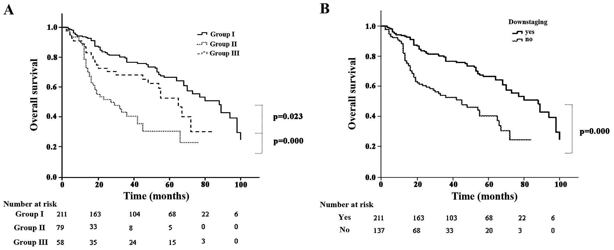
alive with recurrence. The 5-year OS rate was 32.2, 6.3 and 25.9%
in groups I, II and III, respectively (group I vs. III, P=0.023;
and group III vs. II, P<0.001) (Fig.
1A). Patients with persistent N2 disease had a lower 5-year OS
compared with that of patients whose disease was downstaged (14.6
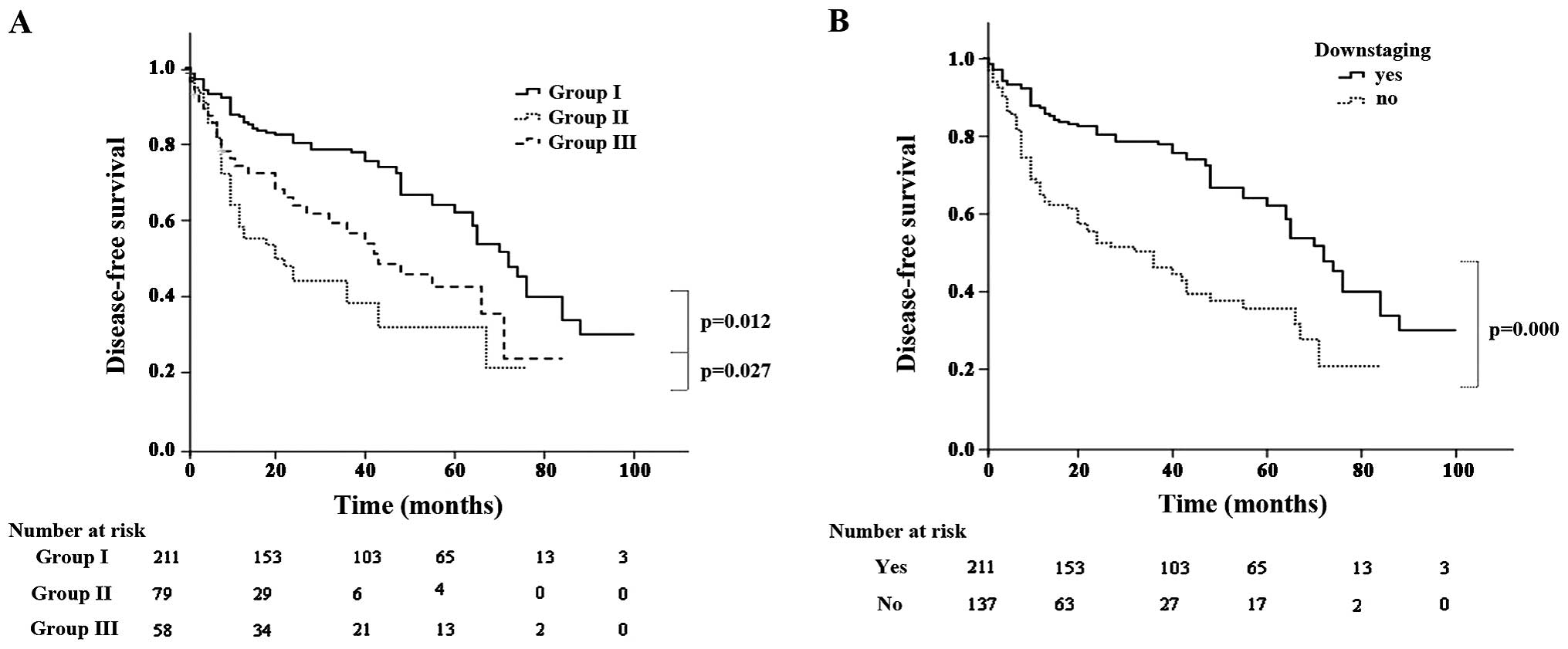
vs. 32.2%, respectively; P<0.001) (Fig. 1B). Recurrence occurred in 166 patients
(47.7%). The pattern of recurrence was locoregional in 69 patients,
distant in 71 and both in 26. The 5-year disease-free survival
(DFS) rate was 30.1, 5.1 and 22.4% in groups I, II and III,
respectively (group I vs. III, P=0.012; and group III vs. II,
P=0.027) (Fig. 2A). Patients with
persistent N2 disease had a lower 5-year DFS compared with that of
patients whose disease was downstaged (12.5. vs. 30.7%,
respectively; P<0.001) (Fig.
2B).
Multivariate analysis of OS and
DFS
All the variables were included in the multivariate
analysis. The results are summarized in Tables III and IV. Grouping (P=0.016), predicted forced
expiratory volume in 1 sec (P=0.035), N downstaging (P=0.013) and
skip N2 metastasis (P=0.003) were identified as independent
predictive factors associated with OS, whereas grouping (P=0.037)
and N downstaging (P=0.032) were found to be independent risk
factors associated with DFS.
| Table III.Multivariate analysis of overall
survival. |
Table III.
Multivariate analysis of overall
survival.
| Variables | Standard error | Hazard ratio | 95% CI | P-value |
|---|
| Grouping | 0.359 | 1.419 | 0.207–0.848 | 0.016 |
| Predicted FEV1 | 0.247 | 1.680 | 1.036–2.724 | 0.035 |
| N downstaging | 0.675 | 2.305 | 1.423–5.074 | 0.013 |
| Skip N2
metastasis | 0.253 | 2.104 | 1.281–3.466 | 0.003 |
| Table IV.Multivariate analysis of disease-free
survival. |
Table IV.
Multivariate analysis of disease-free
survival.
| Variables | Standard error | Hazard ratio | 95% CI | P-value |
|---|
| Grouping | 0.351 | 1.481 | 0.242–0.958 | 0.037 |
| N downstaging | 0.253 | 2.145 | 1.125–3.523 | 0.032 |
Discussion
The optimal treatment for resectable N2 NSCLC
remains controversial. Neoadjuvant chemotherapy was shown to
improve survival compared with surgery alone in certain randomized
trials (7,14,15).
However, these results were not confirmed by other randomized
studies (16,17). Additionally, a potential role for
radiation therapy in the treatment of N2 disease has been
considered. A study by van Meerbeeck et al reported no
differences in survival between surgery and radiation therapy
following induction chemotherapy (18). However, the different selection
criteria determining the optimal approach may be difficult to
evaluate, due to the significant heterogeneity among N2
patients.
Previous studies demonstrated that a mediastinal
pathological complete response was associated with improved
outcomes following resection compared with persistent N2 disease,
with long-term survival rates of patients with persistent N2
disease of <10–15% (6,9). In our study, the 5-year survival of
patients with persistent N2 disease was 14.6%, which was
significantly lower compared with that in patients with N
downstaging (32.2%, P<0.001). This was also consistent with
previous data. Therefore, certain studies concluded that
appropriate adjuvant therapies may be associated with better
outcomes in patients with persistent N2 disease compared with
surgery alone (2,6,7).
CT or PET/CT imaging may yield false-positive or
false-negative results, as they are based on the morphology or FDG
uptake of LNs. Silvestri et al reported the sensitivities of
CT and PET/CT for accurate detection of LN metastasis to be 51 and
74%, respectively, with respective specificities of 86 and 85%
(19). Consequently, false-negative
results may be obtained with imaging examinations, i.e., unexpected
persistent N2 disease following surgery, even when preoperative
imaging suggests N downstaging. Several authors have reported on
the outcomes of unexpected N2 disease (20,21);
however, the number of studies investigating unexpected persistent
N2 disease is limited. In our study, a total of 348 patients with
resectable N2 NSCLC received neoadjuvant chemotherapy. Among these
patients, 58 with persistent N2 disease had been considered to be
N-downstaged on either CT or PET/CT prior to surgery (16.7%).
In the present study, we compared the survival
outcomes among patients with N downstaging (pN0-1, group I), those
with positive imaging findings and pathological N2 disease
(expected persistent N2, group II) and those with negative imaging
findings but positive pathological N2 disease (unexpected
persistent N2, group III). The 5-year OS rate was 32.2 and 25.9% in
groups I and III, respectively (P=0.023), whereas the 5-year DFS
rate was 30.1 and 22.4% in groups I and III, respectively
(P=0.012). OS and DFS in group I were significantly higher compared
with those in group III. This may be attributed to patients in
group III undergoing more extensive resection compared with those
in group I (P<0.001), although the main reason was the disease
stage of the patients in group III. Of note, in our study, there
was a significant difference in OS and DFS between groups II and
III. Alhough both these groups had persistent N2 disease, different
clinical characteristics were associated with different survival.
Furthermore, the 5-year OS rate in group III was significantly
higher compared with that of patients with persistent N2 disease
(22.4 vs. 14.6%, P=0.033), suggesting that surgery was beneficial
for certain patients with persistent N2 disease.
The rate of objective response to chemotherapy was
75.9% (n=264). It was previously reported that objective response
was observed in >50% of cases (4,5,7,22) and
certain studies reported that response to chemotherapy was a strong
prognostic factor (4,7,21).
Accordingly, in our study, grouping and N downstaging were found to
be independent predictive factors associated with OS and DFS,
reflecting response to chemotherapy. Mediastinal node downstaging
on the resected specimen was a powerful and independent prognostic
factor, reflecting the radical resection of systemic disease and
effectiveness of chemotherapy. However, this factor was based on
postoperative specimen examination and patient selection may be
difficult. However, in our study, we demonstrated that patients
with unexpected persistent N2 disease exhibited satisfactory
survival rates. Port reported that certain patients may be cured by
surgery, particularly in case of previous response to chemotherapy
(23).
In patients with persistent N2 disease, mediastinal
LN-related variables, such as the number of N2 levels, may play a
role (3,6). In our series, we did not find N2-related
variables to be associated with survival. All the patients in our
study received neoadjuvant chemotherapy and the variability of
protocols and regimens may be a significant prognostic factor. In
fact, there was no difference regarding clinical response or
pathological nodal status and we did not identify an association
between regimen of chemotherapy and survival. In our study,
mediastinal regional radiotherapy was performed in patients with
persistent N2 disease, provided they were able to tolerate
additional treatments.
Our study had certain limitations. Due to the
retrospective nature of the study, some potentially predictive
clinical variables were not included; therefore, this study had
certain intrinsic drawbacks. Our study was limited by the small
patient sample and important information regarding the optimal
local treatment modality for patients with persistent N2 disease
could not be obtained. Our results suggested that surgery provided
a significant survival benefit for patients with unexpected N2
disease and should be first considered in this population. As
regards the optimal treatment for patients with expected N2
disease, our experience suggests that a definitive course of
radiotherapy (≥60 Gy) is generally recommended, combined with
chemotherapy.
In conclusion, we retrospectively analyzed the
clinical and pathological characteristics of NSCLC patients with N2
disease who underwent surgery following neoadjuvant chemotherapy.
The survival outcomes were compared according to whether persistent
N2 disease was preoperatively expected on the basis of CT or PET/CT
following chemotherapy. Patients with unexpected persistent N2
disease exhibited better survival outcomes compared with those with
expected persistent N2 disease. These findings may be used to
select the optimal therapeutic approach to persistent N2
disease.
References
|
1
|
Groome PA, Bolejack V, Crowley JJ, et al:
The IASLC Lung Cancer Staging Project: validation of the proposals
for the revision of the T, N, and M descriptors and consequent
stage groupings in the forthcoming (seventh) edition of the TNM
classification of malignant tumours. J Thorac Oncol. 2:694–705.
2007. View Article : Google Scholar : PubMed/NCBI
|
|
2
|
Martini N and Flehinger BJ: The role of
surgery in N2 lung cancer. Surg Clin North Am. 67:1037–1049.
1987.PubMed/NCBI
|
|
3
|
Andre F, Grunenwald D, Pignon JP, Dujon A,
Pujol JL, Brichon PY, Brouchet L, Quoix E, Westeel V and Le
Chevalier T: Survival of patients with resected N2 non-small-cell
lung cancer. Evidence for a subclassification and implications. J
Clin Oncol. 18:2981–2989. 2000.PubMed/NCBI
|
|
4
|
Albain KS, Rusch VW, Crowley JJ, et al:
Concurrent cisplatin/etoposide plus chest radiotherapy followed by
surgery for stages IIIA (N2) and IIIB non-small-cell lung cancer:
mature results of Southwest Oncology Group phase II study 8805. J
Clin Oncol. 13:1880–1892. 1995.PubMed/NCBI
|
|
5
|
Martini N, Kris MG, Flehinger BJ, et al:
Preoperative chemotherapy for stage IIIa (N2) lung cancer: The
Sloan-Kettering experience with 136 patients. Ann Thorac Surg.
55:1365–1373; discussion 1373–1374. 1993. View Article : Google Scholar
|
|
6
|
Lorent N, De Leyn P, Lievens Y, Verbeken
E, Nackaerts K, Dooms C, Van Raemdonck D, Anrys B and Vansteenkiste
J: Leuven Lung Cancer Group: Long-term survival of surgically
staged IIIA-N2 non-small-cell lung cancer treated with surgical
combined modality approach: Analysis of a 7-year prospective
experience. Ann Oncol. 15:1645–1653. 2004. View Article : Google Scholar : PubMed/NCBI
|
|
7
|
Rosell R, Gómez-Codina J, Camps C, et al:
A randomized trial comparing preoperative chemotherapy plus surgery
with surgery alone in patients with non-small-cell lung cancer. N
Engl J Med. 330:153–158. 1994. View Article : Google Scholar : PubMed/NCBI
|
|
8
|
Pataer A, Kalhor N, Correa AM, et al:
University of Texas M. Histopathology. J Thorac Oncol. 7:825–832.
2012. View Article : Google Scholar : PubMed/NCBI
|
|
9
|
Betticher DC, Schmitz Hsu SF, Tötsch M, et
al: Mediastinal lymph node clearance after docetaxel-cisplatin
neoadjuvant chemotherapy is prognostic of survival in patients with
stage IIIA pN2 non-small-cell lung cancer: a multicenter phase II
trial. J Clin Oncol. 21:1752–1759. 2003. View Article : Google Scholar : PubMed/NCBI
|
|
10
|
Kim YN, Yi CA, Lee KS, et al: A proposal
for combined MRI and PET/CT interpretation criteria for
preoperative nodal staging in non-small-cell lung cancer. Eur
Radiol. 22:1537–1546. 2012. View Article : Google Scholar : PubMed/NCBI
|
|
11
|
Goldstraw P, Crowley J, Chansky K, et al:
The IASLC Lung Cancer Staging Project: proposals for the revision
of the TNM stage groupings in the forthcoming (seventh) edition of
the TNM Classification of malignant tumours. J Thorac Oncol.
2:706–714. 2007. View Article : Google Scholar : PubMed/NCBI
|
|
12
|
Miller AB, Hoogstraten B, Staquet M and
Winkler A: Reporting results of cancer treatment. Cancer.
47:207–214. 1981. View Article : Google Scholar : PubMed/NCBI
|
|
13
|
Naruke T, Suemasu K and Ishikawa S: Lymph
node mapping and curability at various levels of metastasis in
resected lung cancer. J Thorac Cardiovasc Surg. 76:832–839.
1978.PubMed/NCBI
|
|
14
|
Pass HI, Pogrebniak HW, Steinberg SM,
Mulshine J and Minna J: Randomized trial of neoadjuvant therapy for
lung cancer, Interim analysis. Ann Thorac Surg. 53:992–998. 1992.
View Article : Google Scholar : PubMed/NCBI
|
|
15
|
Roth JA, Fossella F, Komaki R, et al: A
randomized trial comparing perioperative chemotherapy and surgery
with surgery alone in resectable stage IIIA non-small-cell lung
cancer. J Natl Cancer Inst. 86:673–680. 1994. View Article : Google Scholar : PubMed/NCBI
|
|
16
|
Depierre A, Milleron B, Moro-Sibilot D, et
al: French Thoracic Cooperative Group: Preoperative chemotherapy
followed by surgery compared with primary surgery in resectable
stage I (except T1N0), II, and IIIa non-small-cell lung cancer. J
Clin Oncol. 20:247–253. 2002. View Article : Google Scholar : PubMed/NCBI
|
|
17
|
Nagai K, Tsuchiya R, Mori T, Tada H,
Ichinose Y, Koike T and Kato H: Lung Cancer Surgical Study Group of
the Japan Clinical Oncology Group. A randomized trial comparing
induction chemotherapy followed by surgery with surgery alone for
patients with stage IIIA N2 non-small cell lung cancer (JCOG 9209).
J Thorac Cardiovasc Surg. 125:254–260. 2003. View Article : Google Scholar : PubMed/NCBI
|
|
18
|
van Meerbeeck JP, Kramer GW, Van Schil PE,
et al: European Organisation for Research and Treatment of
Cancer-Lung Cancer Group: Randomized controlled trial of resection
versus radiotherapy after induction chemotherapy in stage IIIA-N2
non-small-cell lung cancer. J Natl Cancer Inst. 99:442–450. 2007.
View Article : Google Scholar : PubMed/NCBI
|
|
19
|
Silvestri GA, Gould MK, Margolis ML, et
al: Noninvasive staging of non-small cell lung cancer: ACCP
evidenced-based clinical practice guidelines (2nd edition). Chest.
132((Suppl 3)): 178S–201S. 2007. View Article : Google Scholar : PubMed/NCBI
|
|
20
|
Kim HK, Choi YS, Kim K, Shim YM, Park K,
Ahn YC, Lee KS, Choi JY and Kim J: Outcomes of mediastinoscopy and
surgery with or without neoadjuvant therapy in patients with
non-small cell lung cancer who are N2 negative on positron emission
tomography and computed tomography. J Thorac Oncol. 6:336–342.
2011. View Article : Google Scholar : PubMed/NCBI
|
|
21
|
Al-Sarraf N, Aziz R, Gately K, Lucey J,
Wilson L, McGovern E and Young V: Pattern and predictors of occult
mediastinal lymph node involvement in non-small cell lung cancer
patients with negative mediastinal uptake on positron emission
tomography. Eur J Cardiothorac Surg. 33:104–109. 2008. View Article : Google Scholar : PubMed/NCBI
|
|
22
|
Eberhardt W, Wilke H, Stamatis G, et al:
Preoperative chemotherapy followed by concurrent chemoradiation
therapy based on hyperfractionated accelerated radiotherapy and
definitive surgery in locally advanced non-small-cell lung cancer:
mature results of a phase II trial. J Clin Oncol. 16:622–634.
1998.PubMed/NCBI
|
|
23
|
Port JL, Korst RJ, Lee PC, et al: Surgical
resection for residual N2 disease after induction chemotherapy. Ann
Thorac Surg. 79:1686–1690. 2005. View Article : Google Scholar : PubMed/NCBI
|