Introduction
Due to the involvement of the vocal folds, patients
with glottic cancer often present with hoarseness in the early
stages of the disease (T1–2N0M0). Approximately 75% of patients
with glottic cancer are diagnosed early (1). For early-stage glottic cancer, two
treatment options exist: radiation therapy (RT) and endolaryngeal
surgery (with or without a laser). The two modalities offer
excellent and comparable oncological outcomes (2); therefore, functional outcomes and
toxicity profiles are the predominant considerations used to
develop a therapeutic regimen (3).
Endolaryngeal surgery involves the en bloc
resection of a tumor with a safety margin (4). In contrast, the target volume in RT
encompasses the entire larynx, even for T1a glottic cancer, in
which the entire tumor is limited to one vocal cord (5). To decrease the irradiation of adjacent
non-cancerous tissues, the use of intensity-modulated RT (IMRT) for
early-stage glottic cancer patients has been recommended (6–8). However,
to date, few studies have investigated the possibility of changing
the target volume. Only one institution in the Netherlands has
investigated limiting the IMRT target volume to a single affected
vocal cord for T1a glottic cancer (9–14); their
studies revealed that IMRT with a limited target volume was
associated with a reduction in the irradiation of adjacent normal
tissue. However, the oncological outcomes of limiting the target
volume to a single vocal cord throughout the course of RT remain to
be elucidated.
In a similar manner to routine fractionated RT for
other tumor entities (15), it was
hypothesized that limiting the target volume to a single vocal cord
for patients with T1a glottic cancer may be appropriate if it is
performed following the completion of irradiation for subclinical
microscopic disease in the larynx. In the present study, the
results of a dosimetric analysis of a patient with T1a glottic
cancer are shown, whose IMRT target volume was reduced from the
whole larynx to the single vocal cord affected in the second phase
of treatment.
Case report
A 59-year-old male was admitted to hospital
complaining of hoarseness. A laryngoscopic examination revealed a
luminal protruding mass on the left vocal cord. Neck computed
tomography (CT) revealed a contrast-enhancing mass on the left
vocal cord without cervical lymph node enlargement. The tumor was
limited to the left vocal cord, and the vocal cord mobility
remained intact. The pathological analysis of a laryngoscopy biopsy
specimen revealed a moderately differentiated squamous cell
carcinoma. No evidence of distant metastasis was identified using
18F-fluorodeoxyglucose positron emission tomography-CT.
The clinical tumor-lymph node-metastasis stage was defined as
T1aN0M0 glottic cancer, according to the American Joint Committee
on Cancer Staging Manual Seventh Edition (16). The present study was performed in
accordance with the guidelines of our institutional review board;
written informed consent was waived, since this is a case report
with retrospective dosimetric analysis.
For RT simulation, the patient was immobilized in
the supine position with a thermoplastic head and neck mask. A
contrast-enhanced planning CT scan was performed using a 16-slice
CT scanner (Brilliance CT Big Bore; Philips Medical Systems,
Cleveland, OH, USA) with a 0.2-cm slice thickness. The initial
clinical target volume (CTV) included the false and true vocal
cords, anterior and posterior commissures, arytenoids and
subglottic region, extending from the superior thyroid notch to the
bottom of the cricoid cartilage. The planning target volume (PTV)
was created by adding a 0.5-cm isotropic margin around the CTV. The
contoured organs at risk (OAR) included the carotid artery, whole
thyroid gland and spinal cord. The right and left common, internal
and external carotid arteries were delineated from the
sternoclavicular joints, and upward to the base of the skull.
A volumetric modulated arc therapy (VMAT) plan was
created using the Eclipse Treatment Planning system (Varian Medical
Systems, Inc., Palo Alto, CA, USA), as previously described
(17). A double-arc plan was
generated using 6-MV photons and a high-definition multileaf
collimator. The plan was normalized in order that ≥95% of the PTV
received 100% of the prescribed dose. The prescribed dose in the
initial plan was 40.5 Gy at 2.25 Gy per fraction. Prior to the end
of the initial treatment session, a second planning CT scan was
obtained, and only the affected left vocal cord was contoured as
the CTV. The PTV was defined as the CTV plus a 0.5-cm margin. The
prescribed dose of the reduced field (RF) volumetric modulated
double-arc plan was 22.5 Gy in 10 fractions. Overall, the total
prescribed dose was 63 Gy in 28 fractions. The treatment was
performed using the Novalis Tx platform (Varian Medical Systems,
Inc./BrainLab, Feldkirchen, Germany). The set-up was verified daily
prior to treatment with ExacTrac X-ray monitoring (BrainLab), and
weekly by cone-beam CT. The patient completed the scheduled
treatment without interruption. During the course of treatment, the
patient developed acute mild dysphagia, which subsided
spontaneously.
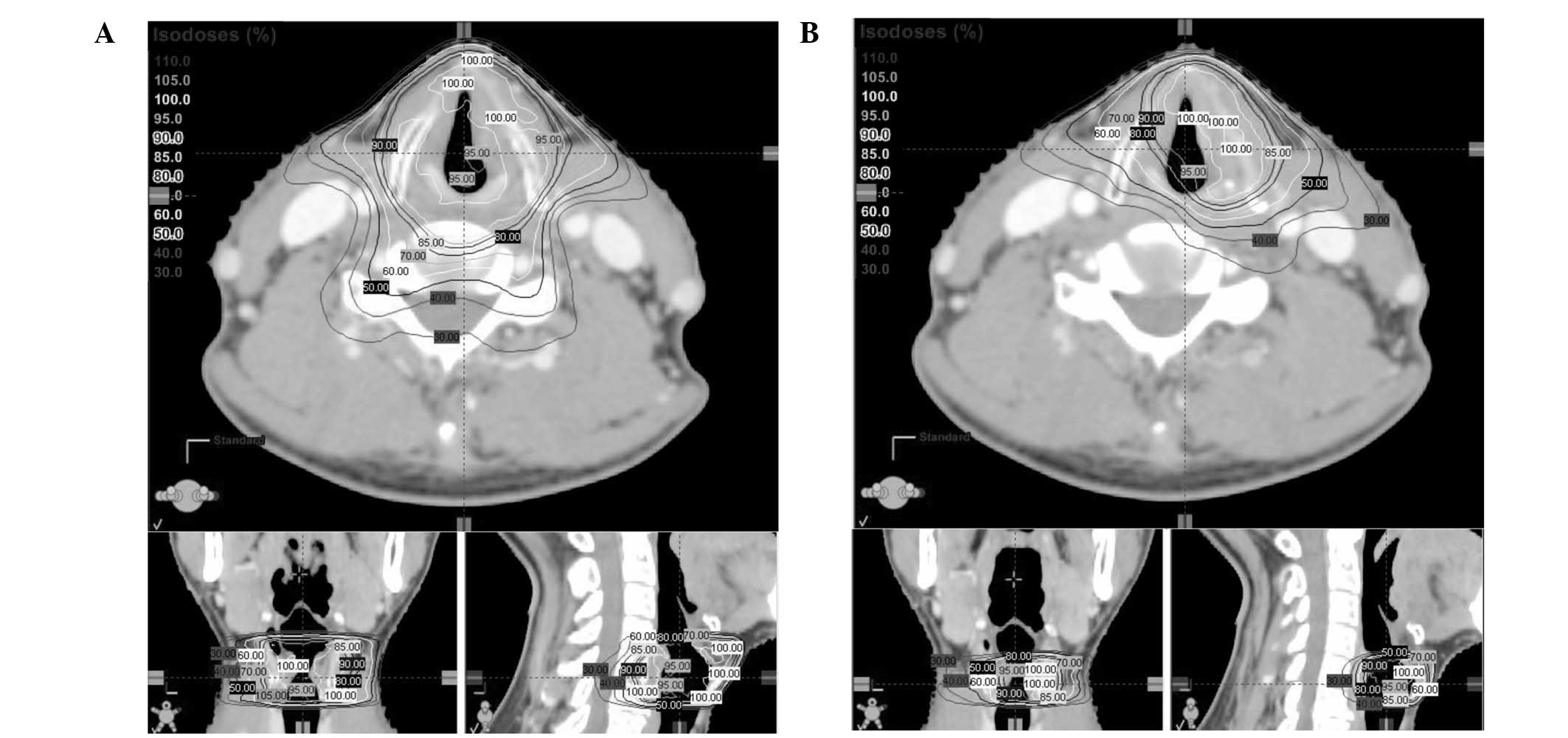
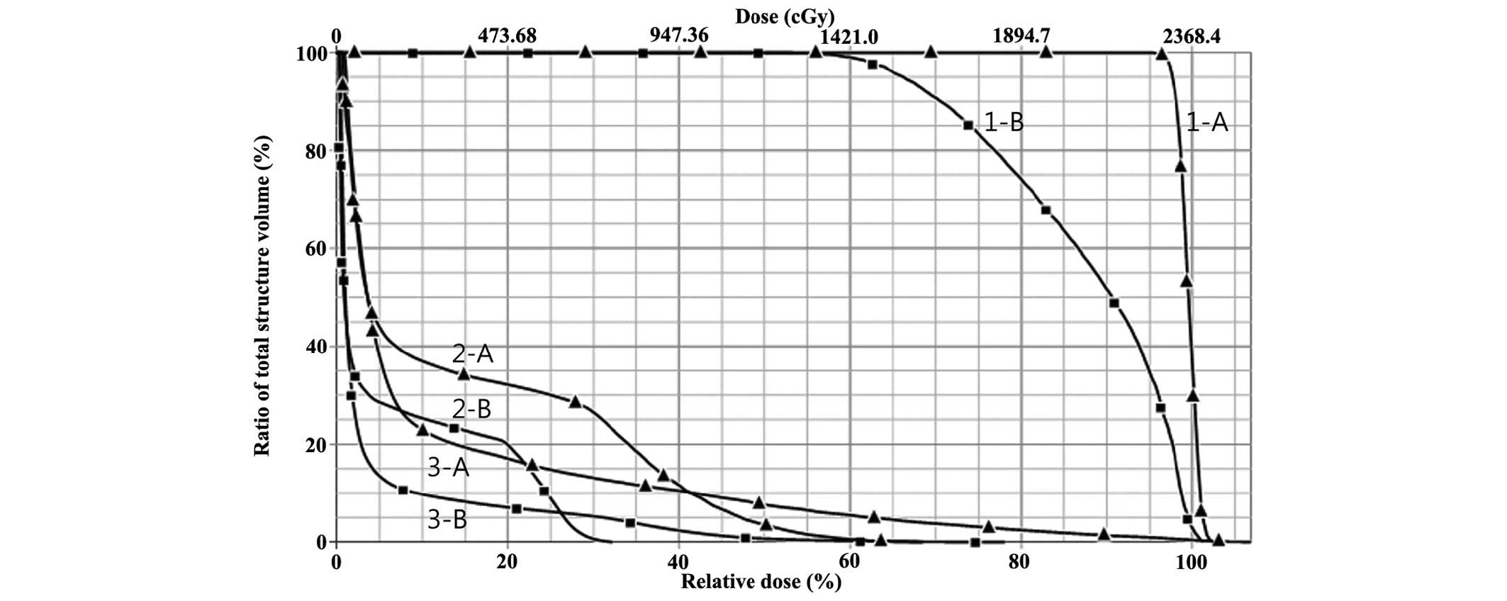
Figs. 1 and 2 show the isodose distribution and the
dose-volume histogram of the OAR, respectively, according to the
target volume (initial or RF plan). To analyze the dosimetric
advantages of the RF plan, the prescribed dose in the initial plan
was modified to be identical with that of the RF plan (22.5 Gy/10
fractions). Table I shows that the
adoption of the RF plan, compared with the initial plan, lowered
the mean and maximum doses to the OAR.
 | Table I.Dosimetric comparison by target volume
(planning target volume dose prescription=22.5 Gy/10
fractions). |
Table I.
Dosimetric comparison by target volume
(planning target volume dose prescription=22.5 Gy/10
fractions).
|
|
| Target volume |
|---|
|
|
|
|
|---|
| Tissue/gland | Dose (Gy) | Whole larynx | The involved left
vocal cord |
|---|
| Vocal cord,
right | Mean | 23.6 | 20.7 |
|
| Maximum | 24.3 | 24.0 |
| Carotid artery,
right | Mean | 3.4 | 1.6 |
|
| Maximum | 16.3 | 7.6 |
| Thyroid gland | Mean | 2.9 | 1.0 |
|
| Maximum | 25.3 | 18.5 |
Discussion
Unlike other head and neck cancers (18), the application of IMRT (including
VMAT) is not generalized in early-stage glottic cancer.
Conventional RT delivered with lateral opposed beams is more
commonly used for early-stage glottic cancer due to the small RT
field and the lack of requirement for elective cervical node
irradiation (17). However, several
institutions have demonstrated that IMRT is able to reduce
unnecessary irradiation of the carotid artery, which may cause
carotid artery stenosis and ischemic stroke (6–8).
Furthermore, it was demonstrated that IMRT is able to reduce
irradiation not only to the carotid artery, but also to the thyroid
gland, which is able to induce hypothyroidism (17). It was proposed that an increased
conformality with IMRT and organ motion may increase the risk of
marginal failures (19). However, a
previous clinical study of IMRT for early-stage glottic cancer
reported that the local control outcomes were comparable with those
with conventional RT (20,21).
Another aspect of RT that should be investigated is
the target volume range used for early-stage glottic cancer.
Endolaryngeal surgery is a highly focused treatment for a gross
tumor with safety margins (4),
whereas the RT field encompasses the whole larynx (5). Targeting only the involved vocal cord
with RT could further improve voice quality, and reduce the
radiation exposure of the surrounding OAR. Osman et al
(9–12), Levendag et al (13) and Kwa et al (14) explored the technical feasibility, and
dosimetric advantages, of targeting only a single affected vocal
cord with IMRT for T1a glottic cancer.
However, the clinical outcomes resulting from the
adoption of this major change in the target volume remain to be
elucidated. Janssen et al (22) reported a three-year local control rate
of 95% for 41 patients with T1–2 glottic cancer, among whom 31 had
received IMRT in which the target was restricted to the involved
laryngeal side. The extent of local control appears to be high,
although the present study did not report the local recurrence rate
according to the target volume range, or the site of local
recurrence. In addition, concurrent systemic chemotherapy has been
administered to several patients with T2 tumors. Zumsteg et
al (20) reported conventional RT
(n=282) and IMRT (n=48) outcomes for patients with T1–2N0M0 glottic
cancer. The IMRT target volume was the entire larynx. In total, 14
of 41 patients had recurrent bilateral vocal cord disease, although
the tumor presentation (single or bilateral) was not specified. Lim
et al (5) analyzed patterns of
local failure following conventional RT for 222 patients with
T1–2N0M0 glottic cancer. The local relapse sites of 29 patients who
presented with a tumor confined to a single vocal cord included 28
cases of a tumor of the ipsilateral vocal cord, and one case of a
tumor in the subglottis. The authors speculated that the absence of
recurrence involving the contralateral vocal cord was due to the
fact that the target volume involved the whole larynx.
Until the clinical feasibility of single vocal cord
RT is verified, reducing the RT target volume to a single vocal
cord may be permissible if performed subsequently to completion of
the irradiation necessary to sterilize subclinical disease in the
larynx. Generally, a dose of 45–50 Gy in 2 Gy fractions is
necessary to sterilize subclinical microscopic tumor cells
(15). In the present study, the
patient began RF treatment following initial treatments of 40.5
Gy/18 fractions, considering a fraction dose of 2.25 Gy and
incidental irradiation of the contralateral vocal cord during
single vocal cord RT. During RF treatment, the radiation dose
administered to the surrounding normal tissues, including the
contralateral vocal cord, carotid artery and thyroid gland, was
reduced compared with continuation of the initial IMRT plan
(Figs. 1 and 2, and Table
I). The normal tissue complication probability model of
radiation-induced hypothyroidism indicated that the probability of
hypothyroidism increases with a higher mean dose to the thyroid
gland (odds ratio, 1.064/Gy) (23).
In a previous study, VMAT exhibited a median 8 Gy reduction in mean
thyroid dose compared with conventional RT (17). The RF plan used in the present study
decreased the mean thyroid dose by an additional 2 Gy, and thus may
have helped to minimize the risk of hypothyroidism. Furthermore,
dose reduction to the vocal cord and the carotid artery is
anticipated to improve voice quality and decrease the risk of
cerebrovascular events, respectively.
A second planning CT scan and re-planning for RF
treatment is also beneficial with respect to the concept of
adaptive IMRT (24). The continuous
use of an initial, highly conformal IMRT plan with no adjustment
may increase the risk of marginal failure and toxicity, when the
morphology of the target and the OAR change markedly during a
treatment course with a duration of several weeks.
In conclusion, the present case study suggested that
RF re-planning with a change in target volume from the whole larynx
to the affected vocal cord during the latter course of IMRT for T1a
glottic cancer may successfully be adopted. By following this
scheme, functional outcomes ought to be improved, while exerting no
detrimental effects on the already excellent oncological
outcome.
Acknowledgements
This study was supported by the Soonchunhyang
University Research Fund.
References
|
1
|
Warner L, Chudasama J, Kelly CG, Loughran
S, McKenzie K, Wight R and Dey P: Radiotherapy versus open surgery
versus endolaryngeal surgery (with or without laser) for early
laryngeal squamous cell cancer. Cochrane Database Systematic Rev.
12:CD0020272014.
|
|
2
|
Yoo J, Lacchetti C, Hammond JA and Gilbert
RW: Head and Neck Cancer Disease Site Group: Role of endolaryngeal
surgery (with or without laser) versus radiotherapy in the
management of early (T1) glottic cancer: A systematic review. Head
Neck. 36:1807–1819. 2014. View Article : Google Scholar : PubMed/NCBI
|
|
3
|
van Loon Y, Sjügren EV, Langeveld TP, de
Jong Baatenburg RJ, Schoones JW and van Rossum MA: Functional
outcomes after radiotherapy or laser surgery in early glottic
carcinoma: A systematic review. Head Neck. 34:1179–1189. 2012.
View Article : Google Scholar : PubMed/NCBI
|
|
4
|
Remacle M, Eckel HE, Antonelli A, Brasnu
D, Chevalier D, Friedrich G, Olofsson J, Rudert HH, Thumfart W, de
Vincentiis M and Wustrow TP: Endoscopic cordectomy. A proposal for
a classification by the working committee, European laryngological
society. Eur Arch Otorhinolaryngol. 257:227–231. 2000. View Article : Google Scholar : PubMed/NCBI
|
|
5
|
Lim YJ, Wu HG, Kwon TK, Hah JH, Sung MW,
Kim KH and Park CI: Long-term outcome of definitive radiotherapy
for early glottic cancer: Prognostic factors and patterns of local
failure. Cancer Res Treat. 47:862–870. 2015. View Article : Google Scholar : PubMed/NCBI
|
|
6
|
Gomez D, Cahlon O, Mechalakos J and Lee N:
An investigation of intensity-modulated radiation therapy versus
conventional two-dimensional and 3D-conformal radiation therapy for
early stage larynx cancer. Radiat Oncol. 5:742010. View Article : Google Scholar : PubMed/NCBI
|
|
7
|
Rosenthal DI, Fuller CD, Barker JL Jr,
Mason B, Garcia JA, Lewin JS, Holsinger FC, Stasney CR, Frank SJ,
Schwartz DL, et al: Simple carotid-sparing intensity-modulated
radiotherapy technique and preliminary experience for T1-2 glottic
cancer. Int J Radiat Oncol Biol Phys. 77:455–461. 2010. View Article : Google Scholar : PubMed/NCBI
|
|
8
|
Chera BS, Amdur RJ, Morris CG and
Mendenhall WM: Carotid-sparing intensity-modulated radiotherapy for
early-stage squamous cell carcinoma of the true vocal cord. Int J
Radiat Oncol Biol Phys. 77:1380–1385. 2010. View Article : Google Scholar : PubMed/NCBI
|
|
9
|
Osman SO, de Boer HC, Heijmen BJ and
Levendag PC: Four-dimensional CT analysis of vocal cords mobility
for highly focused single vocal cord irradiation. Radiother Oncol.
89:19–27. 2008. View Article : Google Scholar : PubMed/NCBI
|
|
10
|
Osman SO, de Boer HC, Astreinidou E,
Gangsaas A, Heijmen BJ and Levendag PC: On-line cone beam CT image
guidance for vocal cord tumor targeting. Radiother Oncol. 93:8–13.
2009. View Article : Google Scholar : PubMed/NCBI
|
|
11
|
Osman SO, Astreinidou E, de Boer HC,
Keskin-Cambay F, Breedveld S, Voet P, Al-Mamgani A, Heijmen BJ and
Levendag PC: IMRT for image-guided single vocal cord irradiation.
Int J Radiat Oncol Biol Phys. 82:989–997. 2012. View Article : Google Scholar : PubMed/NCBI
|
|
12
|
Osman SO, Astreinidou E, Levendag PC and
Heijmen BJ: Impact of geometric variations on delivered dose in
highly focused single vocal cord IMRT. Acta Oncol. 53:278–285.
2014. View Article : Google Scholar : PubMed/NCBI
|
|
13
|
Levendag PC, Teguh DN, Keskin-Cambay F,
Al-Mamgani A, van Rooij P, Astreinidou E, Kwa SL, Heijmen B,
Monserez DA and Osman SO: Single vocal cord irradiation: A
competitive treatment strategy in early glottic cancer. Radiother
Oncol. 101:415–419. 2011. View Article : Google Scholar : PubMed/NCBI
|
|
14
|
Kwa SL, Al-Mamgani A, Osman SO, Gangsaas
A, Levendag PC and Heijmen BJ: Inter- and intrafraction target
motion in highly focused single vocal cord irradiation of T1a
larynx cancer patients. Int J Radiat Oncol Biol Phys. 93:190–195.
2015. View Article : Google Scholar : PubMed/NCBI
|
|
15
|
Withers HR, Peters LJ and Taylor JM:
Dose-response relationship for radiation therapy of subclinical
disease. Int J Radiat Oncol Biol Phys. 31:353–359. 1995. View Article : Google Scholar : PubMed/NCBI
|
|
16
|
Edge SB, Byrd DR, Compton CC, Fritz AG,
Greene FL and Trotti A: AJCC cancer staging manual (7th). New York,
NY: Springer. 2010.
|
|
17
|
Kim ES and Yeo SG: Volumetric modulated
arc radiotherapy sparing the thyroid gland for early-stage glottic
cancer: A dosimetrical analysis. Oncology Lett. 7:1987–1991.
2014.
|
|
18
|
Kong M, Hong SE, Choi J and Kim Y:
Comparison of survival rates between patients treated with
conventional radiotherapy and helical tomotherapy for head and neck
cancer. Radiat Oncol J. 31:1–11. 2013. View Article : Google Scholar : PubMed/NCBI
|
|
19
|
Feigenberg SJ, Lango M, Nicolaou N and
Ridge JA: Intensity-modulated radiotherapy for early larynx cancer:
Is there a role? Int J Radiat Oncol Biol Phys. 68:2–3. 2007.
View Article : Google Scholar : PubMed/NCBI
|
|
20
|
Zumsteg ZS, Riaz N, Jaffery S, Hu M,
Gelblum D, Zhou Y, Mychalczak B, Zelefsky MJ, Wolden S, Rao S and
Lee NY: Carotid sparing intensity-modulated radiation therapy
achieves comparable locoregional control to conventional
radiotherapy in T1-2N0 laryngeal carcinoma. Oral Oncol. 51:716–723.
2015. View Article : Google Scholar : PubMed/NCBI
|
|
21
|
Berwouts D, Swimberghe M, Duprez F,
Boterberg T, Bonte K, Deron P, De Gersem W, De Neve W and Madani I:
Intensity-modulated radiotherapy for early-stage glottic cancer.
Head Neck. 2014.(Epub ahead of print). PubMed/NCBI
|
|
22
|
Janssen S, Glanzmann C, Huber G and Studer
G: Risk-adapted partial larynx and/or carotid artery sparing
modulated radiation therapy of glottic cancer. Radiat Oncol.
9:1362014. View Article : Google Scholar : PubMed/NCBI
|
|
23
|
Boomsma MJ, Bijl HP, Christianen ME, Beetz
I, Chouvalova O, Steenbakkers RJ, van der Laan BF, Wolffenbuttel
BH, Oosting SF, Schilstra C and Langendijk JA: A prospective cohort
study on radiation-induced hypothyroidism: Development of an NTCP
model. Int J Radiat Oncol Biol Phys. 84:e351–e356. 2012. View Article : Google Scholar : PubMed/NCBI
|
|
24
|
Nishi T, Nishimura Y, Shibata T, Tamura M,
Nishigaito N and Okumura M: Volume and dosimetric changes and
initial clinical experience of a two-step adaptive intensity
modulated radiation therapy (IMRT) scheme for head and neck cancer.
Radiother Oncol. 106:85–89. 2013. View Article : Google Scholar : PubMed/NCBI
|