Introduction
Lung cancer is the most common type of cancer
worldwide (1). Concurrent
chemoradiotherapy (CCRT) is the standard treatment strategy for
locally advanced non-resectable non-small-cell lung cancer (NSCLC)
(2). However, the clinical outcome is
disappointing, with a 5-year survival rate of only ~20% (range,
15–40%) (3,4). Recently, combined treatment with
anti-angiogenic therapy and chemotherapy or radiotherapy has shown
a survival advantage (5). Among all
anti-angiogenic drugs, recombinant human endostatin (Endostar),
when administered with traditional CRT in patients with locally
advanced NSCLC, showed promising results (6–10). we
previously conducted a phase II clinical trial of endostatin
combined with CCRT in patients with locally advanced NSCLC,
achieving improved local control and survival rate (11). However, previous research has
demonstrated that not all patients benefit from anti-angiogenic
therapy (11). Thus, suitable case
selection for endostar treatment is crucial to avoid excessive
medical treatment.
It has been demonstrated that inflammation-mediated
interaction between endothelial cells and the innate immune system
plays an important role in resistance to anti-angiogenic therapy
(12,13). Neutrophil-to-lymphocyte ratio (NLR),
as estimated by the number of neutrophils/lymphocytes in the
peripheral blood, is a hallmark of systemic inflammation. It was
found that, in sunitinib-treated metastatic renal cell carcinoma
patients, NLR was associated with response rate (RR),
progression-free survival (PFS) and overall survival (OS) (14). Furthermore, previously published
studies indicate that inflammatory markers in bevacizumab-treated
NSCLC patients may be used as prognostic indicator (15). However, no reports have been published
regarding the prognostic value of inflammation-based factors in
endostar-based first-line treatment in locally advanced NSCLC.
Therefore, a retrospective analysis was conducted on
the basis of clinical trials (recombinant human endostatin in
combination with CCRT in patients with stage III NSCLC), to
determine the association of NLR with RR, PFS and OS in patients
treated with endostar plus CCRT for locally advanced NSCLC.
Patients and methods
Patients and therapy
Between 2009/03/11 and 2015/06/10, 82 patients with
untreated pathologically confirmed inoperable stage IIIA or IIIB
NSCLC were enrolled in this study. The patients received a combined
endostar and CCRT regimen at the Sun Yat-Sen University Cancer
Center and the Zhejiang cancer hospital. endostar (Simcere
Pharmaceutical, Nanjing, China) was administered according to the
protocol of the clinical trials (11). Each patient received four cycles of
endostar, with each cycle lasting for 1 week. The patients received
an intravenous (i.v.) loading dose of endostar (7.5
mg/m2/day) over 4 h for 7 days, or the same dose
continuously pumped for 120 h on weeks 1, 3, 5 and 7. endostar was
administered 1 week prior to the administration of chemotherapy and
thoracic radiotherapy, during the concurrent part of the treatment.
Patients received two cycles of platinum-based chemotherapy with
concurrent thoracic three-dimensional conformal radiation or
intensity-modulated radiotherapy at 60–66 Gy in 30–33 fractions for
6–7 weeks. For chemotherapy, docetaxel (65 mg/m2) and
cisplatin (65 mg/m2) on days 8 and 36 (TP regimen), or
etoposide (50 mg/m2) on days 1–5 and 29–33 and ciplatin
(50 mg/m2) on days 1, 8, 29 and 36 (EP regimen) were
used. Peripheral blood samples were collected at baseline, prior to
any treatment initiation. Of the 82 patients, 4 did not complete
treatment; thus, a total of 78 cases qualified for analysis. Data
were extracted from patient medical records. The data collected for
our analysis included age, gender, pathological type, performance
status (PS), pre-treatment laboratory findings (in particular
neutrophil, lymphocyte, monocyte and platelet counts), and
treatment outcome in terms of RR, PFS and OS. The responses were
assessed according to Response Evaluation Criteria in Solid Tumors,
version 1.1 (16). The study was
performed according to bioethics standards of the participating
institutions and the patients provided written informed consent to
data management at trial enrollment.
Statistical analysis
A total of 78 patients were evaluated for OS and
PFS. OS was defined as the time from the initiation of endostar
treatment to death, while PFS was defined as the time from the
initiation of endostar to disease progression or death. OS and PFS
were considered as the primary endpoints. All potential prognostic
factors were transformed into categorical variables. The patients
were grouped as male vs. female, squamous vs. non-squamous cell
carcinoma, neutrophil count >7,000 vs. ≤7,000/mm3
(upper limit of normal), lymphocyte count ≥1,100 vs.
<1,100/mm3 (lower limit of normal), monocyte count
>600 vs. ≤600/mm3 (upper limit of normal), platelet
count >350×103 vs. ≤350×103/mm3
(upper limit of normal), NLR <5 vs. ≥5 (ratio between neutrophil
and lymphocyte counts at baseline) and platelet-to-lymphocyte ratio
(PLR) <300 vs. ≥300 (ratio between platelet and lymphocyte
counts at baseline). The cut-off values of PLR and NLR were
determined as previously described (17). Survival curves and medians were
estimated with the Kaplan-Meier method and the association between
each variable and survival was assessed by the log-rank test in the
univariate analysis. The Cox proportional hazards model was used
subsequently in the multivariate analysis to assess the
contribution of each potential prognostic factor to survival. The
most significant variables were entered in the model through a
stepwise method. Two-category comparisons were performed with the
χ2 test. The analysis was performed using the SPSS
statistical package, version 16.0 (SPSS Inc., Chicago, IL,
USA).
Results
Patient characteristics
The patients' characteristics at baseline are
summarized in Table I. A total of 78
patients who received treatment with combined endostar and CCRT
were analyzed in our study, including 64 men and 14 women, with a
median age of 57 years (range, 31–71 years). A total of 25 patients
had stage IIIA and 53 patients stage IIIB disease; 25 patients had
a performance status (PS) of 0 and 53 patients had a PS of ≥1. As
regards pathological type, 47 patients had squamous cell carcinoma
and 31 patients non-squamous cell carcinoma. We observed complete
response (CR) after treatment in 13 patients, partial response (PR)
in 49 patients, stable disease (SD) in 6 patients and progressive
disease (PD) in 8 patients, whereas 2 patients could not be
evaluated. An NLR of ≥5 was found in 23 patients (29.48%), while
only 7 patients (8.97%) had a PLR of ≥300. The RR in the NLR
<5.0 group was significantly higher compared with that in the
NLR ≥5 group (87.27 vs. 60.87%, respectively; P=0.048).
 | Table I.Baseline characteristics of the
patients (n=78). |
Table I.
Baseline characteristics of the
patients (n=78).
| Characteristics | Values |
|---|
| Age, years [median
(range)] | 57 (31–71) |
| Gender
(male/female) | 64/14 |
| PS (0/1/2) | 25/52/1 |
| Histology
(Sq/non-Sq) | 47/31 |
| Stagea (IIIA/IIIB) | 25/53 |
| Neutrophil count
(mean ± SD/mm3) | 6,453±3,394
(1,200–2,000) |
| Lymphocyte count
(mean ± SD/mm3) | 1,454±585
(260–3,380) |
| Monocyte count (mean
± SD/mm3) | 541±295
(100–1,820) |
| Platelet count (mean
× 103 ± SD/mm3) | 267±92 (111–534) |
| NLR
(≥5.0/<5.0) | 23/55 |
| PLR
(≥300/<300) | 7/71 |
Inflammation-based indices and
survival
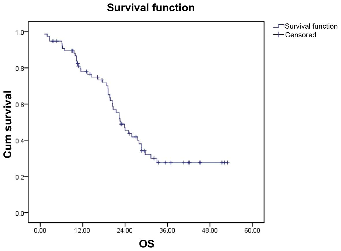
The median follow-up time was 21.48 months (range,
0.20–52.97 months). The overall median PFS of the entire group was
10.50 months (95% CI: 6.298–14.702), while the median OS was 22.83
months (95% CI:19.156–26.504) (Fig.
1).
We analyzed the effect of potential clinical
prognostic variables (including age, gender, PS, histology, stage,
neutrophil count, lymphocyte count, monocyte count, platelet count
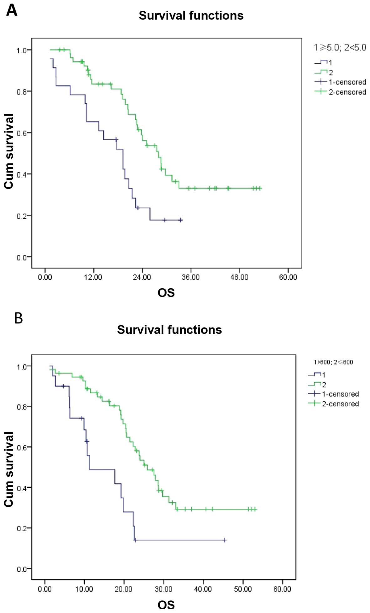
and NLR) on OS (Table II). In the
univariate analysis, NLR and monocyte count were found to be of
significant prognostic value (P=0.004 and P=0.001, respectively).
The OS of the NLR ≥5.0 and the NLR <5.0 groups was 19.23±3.563
months (95% CI: 12.246–26.214) and 27.93±2.654 months (95% CI:
22.727–33.133). In patients with a monocyte count
>600/mm3 and those with a monocyte count
≤600/mm3, the OS was 11.3±6.166 months (95% CI:
0.000–23.386) and 25.87±2.50 months (95% CI: 20.97–30.77) (Fig. 2). Other factors, including age
(P=0.712), gender (P=0.804), PS (P=0.492), stage (P=0.498),
histology (P=0.955), neutrophil count (P=0.157), lymphocyte count
(P=0.867), platelet count (P=0.990) and PLR (P=0.962), exerted no
significant effect on OS. The multivariate analysis confirmed NLR
(P=0.043, HR=0.502) and monocyte count (P=0.011, HR=0.387) as
independent risk factors predicting patient mortality. As regards T
and N stage, the patient sample was too small to perform a
statistical analysis (data not shown). In addition, NLR also
exerted a significant effect on PFS (P<0.001), with a PFS of
6.17±1.23 months (95% Cl:3.76–858) and 13.27±2.113 months (95%
Cl:9.129–17.411) in the NLR ≥5.0 and NLR <5.0 groups,
respectively.
| Table II.Univariate analysis associated for
OS. |
Table II.
Univariate analysis associated for
OS.
| Variables | OS (months) | 95% CI | P-value |
|---|
| Age, years |
|
| 0.712 |
|
<65 | 22.57±2.53 | 17.612–27.528 |
| ≥65 | 23.83±2.829 | 18.285–29.375 |
| Gender |
|
| 0.804 |
| Male | 22.83±2.159 | 18.598–27.062 |
|
Female | 25.00±2.690 | 19.727–30.273 |
| PS |
|
| 0.492 |
| 0 | 27.53 ±3.768 | 20.145–34.915 |
| ≥1 | 20.50 ±1.770 | 17.030–23.970 |
| Histology |
|
| 0.955 |
| Sq | 22.57±2.308 | 18.046–27.094 |
|
Non-Sq | 22.30±1.306 | 19.741–24.895 |
| Stage |
|
| 0.498 |
|
IIIA | 25.87±8.203 | 9.791–41.949 |
|
IIIB | 22.57±1.472 | 19.684–25.456 |
| Neutrophil
count,/mm3 |
|
| 0.157 |
|
≤7,000 | 24.00±3.438 | 17.261–30.739 |
|
>7,000 | 20.63±4.579 | 11.655–29.605 |
| Lymphocyte
count,/mm3 |
|
| 0.867 |
|
<1,100 | 22.330±1.575 | 19.243–25.417 |
|
≥1,100 | 23.83±2.144 | 19.628–28.032 |
| Monocyte
count,/mm3 |
|
| 0.001 |
| ≤600
cells | 25.87±2.50 | 20.97–30.77 |
| >600
cells | 11.30±6.166 | 0.000–23.386 |
| Platelet
count,/mm3 |
|
| 0.990 |
|
≤350×103 | 23.830±3.287 | 17.384–30.273 |
|
>350×103 | 22.33±2.082 | 18.249–26.411 |
| NLR |
|
| 0.004 |
|
<5.0 | 27.93±2.654 | 22.727–33.133 |
|
≥5.0 | 19.23±3.563 | 12.246–26.214 |
| PLR |
|
| 0.962 |
|
≥300 | 19.70±1.898 | 15.979–23.421 |
|
<300 | 22.57±2.152 | 18.352–26.788 |
Discussion
In the present study, we analyzed the potential
correlation between clinical variables and clinical outcome in
patients with locally advanced NSCLC treated with combined
first-line therapy with the anti-angiogenic drug endostar and CCRT.
A total of 82 patients with locally advanced NSCLC were enrolled in
the study; 4 patients did not complete treatment and, finally, 78
patients qualified for data analysis. The median PFS of the entire
group was 10.50 months, while the median OS was 22.83 months. A
total of 13 patients achieved CR after treatment, 49 achieved PR, 6
had SD, 8 developed PD, and 2 patients could not be evaluated. On
χ2test analysis, NLR exerted a significant effect on RR
(P=0.048). The univariate analysis revealed a correlation of low
NLR and low monocyte count with longer OS. The multivariate
analysis confirmed NLR and monocyte count as independent prognostic
factors predicting patient mortality. Keizman et al reported
a correlation between low NLR and higher PFS, OS and RR in
sunitinib treatment of metastatic renal cell carcinoma (14). In addition, Botta et al also
found that a systemic inflammatory status at baseline is an
important prognostic factor for PFS and OS in bevacizumab-treated
patients with advanced NSCLC (15).
These results all suggest that systemic inflammation is correlated
with resistance to anti-angiogenesis.
Mononuclear cells differentiate into
tumor-associated macrophages (TAMs) in the tumor tissue. Under
certain conditions, TAMs undergo tumor-promoting M2-like macrophage
polarization, and secrete angiogenic factors, such as vascular
endothelial growth factor, interleukin-8 and fibroblast growth
factor, to induce vascular formation, thus promoting angiogenesis
(18,19).
A high level of NLR has been previously reported to
be associated with prognosis in various tumor types, including
renal cell carcinoma, malignant mesothelioma, colorectal liver
metastases, advanced pancreatic cancer, ovarian cancer and gastric
cancer (20–26). Previously published studies reported
the prognostic value of pretreatment NLR in NSCLC patients
(27–30), although other studies reported
negative results (31–33). The opposite conclusions may have
resulted from the variability of the investigated cases. Thus,
although our study demonstrated that NLR is a prognostic factor in
stage III NSCLC patients treated with endostar combined with CCRT,
this conclusion requires validation by further clinical
studies.
The association between NLR and clinical outcome is
complex and remains to be elucidated. NLR reflects systemic
inflammation status to a certain extent. A high NLR reflects an
increased neutrophil-dependent inflammatory response and a reduced
lymphocyte-mediated antitumor immune response reaction, in turn
leading to enhanced tumor invasiveness, thus resulting in tumor
progression and poor prognosis (12).
Anti-angiogenic therapy results in vascular normalization, but
increased local hypoxia recruits bone marrow-derived cells,
including dendritic cells, endothelial cells and pericyte
progenitor cells and tams. These cells may produce a number of
different angiogenic factors, and acquire drug resistance by
hypoxic environment adaptation (34,35).
The findings of this study should be interpreted
with caution. First, this retrospective analysis did not
investigate other indices of inflammation, such as C-reactive
protein, erythrocyte sedimentation rate, or plasma inflammatory
cytokines. These indicators are not conventional items for clinical
detection and, therefore, their effect on treatment cannot be
evaluated. In addition, considering patient diversity and sample
size, we did not identify other statistically significant
clinicopathological factors. Finally, neutrophil and lymphocyte
count may also be affected by inflammation and drugs; however,
these factors were not taken into account.
Despite these considerations, we consider high
pretreatment NLR and monocyte count as indicators that patients
with locally advanced NSCLC may benefit less from combined therapy
with endostar and CCRT. These markers may provide useful
information for treatment decision-making, patient selection and
the design of clinical trials. However, our results require
validation by further prospective studies.
Acknowledgements
We would like to thank Dr Yan Xu for the collection
of clinical data.
References
|
1
|
Siegel RL, Miller KD and Jemal A: Cancer
statistics, 2015. CA Cancer J Clin. 65:5–29. 2015. View Article : Google Scholar : PubMed/NCBI
|
|
2
|
Pfister DG, Johnson DH, Azzoli CG, Sause
W, Smith TJ, Baker S Jr, Olak J, Stover D, Strawn JR, Turrisi AT,
et al: American society of clinical oncology treatment of
unresectable non-small-cell lung cancer guideline: Update 2003. J
Clin Oncol. 22:330–353. 2004. View Article : Google Scholar : PubMed/NCBI
|
|
3
|
Rengan R, Maity AM, Stevenson JP and Hahn
SM: New strategies in non-small cell lung cancer: Improving
outcomes in chemoradiotherapy for locally advanced disease. Clin
Cancer Res. 17:4192–4199. 2011. View Article : Google Scholar : PubMed/NCBI
|
|
4
|
Curran WJ Jr, Paulus R, Langer CJ, Komaki
R, Lee JS, Hauser S, Movsas B, Wasserman T, Rosenthal SA, Gore E,
et al: Sequential vs. concurrent chemoradiation for stage III
non-small cell lung cancer: Randomized phase III trial RTOG 9410. J
Natl Cancer Inst. 103:1452–1460. 2011. View Article : Google Scholar : PubMed/NCBI
|
|
5
|
Sandler A, Gray R, Perry MC, Brahmer J,
Schiller JH, Dowlati A, Lilenbaum R and Johnson DH:
Paclitaxel-carboplatin alone or with bevacizumab for non-small-cell
lung cancer. N Engl J Med. 355:2542–2550. 2006. View Article : Google Scholar : PubMed/NCBI
|
|
6
|
Han B, Xiu Q, Wang H, Shen J, Gu A, Luo Y,
Bai C, Guo S, Liu W, Zhuang Z, et al: A multicenter, randomized,
double-blind, placebo-controlled study to evaluate the efficacy of
paclitaxel-carboplatin alone or with endostar for advanced
non-small cell lung cancer. J Thorac Oncol. 6:1104–1109. 2011.
View Article : Google Scholar : PubMed/NCBI
|
|
7
|
Mu H, Shen C and Feng Y: Clinical
observation on the target therapy of rh-endostatin, combined with
chemotherapy in advanced non-small cell lung cancer. Chin J Lung
Cancer. 12:780–784. 2009.(In Chinese).
|
|
8
|
Rong B, Yang S, Li W, Zhang W and Ming Z:
Systematic review and meta-analysis of Endostar (rh-endostatin)
combined with chemotherapy versus chemotherapy alone for treating
advanced non-small cell lung cancer. World J Surg Oncol.
10:1702012. View Article : Google Scholar : PubMed/NCBI
|
|
9
|
Zhao X, Mei K, Cai X, Chen J, Yu J, Zhou C
and Li Q: A randomized phase II study of recombinant human
endostatin plus gemcitabine/cisplatin compared with
gemcitabine/cisplatin alone as first-line therapy in advanced
non-small-cell lung cancer. Invest New Drugs. 30:1144–1149. 2012.
View Article : Google Scholar : PubMed/NCBI
|
|
10
|
Jiang XD, Dai P, Wu J, Song DA and Yu JM:
Effect of recombinant human endostatin on radiosensitivity in
patients with non-small-cell lung cancer. Int J Radiat Oncol Biol
Phys. 83:1272–1277. 2012. View Article : Google Scholar : PubMed/NCBI
|
|
11
|
Bao Y, Peng F, Zhou QC, Yu ZH, Li JC,
Cheng ZB, Chen L, Hu X, Chen YY, Wang J, et al: Phase II trial of
recombinant human endostatin in combination with concurrent
chemoradiotherapy in patients with stage III non-small-cell lung
cancer. Radiother Oncol. 114:161–166. 2015. View Article : Google Scholar : PubMed/NCBI
|
|
12
|
Ribatti D, Crivellato E and Vacca A:
Inflammation and antiangiogenesis in cancer. Curr Med Chem.
19:955–960. 2012. View Article : Google Scholar : PubMed/NCBI
|
|
13
|
Albini A, Tosetti F, Benelli R and Noonan
DM: Tumor inflammatory angiogenesis and its chemoprevention. Cancer
Res. 65:10637–10641. 2005. View Article : Google Scholar : PubMed/NCBI
|
|
14
|
Keizman D, Ish-Shalom M, Huang P,
Eisenberger MA, Pili R, Hammers H and Carducci MA: The association
of pre-treatment neutrophil to lymphocyte ratio with response rate,
progression free survival and overall survival of patients treated
with sunitinib for metastatic renal cell carcinoma. Eur J Cancer.
48:202–208. 2012. View Article : Google Scholar : PubMed/NCBI
|
|
15
|
Botta C, Barbieri V, Ciliberto D, Rossi A,
Rocco D, Addeo R, Staropoli N, Pastina P, Marvaso G, Martellucci I,
et al: Systemic inflammatory status at baseline predicts
bevacizumab benefit in advanced non-small cell lung cancer
patients. Cancer Biol Ther. 14:469–475. 2013. View Article : Google Scholar : PubMed/NCBI
|
|
16
|
Eisenhauer EA, Therasse P, Bogaerts J,
Schwartz LH, Sargent D, Ford R, Dancey J, Arbuck S, Gwyther S,
Mooney M, Rubinstein L, Shankar L, Dodd L, Kaplan R, Lacombe D and
Verweij J: New response evaluation criteria in solid tumours:
revised RECIST guideline (version 1.1). Eur J Cancer. 45:228–247.
2009. View Article : Google Scholar : PubMed/NCBI
|
|
17
|
Pinato DJ, Shiner RJ, Seckl MJ, Stebbing
J, Sharma R and Mauri FA: Prognostic performance of
inflammation-based prognostic indices in primary operable non-small
cell lung cancer. Br J Cancer. 110:1930–1935. 2014. View Article : Google Scholar : PubMed/NCBI
|
|
18
|
Coffelt SB, Tal AO, Scholz A, De Palma M,
Patel S, Urbich C, Biswas SK, Murdoch C, Plate KH, Reiss Y and
Lewis CE: Angiopoietin-2 regulates gene expression in
TIE2-expressing monocytes and augments their inherent proangiogenic
functions. Cancer Res. 70:5270–5280. 2010. View Article : Google Scholar : PubMed/NCBI
|
|
19
|
Coffelt SB, Chen YY, Muthana M, Welford
AF, Tal AO, Scholz A, Plate KH, Reiss Y, Murdoch C, De Palma M and
Lewis CE: Angiopoietin 2 stimulates TIE2-expressing monocytes to
suppress T cell activation and to promote regulatory T cell
expansion. J Immunol. 186:4183–4190. 2011. View Article : Google Scholar : PubMed/NCBI
|
|
20
|
Kao SC, Pavlakis N, Harvie R, Vardy JL,
Boyer MJ, van Zandwijk N and Clarke SJ: High blood
neutrophil-to-lymphocyte ratio is an indicator of poor prognosis in
malignant mesothelioma patients undergoing systemic therapy. Clin
Cancer Res. 16:5805–5813. 2010. View Article : Google Scholar : PubMed/NCBI
|
|
21
|
Kishi Y, Kopetz S, Chun YS, Palavecino M,
Abdalla EK and Vauthey JN: Blood neutrophil-to-lymphocyte ratio
predicts survival in patients with colorectal liver metastases
treated with systemic chemotherapy. Ann Surg Oncol. 16:614–622.
2009. View Article : Google Scholar : PubMed/NCBI
|
|
22
|
Cho H, Hur HW, Kim SW, Kim SH, Kim JH, Kim
YT and Lee K: Pre-treatment neutrophil to lymphocyte ratio is
elevated in epithelial ovarian cancer and predicts survival after
treatment. Cancer Immunol Immunother. 58:15–23. 2009. View Article : Google Scholar : PubMed/NCBI
|
|
23
|
Liao W, Zhang J, Zhu Q, Qin L, Yao W, Lei
B, Shi W, Yuan S, Tahir SA, Jin J and He S: Preoperative
neutrophil-to-lymphocyte ratio as a new prognostic marker in
hepatocellular carcinoma after curative resection. Transl Oncol.
7:248–255. 2014. View Article : Google Scholar : PubMed/NCBI
|
|
24
|
An X, Ding PR, Li YH, Wang FH, Shi YX,
Wang ZQ, He YJ, Xu RH and Jiang WQ: Elevated neutrophil to
lymphocyte ratio predicts survival in advanced pancreatic cancer.
Biomarkerss. 15:516–522. 2010. View Article : Google Scholar
|
|
25
|
Yamanaka T, Matsumoto S, Teramukai S,
Ishiwata R, Nagai Y and Fukushima M: The baseline ratio of
neutrophils to lymphocytes is associated with patient prognosis in
advanced gastric cancer. Oncology. 73:215–220. 2007. View Article : Google Scholar : PubMed/NCBI
|
|
26
|
Ohno Y, Nakashima J, Ohori M, Hatano T and
Tachibana M: Pretreatment neutrophil-to-lymphocyte ratio as an
independent predictor of recurrence in patients with nonmetastatic
renal cell carcinoma. J Urol. 184:873–878. 2010. View Article : Google Scholar : PubMed/NCBI
|
|
27
|
Yao Y, Yuan D, Liu H, Gu X and Song Y:
Pretreatment neutrophil to lymphocyte ratio is associated with
response to therapy and prognosis of advanced non-small cell lung
cancer patients treated with first-line platinum-based
chemotherapy. Cancer Immunol Immunother. 62:471–479. 2013.
View Article : Google Scholar : PubMed/NCBI
|
|
28
|
Cedrés S, Torrejon D, Martínez A, Martinez
P, Navarro A, Zamora E, Mulet-Margalef N and Felip E: Neutrophil to
lymphocyte ratio (NLR) as an indicator of poor prognosis in stage
IV non-small cell lung cancer. Clin Transl Oncol. 14:864–869. 2012.
View Article : Google Scholar : PubMed/NCBI
|
|
29
|
Sarraf KM, Belcher E, Raevsky E, Nicholson
AG, Goldstraw P and Lim E: Neutrophil/lymphocyte ratio and its
association with survival after complete resection in non-small
cell lung cancer. J Thorac Cardiovasc Surg. 137:425–428. 2009.
View Article : Google Scholar : PubMed/NCBI
|
|
30
|
Unal D, Eroglu C, Kurtul N, Oguz A and
Tasdemir A: Are neutrophil/lymphocyte and platelet/lymphocyte rates
in patients with non-small cell lung cancer associated with
treatment response and prognosis? Asian Pac J Cancer Prev.
14:5237–5242. 2013. View Article : Google Scholar : PubMed/NCBI
|
|
31
|
Zhang J, Huang SH, Li H, Li Y, Chen XL,
Zhang WQ, Chen HG and Gu LJ: Preoperative lymphocyte count is a
favorable prognostic factor of disease-free survival in
non-small-cell lung cancer. Med Oncol. 30:3522013. View Article : Google Scholar : PubMed/NCBI
|
|
32
|
Kobayashi N, Usui S, Kikuchi S, Goto Y,
Sakai M, Onizuka M and Sato Y: Preoperative lymphocyte count is an
independent prognostic factor in node-negative non-small cell lung
cancer. Lung Cancer. 75:223–227. 2012. View Article : Google Scholar : PubMed/NCBI
|
|
33
|
Sakai T, Tsushima T, Kimura D, Hatanaka R,
Yamada Y and Fukuda I: A clinical study of the prognostic factors
for postoperative early recurrence in patients who underwent
complete resection for pulmonary adenocarcinoma. Ann Thorac
Cardiovasc Surg. 17:539–543. 2011. View Article : Google Scholar : PubMed/NCBI
|
|
34
|
Erler JT, Bennewith KL, Cox TR, Lang G,
Bird D, Koong A, Le QT and Giaccia AJ: Hypoxia-induced lysyl
oxidase is a critical mediator of bone marrow cell recruitment to
form the premetastatic niche. Cancer Cell. 15:35–44. 2009.
View Article : Google Scholar : PubMed/NCBI
|
|
35
|
Yang L, DeBusk LM, Fukuda K, Fingleton B,
Green-Jarvis B, Shyr Y, Matrisian LM, Carbone DP and Lin PC:
Expansion of myeloid immune suppressor Gr+CD11b+ cells in
tumor-bearing host directly promotes tumor angiogenesis. Cancer
Cell. 6:409–421. 2004. View Article : Google Scholar : PubMed/NCBI
|