Spandidos Publications Logo
  • About
    • About Spandidos
    • Aims and Scopes
    • Abstracting and Indexing
    • Editorial Policies
    • Reprints and Permissions
    • Job Opportunities
    • Terms and Conditions
    • Contact
  • Journals
    • All Journals
    • Oncology Letters
      • Oncology Letters
      • Information for Authors
      • Editorial Policies
      • Editorial Board
      • Aims and Scope
      • Abstracting and Indexing
      • Bibliographic Information
      • Archive
    • International Journal of Oncology
      • International Journal of Oncology
      • Information for Authors
      • Editorial Policies
      • Editorial Board
      • Aims and Scope
      • Abstracting and Indexing
      • Bibliographic Information
      • Archive
    • Molecular and Clinical Oncology
      • Molecular and Clinical Oncology
      • Information for Authors
      • Editorial Policies
      • Editorial Board
      • Aims and Scope
      • Abstracting and Indexing
      • Bibliographic Information
      • Archive
    • Experimental and Therapeutic Medicine
      • Experimental and Therapeutic Medicine
      • Information for Authors
      • Editorial Policies
      • Editorial Board
      • Aims and Scope
      • Abstracting and Indexing
      • Bibliographic Information
      • Archive
    • International Journal of Molecular Medicine
      • International Journal of Molecular Medicine
      • Information for Authors
      • Editorial Policies
      • Editorial Board
      • Aims and Scope
      • Abstracting and Indexing
      • Bibliographic Information
      • Archive
    • Biomedical Reports
      • Biomedical Reports
      • Information for Authors
      • Editorial Policies
      • Editorial Board
      • Aims and Scope
      • Abstracting and Indexing
      • Bibliographic Information
      • Archive
    • Oncology Reports
      • Oncology Reports
      • Information for Authors
      • Editorial Policies
      • Editorial Board
      • Aims and Scope
      • Abstracting and Indexing
      • Bibliographic Information
      • Archive
    • Molecular Medicine Reports
      • Molecular Medicine Reports
      • Information for Authors
      • Editorial Policies
      • Editorial Board
      • Aims and Scope
      • Abstracting and Indexing
      • Bibliographic Information
      • Archive
    • World Academy of Sciences Journal
      • World Academy of Sciences Journal
      • Information for Authors
      • Editorial Policies
      • Editorial Board
      • Aims and Scope
      • Abstracting and Indexing
      • Bibliographic Information
      • Archive
    • International Journal of Functional Nutrition
      • International Journal of Functional Nutrition
      • Information for Authors
      • Editorial Policies
      • Editorial Board
      • Aims and Scope
      • Abstracting and Indexing
      • Bibliographic Information
      • Archive
    • International Journal of Epigenetics
      • International Journal of Epigenetics
      • Information for Authors
      • Editorial Policies
      • Editorial Board
      • Aims and Scope
      • Abstracting and Indexing
      • Bibliographic Information
      • Archive
    • Medicine International
      • Medicine International
      • Information for Authors
      • Editorial Policies
      • Editorial Board
      • Aims and Scope
      • Abstracting and Indexing
      • Bibliographic Information
      • Archive
  • Articles
  • Information
    • Information for Authors
    • Information for Reviewers
    • Information for Librarians
    • Information for Advertisers
    • Conferences
  • Language Editing
Spandidos Publications Logo
  • About
    • About Spandidos
    • Aims and Scopes
    • Abstracting and Indexing
    • Editorial Policies
    • Reprints and Permissions
    • Job Opportunities
    • Terms and Conditions
    • Contact
  • Journals
    • All Journals
    • Biomedical Reports
      • Information for Authors
      • Editorial Policies
      • Editorial Board
      • Aims and Scope
      • Abstracting and Indexing
      • Bibliographic Information
      • Archive
    • Experimental and Therapeutic Medicine
      • Information for Authors
      • Editorial Policies
      • Editorial Board
      • Aims and Scope
      • Abstracting and Indexing
      • Bibliographic Information
      • Archive
    • International Journal of Epigenetics
      • Information for Authors
      • Editorial Policies
      • Editorial Board
      • Aims and Scope
      • Abstracting and Indexing
      • Bibliographic Information
      • Archive
    • International Journal of Functional Nutrition
      • Information for Authors
      • Editorial Policies
      • Editorial Board
      • Aims and Scope
      • Abstracting and Indexing
      • Bibliographic Information
      • Archive
    • International Journal of Molecular Medicine
      • Information for Authors
      • Editorial Policies
      • Editorial Board
      • Aims and Scope
      • Abstracting and Indexing
      • Bibliographic Information
      • Archive
    • International Journal of Oncology
      • Information for Authors
      • Editorial Policies
      • Editorial Board
      • Aims and Scope
      • Abstracting and Indexing
      • Bibliographic Information
      • Archive
    • Medicine International
      • Information for Authors
      • Editorial Policies
      • Editorial Board
      • Aims and Scope
      • Abstracting and Indexing
      • Bibliographic Information
      • Archive
    • Molecular and Clinical Oncology
      • Information for Authors
      • Editorial Policies
      • Editorial Board
      • Aims and Scope
      • Abstracting and Indexing
      • Bibliographic Information
      • Archive
    • Molecular Medicine Reports
      • Information for Authors
      • Editorial Policies
      • Editorial Board
      • Aims and Scope
      • Abstracting and Indexing
      • Bibliographic Information
      • Archive
    • Oncology Letters
      • Information for Authors
      • Editorial Policies
      • Editorial Board
      • Aims and Scope
      • Abstracting and Indexing
      • Bibliographic Information
      • Archive
    • Oncology Reports
      • Information for Authors
      • Editorial Policies
      • Editorial Board
      • Aims and Scope
      • Abstracting and Indexing
      • Bibliographic Information
      • Archive
    • World Academy of Sciences Journal
      • Information for Authors
      • Editorial Policies
      • Editorial Board
      • Aims and Scope
      • Abstracting and Indexing
      • Bibliographic Information
      • Archive
  • Articles
  • Information
    • For Authors
    • For Reviewers
    • For Librarians
    • For Advertisers
    • Conferences
  • Language Editing
Login Register Submit
  • This site uses cookies
  • You can change your cookie settings at any time by following the instructions in our Cookie Policy. To find out more, you may read our Privacy Policy.

    I agree
Search articles by DOI, keyword, author or affiliation
Search
Advanced Search
presentation
Molecular and Clinical Oncology
Join Editorial Board Propose a Special Issue
Print ISSN: 2049-9450 Online ISSN: 2049-9469
Journal Cover
September-2017 Volume 7 Issue 3

Full Size Image

Sign up for eToc alerts
Recommend to Library

Journals

International Journal of Molecular Medicine

International Journal of Molecular Medicine

International Journal of Molecular Medicine is an international journal devoted to molecular mechanisms of human disease.

International Journal of Oncology

International Journal of Oncology

International Journal of Oncology is an international journal devoted to oncology research and cancer treatment.

Molecular Medicine Reports

Molecular Medicine Reports

Covers molecular medicine topics such as pharmacology, pathology, genetics, neuroscience, infectious diseases, molecular cardiology, and molecular surgery.

Oncology Reports

Oncology Reports

Oncology Reports is an international journal devoted to fundamental and applied research in Oncology.

Experimental and Therapeutic Medicine

Experimental and Therapeutic Medicine

Experimental and Therapeutic Medicine is an international journal devoted to laboratory and clinical medicine.

Oncology Letters

Oncology Letters

Oncology Letters is an international journal devoted to Experimental and Clinical Oncology.

Biomedical Reports

Biomedical Reports

Explores a wide range of biological and medical fields, including pharmacology, genetics, microbiology, neuroscience, and molecular cardiology.

Molecular and Clinical Oncology

Molecular and Clinical Oncology

International journal addressing all aspects of oncology research, from tumorigenesis and oncogenes to chemotherapy and metastasis.

World Academy of Sciences Journal

World Academy of Sciences Journal

Multidisciplinary open-access journal spanning biochemistry, genetics, neuroscience, environmental health, and synthetic biology.

International Journal of Functional Nutrition

International Journal of Functional Nutrition

Open-access journal combining biochemistry, pharmacology, immunology, and genetics to advance health through functional nutrition.

International Journal of Epigenetics

International Journal of Epigenetics

Publishes open-access research on using epigenetics to advance understanding and treatment of human disease.

Medicine International

Medicine International

An International Open Access Journal Devoted to General Medicine.

Journal Cover
September-2017 Volume 7 Issue 3

Full Size Image

Sign up for eToc alerts
Recommend to Library

  • Article
  • Citations
    • Cite This Article
    • Download Citation
    • Create Citation Alert
    • Remove Citation Alert
    • Cited By
  • Similar Articles
    • Related Articles (in Spandidos Publications)
    • Similar Articles (Google Scholar)
    • Similar Articles (PubMed)
  • Download PDF
  • Download XML
  • View XML
Case Report Open Access

Non‑neural granular cell tumor of the uterine corpus mimicking uterine leiomyoma: A case report

  • Authors:
    • Yoko Deguchi
    • Naoyuki Iwahashi
    • Yuko Horiuchi
    • Miwa Ikejima
    • Tomoyuki Tanaka
    • Kazuhiko Ino
    • Kenichi Furukawa
  • View Affiliations / Copyright

    Affiliations: Department of Obstetrics and Gynecology, Hashimoto Municipal Hospital, Hashimoto, Wakayama 648‑0005, Japan, Department of Clinical Pathology, Hidaka General Hospital, Gobo, Wakayama 644‑0002, Japan, Department of Obstetrics and Gynecology, Wakayama Medical University, Wakayama 641‑0012, Japan
    Copyright: © Deguchi et al. This is an open access article distributed under the terms of Creative Commons Attribution License.
  • Pages: 351-354
    |
    Published online on: July 13, 2017
       https://doi.org/10.3892/mco.2017.1324
  • Expand metrics +
Metrics: Total Views: 0 (Spandidos Publications: | PMC Statistics: )
Metrics: Total PDF Downloads: 0 (Spandidos Publications: | PMC Statistics: )
Cited By (CrossRef): 0 citations Loading Articles...

This article is mentioned in:


Abstract

Non‑neural granular cell tumors (GCTs) are clinically rare, whereas cases arising in the uterine corpus are exceedingly rare. Only three uterine cervical cases of GCTs have been reported to date and, to the best of our knowledge, there are no reports of GCT of the uterine corpus in the literature. We herein describe the first case of non‑neural GCT arising from the uterine corpus reported to date. A 55‑year‑old premenopausal woman was referred to the Department of Obstetrics and Gynecology of Hashimoto Municipal Hospital (Wakayama, Japan) with a suspected uterine tumor. The tumor presented as a uterine leiomyoma‑like mass on radiological examinations, but the diagnosis of non‑neural GCT was established based on pathological and immunohistochemical examinations. Microscopically, histological examination of the entire surgical specimen revealed large polygonal cells with abundant eosinophilic granular cytoplasm and round to oval nuclei. Immunohistochemistry revealed positive periodic acid‑Schiff staining of the cytoplasmic granules, which was resistant to diastase. In addition, the tumor cells stained positive for CD68, but negative for S‑100, neuron‑specific enolase, cytokeratin, CD34, α‑smooth muscle actin, desmin, estrogen receptor and progesterone receptor. It is important for gynecologists to be aware of the possibility of non‑neural GCT of the uterine corpus, for which accurate diagnosis, complete resection and long‑term follow‑up are crucial, as it may be easily misdiagnosed as uterine leiomyoma.

Introduction

Granular cell tumor (GCT) is a clinically rare neoplasm composed of characteristic cells that contain numerous eosinophilic cytoplasmic granules, as shown on hematoxylin and eosin (H&E) staining (1). GCT may be divided into the neural type, which exhibits S-100 reactivity (conventional GCT), and non-neural GCT, which does not exhibit S-100 reactivity (2). Although non-neural GCT was first described in 1991 (3), it has not been fully characterized, as it has a rather unique immunophenotype, unlike conventional GCT, and reports of this entity in the literature are scarce (2–4). GCT may only be definitively diagnosed postoperatively based on detailed pathological and immunohistochemical examinations. Complete resection is recommended for malignant or metastatic GCT (1,5,6).

GCTs most commonly arises in dermal or subcutaneous regions, or the tongue (1); they may also develop in other parts of the body, but uterine GCT is extremely rare. To date, only three cases of uterine cervical GCT have been documented, all involving conventional GCTs (6–8); however, to the best of our knowledge, there have been no reports of GCT of the uterine corpus in the literature to date.

We herein describe the first reported case of non-neural GCT arising from the uterine corpus, which mimicked uterine leiomyoma and was treated with complete surgical resection, and discuss other related cases that have been reported in the literature.

Case report

A 55-year-old perimenopausal woman, gravida 1, para 1, was referred to the Department of Obstetrics and Gynecology of Hashimoto Municipal Hospital (Wakayama, Japan) with a suspected uterine tumor in October 2015. The patient had no history of lower abdominal or pelvic discomfort, pelvic surgery, or other relevant 
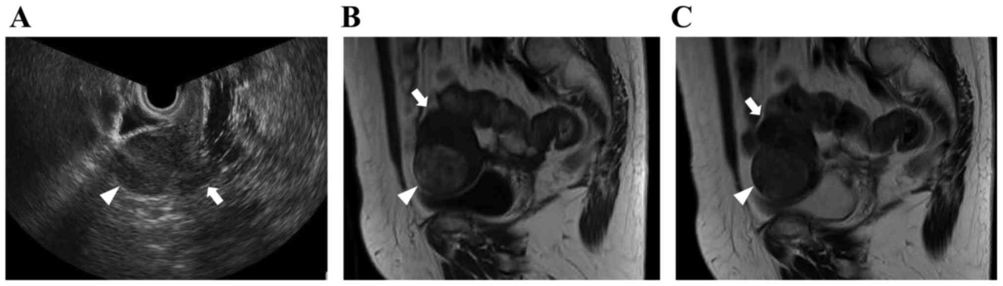
medical conditions. Transvaginal ultrasound examinations revealed a 4-cm well-defined uterine tumor, which exhibited iso-echogenicity (Fig. 1A). On magnetic resonance imaging (MRI), the tumor had a round shape, with heterogeneous signal intensity, including areas of isointensity or hyperintensity on T1-weighted imaging (WI) (Fig. 1B) and isointensity relative to muscle on T2WI (Fig. 1C). Based on these radiological findings, the mass was suspected to be a leiomyoma of the uterine corpus. A laboratory analysis of the patient's peripheral blood revealed normal tumor marker levels [cancer antigen (CA)125, CA19-9 and carcinoembryonic antigen] and an elevated lactate dehydrogenase level (215 IU/l). Total abdominal hysterectomy and bilateral 
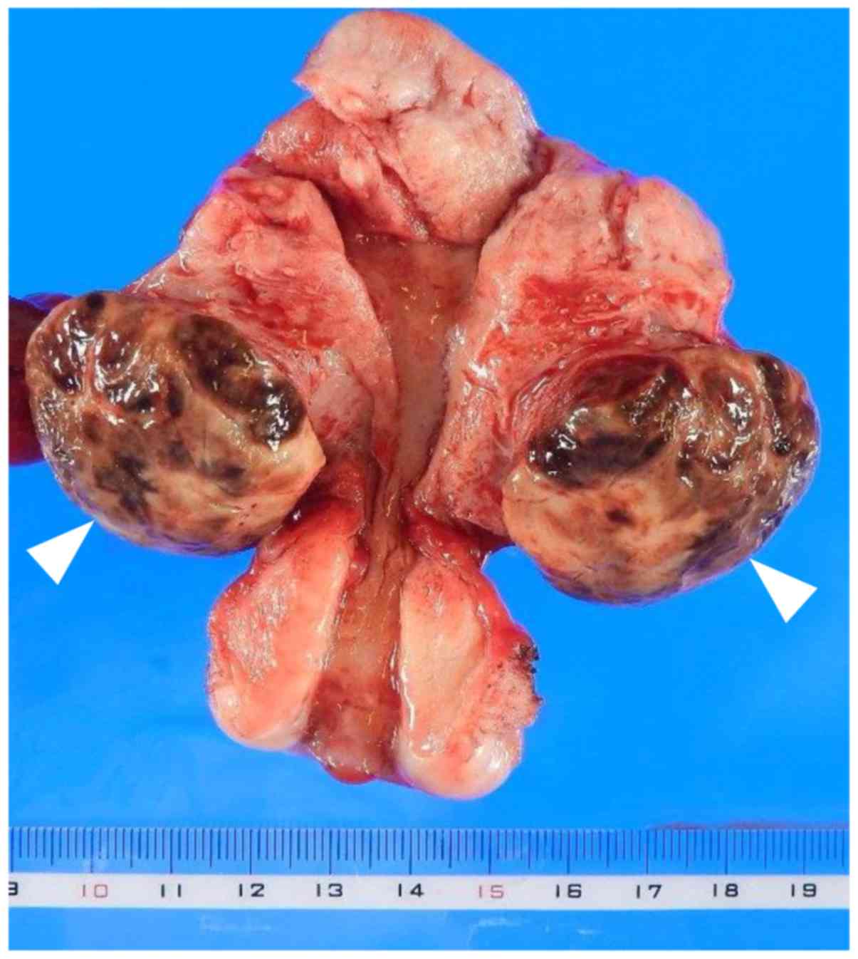
salpingo-oophorectomy were planned. On intraoperative examination, a tumor was identified arising from the anterior part of the uterine corpus, and it was completely resected. The mass was 3.7 cm in greatest diameter and elastic-hard in consistency; the cut 
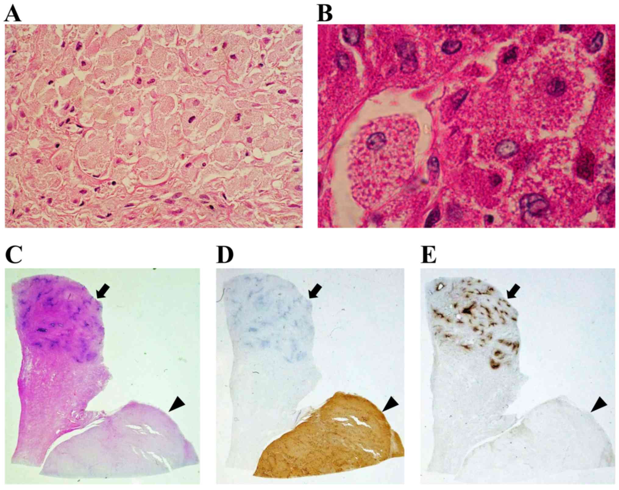
surface of the surgical specimen was yellowish-brown (Fig. 2). Microscopically, histological examination of the surgical specimen revealed large polygonal cells with abundant eosinophilic granular cytoplasm and round to oval nuclei (Fig. 3A). The tumor was located mainly in the muscle layer of the uterine body (Fig. 3C). The mitotic activity of the tumor cells were <5/10 high-power fields (HPFs) and the tumor had not invaded the mucosal layer (Fig. 3C). Immunohistochemistry revealed positive periodic acid-Schiff (PAS) staining of the cytoplasmic granules, which was resistant to diastase (Fig. 3B). In addition, the tumor cells stained positive for CD68 (Fig. 3D), but negative for S-100, neuron-specific enolase (NSE), cytokeratin, CD34, α-smooth muscle actin (SMA), desmin, estrogen receptor (ER) and progesterone receptor (PR) (Fig. 3E). The Ki-67 labeling index was ~6% in 20 random HPFs (data not shown). The immunohistochemistry findings excluded leiomyoma, leiomyosarcoma, malignant schwannoma, gastrointestinal stromal tumor and solitary fibrous tumor. The diagnosis of non-neural GCT of the uterine corpus was confirmed based on the pathological and immunohistochemical findings. The postoperative course was uneventful, and the patient was discharged from the hospital on postoperative day 7. At 12 months after the diagnosis, there was no evidence of local recurrence or systemic disease. Written informed consent was obtained from the patient regarding the publication of this case report and associated images.

Figure 1.

Radiological findings of non-neural granular cell tumor of the uterine corpus (A) Transvaginal ultrasonography; (B) T1-weighted sagittal image; (C) T2-weighted sagittal image (arrowheads, tumor; arrows, uterine corpus).

Figure 2.

Macroscopic findings of non-neural granular cell tumor (arrowheads) of the uterine corpus.

Figure 3.

Microscopic findings of non-neural granular cell tumor of the uterine corpus (A) Hematoxylin and eosin (H&E) staining (magnification, ×200); (B) periodic acid-Schiff staining (magnification, ×400). (C-E) Low magnification (x4) of tissue sections (arrowheads, tumor; arrows, endometrium of the uterine corpus) stained with (C) H&E; (D) anti-CD68; (E) anti-estrogen receptor.

Discussion

GCT is a rare slow-growing neoplasm that accounts for ~0.5% of all soft tissue tumors. Women are twice as likely to develop GCT as men. Furthermore, GCT mainly occurs between 40 and 60 years of age, and generally arises in the subcutaneous tissues of the head and neck region, although it may occur at several other sites (1). The majority of GCTs are considered to have undergone neural crest differentiation and exhibit positivity for S-100. These tumors are referred to as conventional GCTs (1). However, GCTs that do not display S-100 protein expression have been described in certain case series since 1991, and these tumors are referred to as non-neural GCTs (2–4). Therefore, GCT is classified into conventional GCT (S-100-positive) and non-neural GCT (S-100-negative) types. GCT rarely occurs as a gynecological tumor, although when it does it mainly arises in the vulva (6,7). As regards the uterus, only three cases of uterine cervical GCT have been reported to date, all involving conventional GCTs. However, to the best of our knowledge, no cases of non-neural GCT of the uterine corpus have been reported in the English literature (PubMed and MEDLINE databases) to date (6–8).

The diagnosis of non-neural GCT of the uterine corpus is difficult, as surgery is required for a definitive diagnosis. In a previous case of GCT, it was demonstrated that the results of imaging analysis are non-specific (5). On MRI, GCTs are usually located superficially and display a round or oval shape and heterogeneous signal intensity, including areas of isointensity or hyperintensity relative to muscle or suppressed fat signals on T1WI, whereas they frequently exhibited isointensity or hyperintensity relative to muscle on T2WI. In a case of GCT involving a premenopausal female patient, leiomyoma was initially suspected due to the round shape and smooth surface and the fact that it appeared isointense on both T1WI and T2WI, which made the diagnosis of GCT difficult.

Macroscopically, GCT surgical specimens are usually composed of yellowish-brown tissue with a nodular yellow-grey cut surface (1), as in the present case. Microscopically, GCT is composed of ovoid or polygonal cells with abundant eosinophilic granular cytoplasm, as shown on H&E staining. Immunohistochemical examination may be used to confirm the pathological diagnosis. In non-neural GCT, some of the cytoplasmic granules stain positively for PAS and are resistant to diastase, and the tumor cells stain positive for CD68, but are negative for S-100, NSE, cytokeratin, CD34 and other mesenchymal markers (α-SMA and desmin) or sex hormone receptors, namely ER and PR (2–4). In case of gynecological tumors, clinicians must be able to differentiate GCT from granular cell variants of leiomyoma. Such granular cell changes usually only affect part of the lesion, which allows GCT to be diagnosed using conventional morphological and immunohistochemical criteria based on mesenchymal markers and sex hormone receptors. In the present case, immunohistochemistry revealed that the tumor stained positive for PAS and CD68, whereas staining for S-100, NSE, cytokeratin, CD34, α-SMA, desmin, ER and PR was negative. Thus, a final diagnosis of non-neural GCT of the uterine corpus was made.

The most appropriate clinical management strategy for non-neural GCT of the uterine corpus is not always clear, as our experience with such cases is limited. GCT is usually a clinically and histologically benign tumor, but some malignant forms of GCT have been reported (1,5,6). Malignant GCTs are relatively uncommon, constituting only 1–2% of all GCTs. The characteristics of malignant GCT include nuclear pleomorphism, necrosis, and the presence of any mitotic activity combined with an aggressive clinical course and the destruction of neighboring structures. Thus, the differentiation between benign and malignant GCTs is only possible postoperatively based on detailed pathological and immunohistochemical examinations of the tumor. However, even in benign GCTs, local recurrence and secondary lymph node invasion have been reported (5). A wide, complete excisional margin is always preferred due to the lesion's infiltrative pattern and potential for recurrence, and to ensure that a correct pathological diagnosis is obtained. Metastasis and recurrence usually occurs within 2 years; thus, GCT should be followed up for at least 2 years postoperatively. According to three previous reports on GCT of the uterine cervix (6–8), uterine GCT appears to have a better prognosis following complete resection, as in the present case, although one of the cases involved a malignant GCT (6). Further data on uterine GCT are needed, particularly regarding its natural history, diagnosis and treatment.

To the best of our knowledge, this is the first reported case of non-neural GCT arising from the uterine corpus. The tumor was successfully treated with complete surgical resection. It is important for gynecologists to be aware of the existence of non-neural GCT of the uterine corpus, which requires accurate diagnosis, complete resection and long-term follow-up, combined with the findings on clinical presentation and pathology, as it may be misdiagnosed as uterine leiomyoma.

References

1 

Elkousy H, Harrelson J, Dodd L, Martinez S and Scully S: Granular cell tumors of the extremities. Clin Orthop Relat Res. 1–198. 2000.

2 

Lazar AJ and Fletcher CD: Primitive nonneural granular cell tumors of skin: Clinicopathologic analysis of 13 cases. Am J Surg Pathol. 29:927–934. 2005. View Article : Google Scholar : PubMed/NCBI

3 

LeBoit PE, Barr RJ, Burall S, Metcalf JS, Yen TS and Wick MR: Primitive polypoid granular-cell tumor and other cutaneous granular-cell neoplasms of apparent nonneural origin. Am J Surg Pathol. 15:48–58. 1991. View Article : Google Scholar : PubMed/NCBI

4 

Chaudhry IH and Calonje E: Dermal non-neural granular cell tumour (so-called primitive polypoid granular cell tumour): A distinctive entity further delineated in a clinicopathological study of 11 cases. Histopathology. 47:179–185. 2005. View Article : Google Scholar : PubMed/NCBI

5 

Behzatoğlu K and Bahadir B: Malignant granular cell tumor with unusual histological features. Pathol Int. 57:115–119. 2007. View Article : Google Scholar : PubMed/NCBI

6 

Guo N, Peng Z, Yang K and Lou J: Uterine cervical malignant granular cell tumor. J Obstet Gynaecol Res. 38:944–947. 2012. View Article : Google Scholar : PubMed/NCBI

7 

Yang F, He YM, Yao XY and Yang KX: Granular cell tumor of the uterine cervix. A case report. J Reprod Med. 58:177–180. 2013.PubMed/NCBI

8 

Haberal A, Turgut F, Ozbey B, Küçükalï T and Sapmaz M: Granular cell myoblastoma of the cervix in a 14 year old girl. Cent Afr J Med. 41:298–300. 1995.PubMed/NCBI

Related Articles

  • Abstract
  • View
  • Download
  • Twitter
Copy and paste a formatted citation
Spandidos Publications style
Deguchi Y, Iwahashi N, Horiuchi Y, Ikejima M, Tanaka T, Ino K and Furukawa K: Non‑neural granular cell tumor of the uterine corpus mimicking uterine leiomyoma: A case report. Mol Clin Oncol 7: 351-354, 2017.
APA
Deguchi, Y., Iwahashi, N., Horiuchi, Y., Ikejima, M., Tanaka, T., Ino, K., & Furukawa, K. (2017). Non‑neural granular cell tumor of the uterine corpus mimicking uterine leiomyoma: A case report. Molecular and Clinical Oncology, 7, 351-354. https://doi.org/10.3892/mco.2017.1324
MLA
Deguchi, Y., Iwahashi, N., Horiuchi, Y., Ikejima, M., Tanaka, T., Ino, K., Furukawa, K."Non‑neural granular cell tumor of the uterine corpus mimicking uterine leiomyoma: A case report". Molecular and Clinical Oncology 7.3 (2017): 351-354.
Chicago
Deguchi, Y., Iwahashi, N., Horiuchi, Y., Ikejima, M., Tanaka, T., Ino, K., Furukawa, K."Non‑neural granular cell tumor of the uterine corpus mimicking uterine leiomyoma: A case report". Molecular and Clinical Oncology 7, no. 3 (2017): 351-354. https://doi.org/10.3892/mco.2017.1324
Copy and paste a formatted citation
x
Spandidos Publications style
Deguchi Y, Iwahashi N, Horiuchi Y, Ikejima M, Tanaka T, Ino K and Furukawa K: Non‑neural granular cell tumor of the uterine corpus mimicking uterine leiomyoma: A case report. Mol Clin Oncol 7: 351-354, 2017.
APA
Deguchi, Y., Iwahashi, N., Horiuchi, Y., Ikejima, M., Tanaka, T., Ino, K., & Furukawa, K. (2017). Non‑neural granular cell tumor of the uterine corpus mimicking uterine leiomyoma: A case report. Molecular and Clinical Oncology, 7, 351-354. https://doi.org/10.3892/mco.2017.1324
MLA
Deguchi, Y., Iwahashi, N., Horiuchi, Y., Ikejima, M., Tanaka, T., Ino, K., Furukawa, K."Non‑neural granular cell tumor of the uterine corpus mimicking uterine leiomyoma: A case report". Molecular and Clinical Oncology 7.3 (2017): 351-354.
Chicago
Deguchi, Y., Iwahashi, N., Horiuchi, Y., Ikejima, M., Tanaka, T., Ino, K., Furukawa, K."Non‑neural granular cell tumor of the uterine corpus mimicking uterine leiomyoma: A case report". Molecular and Clinical Oncology 7, no. 3 (2017): 351-354. https://doi.org/10.3892/mco.2017.1324
Follow us
  • Twitter
  • LinkedIn
  • Facebook
About
  • Spandidos Publications
  • Careers
  • Cookie Policy
  • Privacy Policy
How can we help?
  • Help
  • Live Chat
  • Contact
  • Email to our Support Team