Spandidos Publications Logo
  • About
    • About Spandidos
    • Aims and Scopes
    • Abstracting and Indexing
    • Editorial Policies
    • Reprints and Permissions
    • Job Opportunities
    • Terms and Conditions
    • Contact
  • Journals
    • All Journals
    • Oncology Letters
      • Oncology Letters
      • Information for Authors
      • Editorial Policies
      • Editorial Board
      • Aims and Scope
      • Abstracting and Indexing
      • Bibliographic Information
      • Archive
    • International Journal of Oncology
      • International Journal of Oncology
      • Information for Authors
      • Editorial Policies
      • Editorial Board
      • Aims and Scope
      • Abstracting and Indexing
      • Bibliographic Information
      • Archive
    • Molecular and Clinical Oncology
      • Molecular and Clinical Oncology
      • Information for Authors
      • Editorial Policies
      • Editorial Board
      • Aims and Scope
      • Abstracting and Indexing
      • Bibliographic Information
      • Archive
    • Experimental and Therapeutic Medicine
      • Experimental and Therapeutic Medicine
      • Information for Authors
      • Editorial Policies
      • Editorial Board
      • Aims and Scope
      • Abstracting and Indexing
      • Bibliographic Information
      • Archive
    • International Journal of Molecular Medicine
      • International Journal of Molecular Medicine
      • Information for Authors
      • Editorial Policies
      • Editorial Board
      • Aims and Scope
      • Abstracting and Indexing
      • Bibliographic Information
      • Archive
    • Biomedical Reports
      • Biomedical Reports
      • Information for Authors
      • Editorial Policies
      • Editorial Board
      • Aims and Scope
      • Abstracting and Indexing
      • Bibliographic Information
      • Archive
    • Oncology Reports
      • Oncology Reports
      • Information for Authors
      • Editorial Policies
      • Editorial Board
      • Aims and Scope
      • Abstracting and Indexing
      • Bibliographic Information
      • Archive
    • Molecular Medicine Reports
      • Molecular Medicine Reports
      • Information for Authors
      • Editorial Policies
      • Editorial Board
      • Aims and Scope
      • Abstracting and Indexing
      • Bibliographic Information
      • Archive
    • World Academy of Sciences Journal
      • World Academy of Sciences Journal
      • Information for Authors
      • Editorial Policies
      • Editorial Board
      • Aims and Scope
      • Abstracting and Indexing
      • Bibliographic Information
      • Archive
    • International Journal of Functional Nutrition
      • International Journal of Functional Nutrition
      • Information for Authors
      • Editorial Policies
      • Editorial Board
      • Aims and Scope
      • Abstracting and Indexing
      • Bibliographic Information
      • Archive
    • International Journal of Epigenetics
      • International Journal of Epigenetics
      • Information for Authors
      • Editorial Policies
      • Editorial Board
      • Aims and Scope
      • Abstracting and Indexing
      • Bibliographic Information
      • Archive
    • Medicine International
      • Medicine International
      • Information for Authors
      • Editorial Policies
      • Editorial Board
      • Aims and Scope
      • Abstracting and Indexing
      • Bibliographic Information
      • Archive
  • Articles
  • Information
    • Information for Authors
    • Information for Reviewers
    • Information for Librarians
    • Information for Advertisers
    • Conferences
  • Language Editing
Spandidos Publications Logo
  • About
    • About Spandidos
    • Aims and Scopes
    • Abstracting and Indexing
    • Editorial Policies
    • Reprints and Permissions
    • Job Opportunities
    • Terms and Conditions
    • Contact
  • Journals
    • All Journals
    • Biomedical Reports
      • Information for Authors
      • Editorial Policies
      • Editorial Board
      • Aims and Scope
      • Abstracting and Indexing
      • Bibliographic Information
      • Archive
    • Experimental and Therapeutic Medicine
      • Information for Authors
      • Editorial Policies
      • Editorial Board
      • Aims and Scope
      • Abstracting and Indexing
      • Bibliographic Information
      • Archive
    • International Journal of Epigenetics
      • Information for Authors
      • Editorial Policies
      • Editorial Board
      • Aims and Scope
      • Abstracting and Indexing
      • Bibliographic Information
      • Archive
    • International Journal of Functional Nutrition
      • Information for Authors
      • Editorial Policies
      • Editorial Board
      • Aims and Scope
      • Abstracting and Indexing
      • Bibliographic Information
      • Archive
    • International Journal of Molecular Medicine
      • Information for Authors
      • Editorial Policies
      • Editorial Board
      • Aims and Scope
      • Abstracting and Indexing
      • Bibliographic Information
      • Archive
    • International Journal of Oncology
      • Information for Authors
      • Editorial Policies
      • Editorial Board
      • Aims and Scope
      • Abstracting and Indexing
      • Bibliographic Information
      • Archive
    • Medicine International
      • Information for Authors
      • Editorial Policies
      • Editorial Board
      • Aims and Scope
      • Abstracting and Indexing
      • Bibliographic Information
      • Archive
    • Molecular and Clinical Oncology
      • Information for Authors
      • Editorial Policies
      • Editorial Board
      • Aims and Scope
      • Abstracting and Indexing
      • Bibliographic Information
      • Archive
    • Molecular Medicine Reports
      • Information for Authors
      • Editorial Policies
      • Editorial Board
      • Aims and Scope
      • Abstracting and Indexing
      • Bibliographic Information
      • Archive
    • Oncology Letters
      • Information for Authors
      • Editorial Policies
      • Editorial Board
      • Aims and Scope
      • Abstracting and Indexing
      • Bibliographic Information
      • Archive
    • Oncology Reports
      • Information for Authors
      • Editorial Policies
      • Editorial Board
      • Aims and Scope
      • Abstracting and Indexing
      • Bibliographic Information
      • Archive
    • World Academy of Sciences Journal
      • Information for Authors
      • Editorial Policies
      • Editorial Board
      • Aims and Scope
      • Abstracting and Indexing
      • Bibliographic Information
      • Archive
  • Articles
  • Information
    • For Authors
    • For Reviewers
    • For Librarians
    • For Advertisers
    • Conferences
  • Language Editing
Login Register Submit
  • This site uses cookies
  • You can change your cookie settings at any time by following the instructions in our Cookie Policy. To find out more, you may read our Privacy Policy.

    I agree
Search articles by DOI, keyword, author or affiliation
Search
Advanced Search
presentation
Molecular and Clinical Oncology
Join Editorial Board Propose a Special Issue
Print ISSN: 2049-9450 Online ISSN: 2049-9469
Journal Cover
September-2017 Volume 7 Issue 3

Full Size Image

Sign up for eToc alerts
Recommend to Library

Journals

International Journal of Molecular Medicine

International Journal of Molecular Medicine

International Journal of Molecular Medicine is an international journal devoted to molecular mechanisms of human disease.

International Journal of Oncology

International Journal of Oncology

International Journal of Oncology is an international journal devoted to oncology research and cancer treatment.

Molecular Medicine Reports

Molecular Medicine Reports

Covers molecular medicine topics such as pharmacology, pathology, genetics, neuroscience, infectious diseases, molecular cardiology, and molecular surgery.

Oncology Reports

Oncology Reports

Oncology Reports is an international journal devoted to fundamental and applied research in Oncology.

Experimental and Therapeutic Medicine

Experimental and Therapeutic Medicine

Experimental and Therapeutic Medicine is an international journal devoted to laboratory and clinical medicine.

Oncology Letters

Oncology Letters

Oncology Letters is an international journal devoted to Experimental and Clinical Oncology.

Biomedical Reports

Biomedical Reports

Explores a wide range of biological and medical fields, including pharmacology, genetics, microbiology, neuroscience, and molecular cardiology.

Molecular and Clinical Oncology

Molecular and Clinical Oncology

International journal addressing all aspects of oncology research, from tumorigenesis and oncogenes to chemotherapy and metastasis.

World Academy of Sciences Journal

World Academy of Sciences Journal

Multidisciplinary open-access journal spanning biochemistry, genetics, neuroscience, environmental health, and synthetic biology.

International Journal of Functional Nutrition

International Journal of Functional Nutrition

Open-access journal combining biochemistry, pharmacology, immunology, and genetics to advance health through functional nutrition.

International Journal of Epigenetics

International Journal of Epigenetics

Publishes open-access research on using epigenetics to advance understanding and treatment of human disease.

Medicine International

Medicine International

An International Open Access Journal Devoted to General Medicine.

Journal Cover
September-2017 Volume 7 Issue 3

Full Size Image

Sign up for eToc alerts
Recommend to Library

  • Article
  • Citations
    • Cite This Article
    • Download Citation
    • Create Citation Alert
    • Remove Citation Alert
    • Cited By
  • Similar Articles
    • Related Articles (in Spandidos Publications)
    • Similar Articles (Google Scholar)
    • Similar Articles (PubMed)
  • Download PDF
  • Download XML
  • View XML
Case Report

Anastomosing haemangioma of liver: A case report

  • Authors:
    • Xiuhua Peng
    • Jin Li
    • Zonghui Liang
  • View Affiliations / Copyright

    Affiliations: Department of Radiology, Shanghai Jing'an District Centre Hospital, Shanghai 200040, P.R. China
  • Pages: 507-509
    |
    Published online on: July 24, 2017
       https://doi.org/10.3892/mco.2017.1341
  • Expand metrics +
Metrics: Total Views: 0 (Spandidos Publications: | PMC Statistics: )
Metrics: Total PDF Downloads: 0 (Spandidos Publications: | PMC Statistics: )
Cited By (CrossRef): 0 citations Loading Articles...

This article is mentioned in:


Abstract

Anastomosing haemangioma (AH) is a recently described, unusual variant of capillary hemangioma that appears to be unique to the genitourinary system, with a particular proclivity for the kidney. AH is a subtype of capillary haemangioma, which is rarely encountered in clinical practice, particularly in the liver. We herein present the case of a 57‑year‑old woman with an incidental finding on magnetic resonance imaging of a local lesion in the liver, sized 3.3x3.0 cm. The patient underwent hepatectomy with a good postoperative recovery. The histopathological diagnosis was AH of the liver. To the best of our knowledge, this is the first case report of hepatic AH.

Introduction

Anastomosing haemangioma (AH) is a subtype of capillary haemangioma that is rarely encountered in clinical practice, particularly in the liver. AH is a recently described and unusual variant of capillary hemangioma that appears to be unique to the genitourinary system, with a particular proclivity for the kidney. Previous studies provide strong evidence supporting the benign behavior of AH, whereas others suggest that less aggressive treatment may be preferable. Thus, the minimally invasive strategy, which is diagnostic biopsy and no treatment, should be weighed against the potential of significant tissue damage due to the growing lesion over time (1,2). We herein present a rare case of pathologically confirmed AH in the liver in a 57-year-old female patient.

Case report

A 57-year-old Chinese woman with no significant medical history was incidentally found to have a hepatic lesion on ultrasound imaging during a routine health check-up on September 20, 2014. A mass was identified on color Doppler imaging, sized ~3.3×2.8 cm. The possibility of malignancy was high, and metastasis was highly unlikely. The lesion was further evaluated with an abdominal contrast-enhanced magnetic resonance imaging (MRI) scan. 
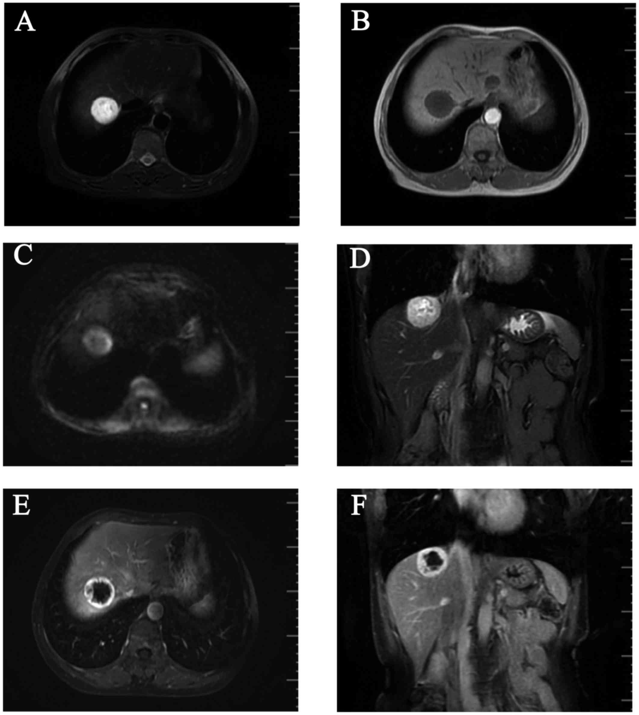
T1-weighted images (WI) on MRI revealed a round, well circumscribed, homogeneously hypointense mass, sized ~3.3×3.0 cm, in the right lobe of the liver (Fig. 1A), which was hyperintense on T2WI (Fig. 1B and D) and diffusion WI (DWI; Fig. 1C). The lesion exhibited a strong contrast enhancement in the periphery during the early phase (Fig. 1E) and homogeneously persistent enhancement with a well-circumscribed boundary during the late phase (Fig. 1F). A positron emission tomography-computed tomography (PET-CT) scan revealed a low-density mass in the right hepatic lobe, with lower glycometabolism compared with the normal liver. The patient consented to receiving a hepatectomy in December 5, 2014, and she remained alive and disease-free during the 12-month postoperative follow-up (last follow-up, December 30, 2015). The patient consented to the publication of the case details.

Figure 1.

Contrast-enhanced magnetic resonance imaging scan. A round, well-circumscribed mass sized ~3.3×3.0 cm was identified in the right hepatic lobe, (A) homogeneously hypointense on T1-weighted images (WI), exhibiting (B) marked hyperintensity on T2WI and (C) slight hyperintensity on diffusion WI. (D) The mass exhibited hyperintensity on coronal T2WI, with (E) strong contrast enhancement in the periphery during the early phase and (F) persistent enhancement with a well-circumscribed boundary during the late phase.

The tumour was located in the VIII segment of the liver in close proximity to the right hepatic vein. There were no lymph node metastases and no tumour thrombi were identified in the liver. On macroscopic inspection, the resected specimen was a well-circumscribed, homogeneous, encapsulated nodular mass, sized 3.5×3×3 cm and the cut surface of the tumour was fleshy and red-gray. On microscopic examination, various numbers of blood cells were observed in the anastomosing vessels and differently sized vascular compartments. The immunohistochemical staining for CD31 was positive. The histopathological appearance together with the immunophenotypic characteristics of this tumour were indicative of AH of the liver.

Discussion

Hemangiomas are more commonly located in the skin and subcutaneous tissues. Visceral hemangiomas are generally not that common and occur mostly in the liver. Histologically, hemangiomas have been broadly classified as cavernous and capillary (3,4). Hepatic hemangioma (HH) is the most common benign tumour of the liver during infancy (5). On the basis of its distribution in the liver, hemangiomas may be classified as focal, multifocal, or diffuse (6,7). The majority of hepatic hemangiomas are of the cavernous type, followed by the capillary type. AH is a rare subtype of hemangioma. AHs are distinguished from their cavernous counterparts by the anastomosing sinusoidal-like pattern of tightly packed capillary channels. Montgomery and Epstein described 6 cases of new variants of benign vascular tumours involving the kidneys, perinephric adipose tissue and testes, which the authors classified as AHs in 2009 (6). Subsequently, more cases of this vascular tumour have been reported in the adrenal gland (7), breast (8), liver and gastrointestinal tract (9). The mean size of this tumour in the liver is reportedly ~3.35 cm, which is larger compared with that in other organs (10). The incidence in male patients suggests a slight male predilection, with a reported male:female ratio of 1.8:1 (3). The association between end-stage renal disease and malignant renal epithelial neoplasms is well-documented (11).

Radiologically, as the number of reported cases of AH of the liver is limited, imaging information on the characteristics of hepatic AHs is also limited. Tao et al reported that the unenhanced axial CT scan showed a mass with a round, well-circumscribed outline, appearing to be heterogeneous. The boundary of the lesion exhibited an obviously annular and nodular enhancement in the arterial phase of the contrast-enhanced CT scan. In the venous phase, the lesion exhibited further intense enhancement. The lesion displayed homogeneously persistent enhancement and a well-circumscribed boundary on delayed images (10). A contrast-enhanced CT of the abdomen and pelvis confirmed a suspicious 3.4-cm renal mass, located between the left and middle part of the liver. A contrast-enhanced MRI scan revealed a round, well-demarcated mass in the upper pole of the right kidney, homogeneously hyperintense, measuring 2.3×2.1 cm (12).

In the present case, a contrast-enhanced abdominal MRI scan revealed a round, well-circumscribed mass sized ~3.3×3.0 cm in the right hepatic lobe, homogeneously hypointense on T1WI and hyperintense on T2WI and DWI. The lesion exhibited a strong contrast enhancement in the periphery during the early phase, with homogeneously persistent enhancement and a well-circumscribed boundary during the late phase.

Macroscopically, AHs are reportedly 0.1-6 cm in diameter and are well-demarcated, always unencapsulated, with a mahogany brown spongy appearance, without grossly evident necrosis or vascular invasion (3,4,8,9,13,14). On pathological examination, the characteristic cytoarchitectural features of AH include numerous thin-walled vascular channels exhibiting a complex anastomosing growth pattern, but no evident endothelial atypia or multilayering (14). Immunohistochemical studies demonstrated that the tumour cells were diffusely positive for CD34 and CD31, the stromal cells were positive for smooth muscle actin, and the Ki-67 indicates a low proliferative activity of the tumour cells (15). In the present case, the tumour was macroscopically fleshy and red-gray. The tumour cells were abundant and formed anastomosing vascular channels. The immunohistochemical staining for CD31 was positive.

The differential diagnosis of AH includes hepatocellular carcinoma (HCC), primary hepatic angiosarcoma (PHA) and hepatic angiomyolipoma (AML). Generally, HCC patients often have a history of chronic liver disease or cirrhosis and high serum α-fetoprotein (AFP) levels. The lesion exhibited strong contrast enhancement in the early phase and weakened enhancement in the late phase. Angiosarcoma, a subtype of soft tissue sarcoma, is an aggressive malignant disease derived from the endothelium of lymphatics or blood vessels (16). The typical imaging manifestation of PHA is moderate to marked enhancement at the peripheral and central area of the tumour during the arterial phase, which persists during the portal and delayed phases; in addition, in the majority of the cases, the central area of the lesion cannot be completely filled with the contrast agent. PHA is often accompanied by bleeding and is enclosed in a capsule. The majority of the PHA cases are accompanied by obvious clinical symptoms and compromised liver function. AML is a unique mesenchymal neoplasm consisting of blood vessels, smooth muscle and adipose cells (17). When the liver lesions exhibit early enhancement, peripheral enhancement, no capsule, the AFP levels are normal and there is no cirrhosis, the diagnosis of hepatic AH should be considered (18).

References

1 

Brown JG, Folpe AL, Rao P, Lazar AJ, Paner GP, Gupta R, Parakh R, Cheville JC and Amin MB: Primary vascular tumors and tumor-like lesions of the kidney: A clinicopathologic analysis of 25 cases. Am J Surg Pathol. 34:942–949. 2010. View Article : Google Scholar : PubMed/NCBI

2 

Wetherell DR, Skene A, Manya K, Manecksha RP, Chan Y and Bolton DM: Anastomosing haemangioma of the kidney: A rare morphological variant of haemangioma characteristic of genitourinary tract location. Pathology. 45:193–196. 2013. View Article : Google Scholar : PubMed/NCBI

3 

Meyers RL: Tumors of the liver in children. Surg Oncol. 16:195–203. 2007. View Article : Google Scholar : PubMed/NCBI

4 

Christison-Lagay ER, Burrows PE, Alomari A, Dubois J, Kozakewich HP, Lane TS, Paltiel HJ, Klement G, Mulliken JB and Fishman SJ: Hepatic hemangiomas: Subtype classification and development of a clinical practice algorithm and registry. J Pediatr Surg. 42:62–68. 2007. View Article : Google Scholar : PubMed/NCBI

5 

Kulungowski AM, Alomari AI, Chawla A, Christison-Lagay ER and Fishman SJ: Lessons from a liver hemangioma registry: Subtype classification. J Pediatr Surg. 47:165–170. 2012. View Article : Google Scholar : PubMed/NCBI

6 

Montgomery E and Epstein JI: Anastomosing hemangioma of the genitourinary tract: A lesion mimicking angiosarcoma. Am J Surg Pathol. 33:1364–1369. 2009. View Article : Google Scholar : PubMed/NCBI

7 

Ross M, Polcari A, Picken M, Sankary H and Milner J: Anastomosing hemangioma arising from the adrenal gland. Urology. 80:e27–e28. 2012. View Article : Google Scholar : PubMed/NCBI

8 

Brehm B, Rauh C, Dankerl P and Schulz-Wendtland R: Anastomosing hemangioma in the male breast-a rarity). Rofo. 186:80–81. 2014.PubMed/NCBI

9 

Lin J, Bigge J, Ulbright TM and Montgomery E: Anastomosing hemangioma of the liver and gastrointestinal tract: An unusual variant histologically mimicking angiosarcoma. Am J Surg Pathol. 37:1761–1765. 2013. View Article : Google Scholar : PubMed/NCBI

10 

Tao LL, Dai Y, Yin W and Chen J: A case report of a renal anastomosing hemangioma and a literature review: An unusual variant histologically mimicking angiosarcoma. Diagn Pathol. 9:1592014. View Article : Google Scholar : PubMed/NCBI

11 

Kryvenko ON, Haley SL, Smith SC, Shen SS, Paluru S, Gupta NS, Jorda M, Epstein JI, Amin MB and Truong LD: Haemangiomas in kidneys with end-stage renal disease: A novel clinicopathological association. Histopathology. 65:309–318. 2014. View Article : Google Scholar : PubMed/NCBI

12 

Zhao M, Li C, Zheng J and Sun K: Anastomosing hemangioma of the kidney: A case report of a rare subtype of hemangioma mimicking angiosarcoma and review of the literature. Int J Clin Exp Pathol. 6:757–765. 2013.PubMed/NCBI

13 

Kryvenko ON, Gupta NS, Meier FA, Lee MW and Epstein JI: Anastomosing hemangioma of the genitourinary system: Eight cases in the kidney and ovary with immunohistochemical and ultrastructural analysis. Am J Clin Pathol. 136:450–457. 2011. View Article : Google Scholar : PubMed/NCBI

14 

Mehta V, Ananthanarayanan V, Antic T, Krausz T, Milner J, Venkataraman G and Picken MM: Primary benign vascular tumors and tumorlike lesions of the kidney: A clinicopathologic analysis of 15 cases. Virchows Arch. 461:669–676. 2012. View Article : Google Scholar : PubMed/NCBI

15 

Omiyale AO: Anastomosing hemangioma of the kidney: A literature review of a rare morphological variant of hemangioma. Ann Transl Med. 3:1512015.PubMed/NCBI

16 

Heidegger I, Pichler R, Schäfer G, Zelger B, Zelger B, Aigner F, Bektic J and Horninger W: Long-term follow up of renal anastomosing hemangioma mimicking renal angiosarcoma. Int J Urol. 21:836–838. 2014. View Article : Google Scholar : PubMed/NCBI

17 

Cai PQ, Wu YP, Xie CM, Zhang WD, Han R and Wu PH: Hepatic angiomyolipoma: CT and MR imaging findings with clinical-pathologic comparison. Abdom Imaging. 38:482–489. 2013. View Article : Google Scholar : PubMed/NCBI

18 

Li T, Wang L, Yu HH, Sun HC, Qin LX, Ye QH, Fan J and Tang ZY: Hepatic angiomyolipoma: A retrospective study of 25 cases. Surg Today. 38:529–535. 2008. View Article : Google Scholar : PubMed/NCBI

Related Articles

  • Abstract
  • View
  • Download
  • Twitter
Copy and paste a formatted citation
Spandidos Publications style
Peng X, Li J and Liang Z: Anastomosing haemangioma of liver: A case report. Mol Clin Oncol 7: 507-509, 2017.
APA
Peng, X., Li, J., & Liang, Z. (2017). Anastomosing haemangioma of liver: A case report. Molecular and Clinical Oncology, 7, 507-509. https://doi.org/10.3892/mco.2017.1341
MLA
Peng, X., Li, J., Liang, Z."Anastomosing haemangioma of liver: A case report". Molecular and Clinical Oncology 7.3 (2017): 507-509.
Chicago
Peng, X., Li, J., Liang, Z."Anastomosing haemangioma of liver: A case report". Molecular and Clinical Oncology 7, no. 3 (2017): 507-509. https://doi.org/10.3892/mco.2017.1341
Copy and paste a formatted citation
x
Spandidos Publications style
Peng X, Li J and Liang Z: Anastomosing haemangioma of liver: A case report. Mol Clin Oncol 7: 507-509, 2017.
APA
Peng, X., Li, J., & Liang, Z. (2017). Anastomosing haemangioma of liver: A case report. Molecular and Clinical Oncology, 7, 507-509. https://doi.org/10.3892/mco.2017.1341
MLA
Peng, X., Li, J., Liang, Z."Anastomosing haemangioma of liver: A case report". Molecular and Clinical Oncology 7.3 (2017): 507-509.
Chicago
Peng, X., Li, J., Liang, Z."Anastomosing haemangioma of liver: A case report". Molecular and Clinical Oncology 7, no. 3 (2017): 507-509. https://doi.org/10.3892/mco.2017.1341
Follow us
  • Twitter
  • LinkedIn
  • Facebook
About
  • Spandidos Publications
  • Careers
  • Cookie Policy
  • Privacy Policy
How can we help?
  • Help
  • Live Chat
  • Contact
  • Email to our Support Team