Spandidos Publications Logo
  • About
    • About Spandidos
    • Aims and Scopes
    • Abstracting and Indexing
    • Editorial Policies
    • Reprints and Permissions
    • Job Opportunities
    • Terms and Conditions
    • Contact
  • Journals
    • All Journals
    • Oncology Letters
      • Oncology Letters
      • Information for Authors
      • Editorial Policies
      • Editorial Board
      • Aims and Scope
      • Abstracting and Indexing
      • Bibliographic Information
      • Archive
    • International Journal of Oncology
      • International Journal of Oncology
      • Information for Authors
      • Editorial Policies
      • Editorial Board
      • Aims and Scope
      • Abstracting and Indexing
      • Bibliographic Information
      • Archive
    • Molecular and Clinical Oncology
      • Molecular and Clinical Oncology
      • Information for Authors
      • Editorial Policies
      • Editorial Board
      • Aims and Scope
      • Abstracting and Indexing
      • Bibliographic Information
      • Archive
    • Experimental and Therapeutic Medicine
      • Experimental and Therapeutic Medicine
      • Information for Authors
      • Editorial Policies
      • Editorial Board
      • Aims and Scope
      • Abstracting and Indexing
      • Bibliographic Information
      • Archive
    • International Journal of Molecular Medicine
      • International Journal of Molecular Medicine
      • Information for Authors
      • Editorial Policies
      • Editorial Board
      • Aims and Scope
      • Abstracting and Indexing
      • Bibliographic Information
      • Archive
    • Biomedical Reports
      • Biomedical Reports
      • Information for Authors
      • Editorial Policies
      • Editorial Board
      • Aims and Scope
      • Abstracting and Indexing
      • Bibliographic Information
      • Archive
    • Oncology Reports
      • Oncology Reports
      • Information for Authors
      • Editorial Policies
      • Editorial Board
      • Aims and Scope
      • Abstracting and Indexing
      • Bibliographic Information
      • Archive
    • Molecular Medicine Reports
      • Molecular Medicine Reports
      • Information for Authors
      • Editorial Policies
      • Editorial Board
      • Aims and Scope
      • Abstracting and Indexing
      • Bibliographic Information
      • Archive
    • World Academy of Sciences Journal
      • World Academy of Sciences Journal
      • Information for Authors
      • Editorial Policies
      • Editorial Board
      • Aims and Scope
      • Abstracting and Indexing
      • Bibliographic Information
      • Archive
    • International Journal of Functional Nutrition
      • International Journal of Functional Nutrition
      • Information for Authors
      • Editorial Policies
      • Editorial Board
      • Aims and Scope
      • Abstracting and Indexing
      • Bibliographic Information
      • Archive
    • International Journal of Epigenetics
      • International Journal of Epigenetics
      • Information for Authors
      • Editorial Policies
      • Editorial Board
      • Aims and Scope
      • Abstracting and Indexing
      • Bibliographic Information
      • Archive
    • Medicine International
      • Medicine International
      • Information for Authors
      • Editorial Policies
      • Editorial Board
      • Aims and Scope
      • Abstracting and Indexing
      • Bibliographic Information
      • Archive
  • Articles
  • Information
    • Information for Authors
    • Information for Reviewers
    • Information for Librarians
    • Information for Advertisers
    • Conferences
  • Language Editing
Spandidos Publications Logo
  • About
    • About Spandidos
    • Aims and Scopes
    • Abstracting and Indexing
    • Editorial Policies
    • Reprints and Permissions
    • Job Opportunities
    • Terms and Conditions
    • Contact
  • Journals
    • All Journals
    • Biomedical Reports
      • Information for Authors
      • Editorial Policies
      • Editorial Board
      • Aims and Scope
      • Abstracting and Indexing
      • Bibliographic Information
      • Archive
    • Experimental and Therapeutic Medicine
      • Information for Authors
      • Editorial Policies
      • Editorial Board
      • Aims and Scope
      • Abstracting and Indexing
      • Bibliographic Information
      • Archive
    • International Journal of Epigenetics
      • Information for Authors
      • Editorial Policies
      • Editorial Board
      • Aims and Scope
      • Abstracting and Indexing
      • Bibliographic Information
      • Archive
    • International Journal of Functional Nutrition
      • Information for Authors
      • Editorial Policies
      • Editorial Board
      • Aims and Scope
      • Abstracting and Indexing
      • Bibliographic Information
      • Archive
    • International Journal of Molecular Medicine
      • Information for Authors
      • Editorial Policies
      • Editorial Board
      • Aims and Scope
      • Abstracting and Indexing
      • Bibliographic Information
      • Archive
    • International Journal of Oncology
      • Information for Authors
      • Editorial Policies
      • Editorial Board
      • Aims and Scope
      • Abstracting and Indexing
      • Bibliographic Information
      • Archive
    • Medicine International
      • Information for Authors
      • Editorial Policies
      • Editorial Board
      • Aims and Scope
      • Abstracting and Indexing
      • Bibliographic Information
      • Archive
    • Molecular and Clinical Oncology
      • Information for Authors
      • Editorial Policies
      • Editorial Board
      • Aims and Scope
      • Abstracting and Indexing
      • Bibliographic Information
      • Archive
    • Molecular Medicine Reports
      • Information for Authors
      • Editorial Policies
      • Editorial Board
      • Aims and Scope
      • Abstracting and Indexing
      • Bibliographic Information
      • Archive
    • Oncology Letters
      • Information for Authors
      • Editorial Policies
      • Editorial Board
      • Aims and Scope
      • Abstracting and Indexing
      • Bibliographic Information
      • Archive
    • Oncology Reports
      • Information for Authors
      • Editorial Policies
      • Editorial Board
      • Aims and Scope
      • Abstracting and Indexing
      • Bibliographic Information
      • Archive
    • World Academy of Sciences Journal
      • Information for Authors
      • Editorial Policies
      • Editorial Board
      • Aims and Scope
      • Abstracting and Indexing
      • Bibliographic Information
      • Archive
  • Articles
  • Information
    • For Authors
    • For Reviewers
    • For Librarians
    • For Advertisers
    • Conferences
  • Language Editing
Login Register Submit
  • This site uses cookies
  • You can change your cookie settings at any time by following the instructions in our Cookie Policy. To find out more, you may read our Privacy Policy.

    I agree
Search articles by DOI, keyword, author or affiliation
Search
Advanced Search
presentation
Molecular and Clinical Oncology
Join Editorial Board Propose a Special Issue
Print ISSN: 2049-9450 Online ISSN: 2049-9469
Journal Cover
December-2017 Volume 7 Issue 6

Full Size Image

Sign up for eToc alerts
Recommend to Library

Journals

International Journal of Molecular Medicine

International Journal of Molecular Medicine

International Journal of Molecular Medicine is an international journal devoted to molecular mechanisms of human disease.

International Journal of Oncology

International Journal of Oncology

International Journal of Oncology is an international journal devoted to oncology research and cancer treatment.

Molecular Medicine Reports

Molecular Medicine Reports

Covers molecular medicine topics such as pharmacology, pathology, genetics, neuroscience, infectious diseases, molecular cardiology, and molecular surgery.

Oncology Reports

Oncology Reports

Oncology Reports is an international journal devoted to fundamental and applied research in Oncology.

Experimental and Therapeutic Medicine

Experimental and Therapeutic Medicine

Experimental and Therapeutic Medicine is an international journal devoted to laboratory and clinical medicine.

Oncology Letters

Oncology Letters

Oncology Letters is an international journal devoted to Experimental and Clinical Oncology.

Biomedical Reports

Biomedical Reports

Explores a wide range of biological and medical fields, including pharmacology, genetics, microbiology, neuroscience, and molecular cardiology.

Molecular and Clinical Oncology

Molecular and Clinical Oncology

International journal addressing all aspects of oncology research, from tumorigenesis and oncogenes to chemotherapy and metastasis.

World Academy of Sciences Journal

World Academy of Sciences Journal

Multidisciplinary open-access journal spanning biochemistry, genetics, neuroscience, environmental health, and synthetic biology.

International Journal of Functional Nutrition

International Journal of Functional Nutrition

Open-access journal combining biochemistry, pharmacology, immunology, and genetics to advance health through functional nutrition.

International Journal of Epigenetics

International Journal of Epigenetics

Publishes open-access research on using epigenetics to advance understanding and treatment of human disease.

Medicine International

Medicine International

An International Open Access Journal Devoted to General Medicine.

Journal Cover
December-2017 Volume 7 Issue 6

Full Size Image

Sign up for eToc alerts
Recommend to Library

  • Article
  • Citations
    • Cite This Article
    • Download Citation
    • Create Citation Alert
    • Remove Citation Alert
    • Cited By
  • Similar Articles
    • Related Articles (in Spandidos Publications)
    • Similar Articles (Google Scholar)
    • Similar Articles (PubMed)
  • Download PDF
  • Download XML
  • View XML
Case Report Open Access

Mucinous tubular and spindle cell carcinoma with a high nuclear grade and micropapillary pattern: A case report

  • Authors:
    • Toru Sakatani
    • Yoshinaga Okumura
    • Naoto Kuroda
    • Toshihiro Magaribuchi
    • Yorika Nakano
    • Toshiaki Shirahase
    • Jun Watanabe
    • Yoji Taki
    • Mitsuhiko Okigaki
    • Susumu Ikehara
    • Yasushi Adachi
  • View Affiliations / Copyright

    Affiliations: Department of Urology, Toyooka Hospital, Toyooka, Hyogo 668‑8501, Japan, Department of Diagnostic Pathology, Kochi Red Cross Hospital, Kochi 780‑8562, Japan, Department of Histopathology and Cytology, Japanese Red Cross Kyoto Daini Hospital, Kyoto 602‑8026, Japan, Department of Internal Medicine, Otokoyama Hospital, Yawata, Kyoto 614‑8366, Japan, Professor Emeritus, Kansai Medical University, Hirakata, Osaka 573‑1010, Japan, Department of Diagnostic Pathology, Toyooka Hospital, Toyooka, Hyogo 668‑8501, Japan
    Copyright: © Sakatani et al. This is an open access article distributed under the terms of Creative Commons Attribution License.
  • Pages: 976-980
    |
    Published online on: September 29, 2017
       https://doi.org/10.3892/mco.2017.1430
  • Expand metrics +
Metrics: Total Views: 0 (Spandidos Publications: | PMC Statistics: )
Metrics: Total PDF Downloads: 0 (Spandidos Publications: | PMC Statistics: )
Cited By (CrossRef): 0 citations Loading Articles...

This article is mentioned in:


Abstract

Mucinous tubular and spindle cell carcinoma (MTSCC) is rare in renal cell carcinoma (RCC) and usually demonstrates a low nuclear grade and a better prognosis compared with other RCCs. The authors present a case report of MTSCC containing an area of Fuhrman nuclear grade 3, in addition to an area with a micropapillary pattern. An 82‑year‑old man consulted a hospital due to macrohematuria, and a tumor in the right kidney was detected. The tumor was resected and histologically examined. The tumor consisted of various growth patterns: Elongated tubular structure, a papillary structure containing a micropapillary pattern and solid pattern with spindle cells. The tumor cells demonstrated Fuhrman nuclear grades 2 and 3. Invasion into the lymph vessel and metastasis into the regional lymph node were observed. Thus, the tumor was diagnosed as a high grade MTSCC. Five months following resection, a computed tomography scan suggested metastasis of the tumor into the para‑aortic lymph nodes and liver, and the patient succumbed to brain metastasis. When MTSCC of kidney is observed, careful histological observation is important to avoid missing a high nuclear grade area.

Introduction

Mucinous tubular and spindle cell carcinoma (MTSCC) of the kidney is a rare tumor, which has been integrated into the World Health Organization (WHO) classification since 2004 (1–3). MTSCC is histologically characterized by elongated tubular, cord-like architecture and cuboidal to spindle cells with low nuclear grade and myxoid/mucinous stroma (1,3). MTSCC usually consists of low nuclear grade tumor cells, and MTSCC with high nuclear grade (more than Fuhrman grade 3) is extremely rare (4,5).

A micropapillary growth pattern in carcinoma was first reported for breast cancer (6). Currently, micropapillary growth pattern is well known in various carcinomas, such as lung adenocarcinoma (7), colorectal adenocarcinoma (8), gastric adenocarcinoma (9), and urothelial carcinoma (10). However, micropapillary growth pattern in renal cell carcinoma (RCC) is extremely rare (11).

In this study, we described a case of high nuclear grade MTSCC with micropapillary pattern and poor prognosis.

Case report

An 82-year-old man consulted a hospital because of macrohematuria. The patient had brain infarction, diabetes mellitus, and hypertension. Τhe patient's family provided written informed consent to the publication of the case details and associated images. The Ethics Committee of the Toyooka Hospital (Hyogo, Japan) approved the study.

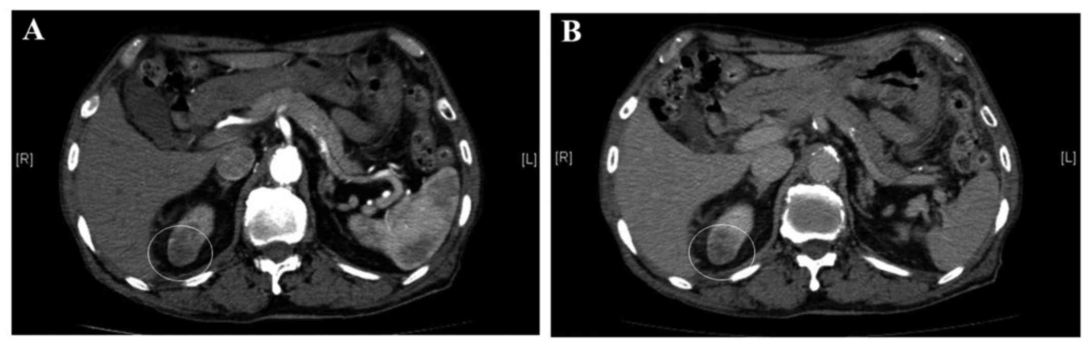
A dynamic computed tomography (CT) scan revealed a 21-mm diameter tumor in the superior pole of the right kidney (Fig. 1). The inside of the tumor was slightly enhanced in the arterial and venous phases on the dynamic CT, but did not show strong enhancement in the arterial phase as observed in clear cell RCC. The right kidney was resected, and the tumor was macroscopically and histologically examined.

Figure 1.

Dynamic computed tomography (CT) revealed a 21-mm diameter tumor in the superior pole of the right kidney. (A) Arterial phase image and (B) venous phase images of dynamic CT are shown. White-line circles show the tumor.

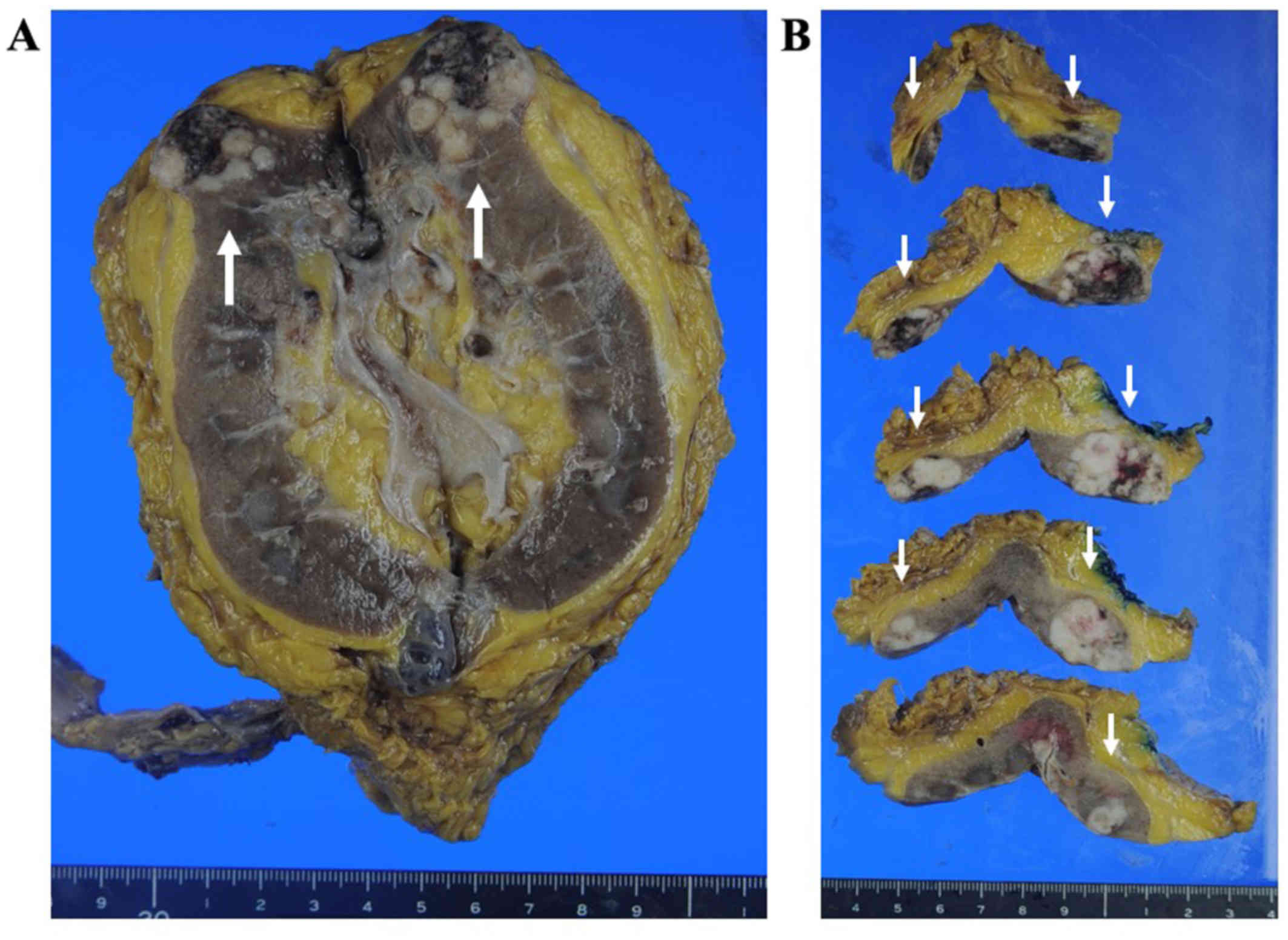
Macroscopically, a 27×25×25 mm-sized tumor residing in the cortex, medulla, and pelvis of the superior pole of the right kidney was revealed (Fig. 2). The tumor consisted of whitish multi-nodules without fibrous 
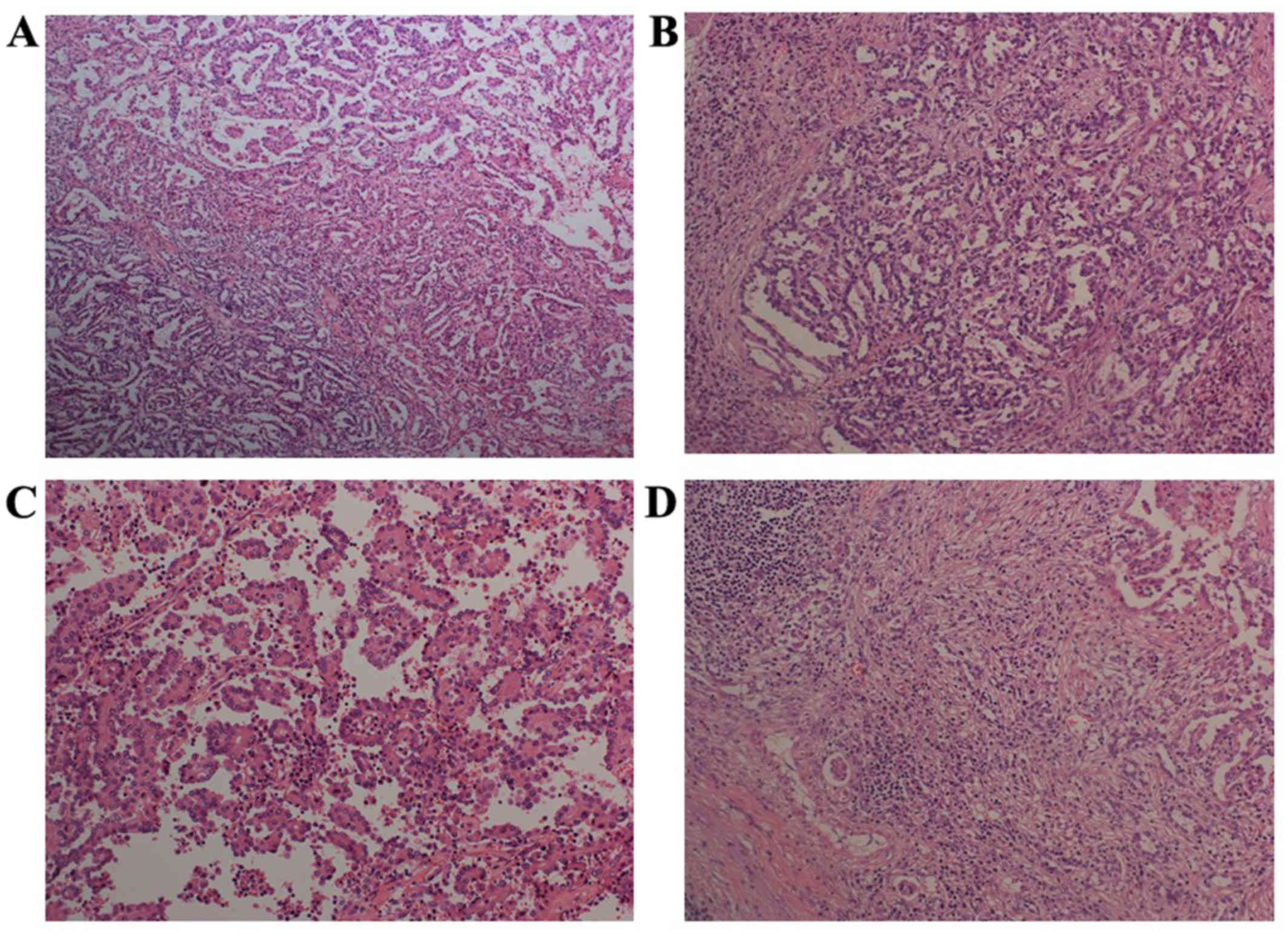
capsule, and showed necrotic area and bleeding. Microscopically, the tumor consisted of various growth patterns: a papillary structure containing a micropapillary pattern, an elongated tubular structure and, a solid pattern with spindle cells (Figs. 3 and 4). The tumor cells had eosinophilic cytoplasm. In the papillary area, two growth patterns of the tumor were observed: papillary growth with fibrovascular core and 
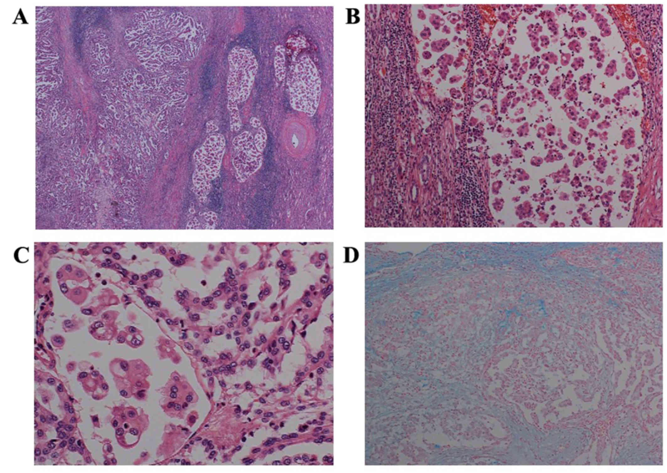
micropapillary pattern. We also found lymph vessel invasion of the MTSCC (Fig. 5). Invasion of the tumor into the lymph vessels was prominent in the micropapillary area in the MTSCC. In the small area, psammoma bodies were observed (data not shown). Small amounts of mucin were observed in the interstitium of the tumor using alcian blue stain. The tumor cells showed not only Fuhrman nuclear grade 2, but also Fuhrman nuclear 
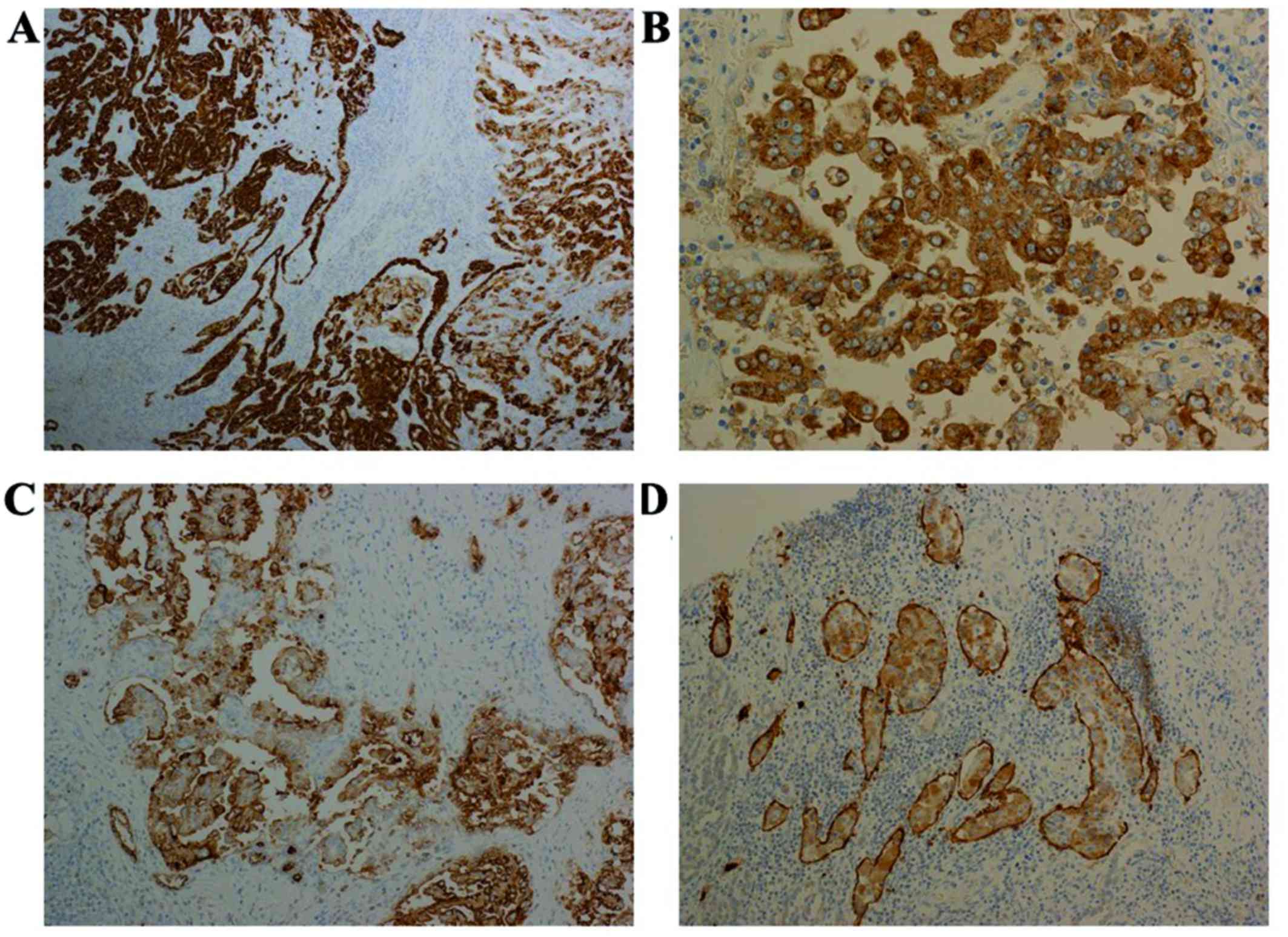
grade 3. On immunohistological staining, the tumor was diffuse positive for cytokeratin 7, AMACR, and PAX8, partially positive for cytokeratin 19, PAX2, RCC Ma, and CD10, and negative for melanosome (HMB45), melan A, catepsin K, ALK, TFE3, and uroplakin II (Fig. 5, data not shown). In the micropapillary area, the tumor cells were positive for MUC-1 on the outside of the cell membrane (inside-out pattern), and EMA was positive on the cell membrane and cytoplasm of the tumor cells. Around the tumor were inflammatory cells mainly containing lymphoid cells and plasma cells, infiltrated with mild fibrosis. Solid growth with spindle and cuboidal cells with Fuhrman nuclear grade 3 were found in the regional lymph node, suggesting metastasis of MTSCC into the lymph node. Based on the morphological features, special staining and immunohistological staining, we diagnosed the tumor as MTSCC, mucin-poor variant with metastasis into the regional lymph node, and we also found a high nuclear grade area and micropapillary growth in the tumor.

Figure 2.

Macroscopical findings. (A) The sagittal cut surface of formalin-fixed resected kidney. (B) Horizontal cut surfaces of the kidney. Arrows show the tumor.

Figure 3.

Microscopic findings of the tumor show various patterns of histology in hematoxylin and eosin staining. (A) Findings in the low power view (original objective lens: ×4), (B) Tubular architecture, (C) papillary architecture, and (D) a solid pattern with spindle cells (original objective lens of B, C, and D: ×20).

Figure 4.

Micropapillary architecture in the tumor and mucin production of the tumor. (A) Papillary and micropapillary architecture of the tumor (objective lens: ×4). (B) A middle-powered view of the micropapillary pattern of the tumor (objective lens: ×20). (C) Tumor cells showing Fuhrman nuclear grades 2 or 3 (original objective lens: ×40). (D) Mucin production of tumor cells detected by alcian blue staining (original magnification: ×20).

Figure 5.

Immunohistochemical findings of the tumor. (A) The tumor is positive for CK7 (A) (original objective lens: ×10). (B) In the area of micropapillary architecture, EMA was positive in the cytoplasm and cell membrane of the tumor cells. (C) MUC-1 was positive on the outside of the cell membrane of the tumor cells (original objective lens: ×40). (D) Lymph vessels were positive for podoplanin, indicating invasion of the tumor into the lymph vessels (original objective lens: ×40).

Even after resection of the kidney, cytological examination of the patient's urine suggested malignancy. Four months after the operation, his urinary bladder was examined by cystoscopy, and transuretheral resection (TUR) was carried out. Urothelial carcinoma in situ was found in the TUR specimen.

Five months after resection of the kidney, follow-up positron-emission tomography (PET)-CT revealed swelling of the para-aortic and para-common iliac arterial lymph nodes and a hepatic tumor, suggesting metastatic lesions of the MTSCC in the lymph nodes and liver. As another malignant tumor of the patient, urothelial carcinoma, was a carcinoma in situ, which usually do not metastasize. Administration of sunitinib was initiated to treat the metastatic lesions. Twelve days after the start of medication, cardiac and pulmonary arrest in the patient suddenly occurred. Cardiopulmonary resuscitation (CPR) was carried out on the patient. Brain CT revealed a brain hemorrhage and hydrocephalus, probably induced by intra-brain hemorrhage in the patient. The hemorrhage was thought to have occurred because of the metastatic lesion of the MTSCC. Although emergency external ventricular drainage was carried out, the patient succumbed to heart failure on the same day.

Discussion

MTSCC is a rare type of renal tumor, and MTSCC is believed to have been first reported in 1997 as ‘low-grade collecting duct carcinoma’ by MacLennan et al (12). The reported age range of MTSCC is between 17 and 82 years, with a mean age of 53 years and is dominant in female individuals (13,14). In our case, the patient was an 82-year-old man, which is relatively rare in MTSCC cases with regard to age and gender. Although MTSCC is histologically characterized by elongated tubules and low-grade spindle cell components separated by pale mucinous stroma, mucin-poor variant of MTSCC has been also reported (13,15–17). In our case, elongated tubules lined by cuboidal cells and a papillary pattern were prominent. However, spindle cells existed in certain areas containing a small amount of mucin. Therefore, we diagnosed our case as consistent with mucin-poor variant of MTSCC.

It has been reported that MTSCC is usually of a low nuclear grade and has a better prognosis than other RCCs (1). Rakozy et al have reported that patients with MTSCC show recurrences, but do not show distant metastasis or death by the tumor (2). However, MTSCC with a high nuclear grade (more than Fuhrman nuclear grade 3) has been reported, and it has also been reported that the prognosis for this minor MTSCC is not good (4,5,18).

In our case, invasion of the MTSCC into the lymph vessels and metastasis into the regional lymph node were found histologically. The metastasis of the MTSCC into the para-aortic lymph nodes, para-common iliac arterial lymph nodes, liver, and brain were suggested by diagnostic imaging, and the patient finally succumbed probably due to brain metastasis. These findings suggest that the MTSCC in our case was aggressive and had a poor prognosis. The MTSCC in our case contained not only a low nuclear grade area, but also a high nuclear grade one, suggesting that transformation from low to high grade could occur in MTSCC.

In our case, a part of the tumor showed micropapillary growth and lymph vessel-invasion of the micropapillary component. To the best of our knowledge, there has been only one report showing micropapillary growth in RCC (11). It has been reported that carcinomas showing micropapillary structure lead to a high incidence of lymph node metastasis (19) and poor prognosis (20–22). In our case, invasion into the lymph vessels in MTSCC was prominent in the area showing micropapillary structure. Moreover, we found that MTSCC showing micropapillary structure existed in the lymph vessels, suggesting that the micropapillary component could easily metastasize. Our patient also finally succumbed probably due to brain metastasis, and his MTSCC also showed high nuclear grade and micropapillary structure, suggesting a poor prognosis.

In this study, we have presented a case of mucin-poor MTSCC with high nuclear grade, micropapillary structure and metastasis into the lymph nodes and other organs. When diagnosing MTSCC, sufficient micro-slides should be prepared and observed carefully to detect the area of high nuclear grade.

Acknowledgements

We would like to thank Ms. H. Ogaki, Mr. K Nagaoka, Mr. T. Kuge, Mr. H. Takenaka and Ms. S. Eriguchi of the Toyooka Hospital for their expert technical assistance.

References

1 

Hes O, Hora M, Perez-Montiel DM, Suster S, Curík R, Sokol L, Ondic O, Mikulástík J, Betlach J, Peychl L, et al: Spindle and cuboidal renal cell carcinoma, a tumour having frequent association with nephrolithiasis: Report of 11 cases including a case with hybrid conventional renal cell carcinoma/spindle and cuboidal renal cell carcinoma components. Histopathology. 41:549–555. 2002. View Article : Google Scholar : PubMed/NCBI

2 

Rakozy C, Schmahl GE, Bogner S and Störkel S: Low-grade tubular-mucinous renal neoplasms: Morphologic, immunohistochemical, and genetic features. Mod Pathol. 15:1162–1171. 2002. View Article : Google Scholar : PubMed/NCBI

3 

Srigley J: Mucinous tubular and spindle cell carcinoma. IRAC Press; Lyon: 2004

4 

Shen SS, Ro JY, Tamboli P, Truong LD, Zhai Q, Jung SJ, Tibbs RG, Ordonez NG and Ayala AG: Mucinous tubular and spindle cell carcinoma of kidney is probably a variant of papillary renal cell carcinoma with spindle cell features. Ann Diagn Pathol. 11:13–21. 2007. View Article : Google Scholar : PubMed/NCBI

5 

Kuroda N, Hes O, Michal M, Nemcova J, Gal V, Yamaguchi T, Kawada T, Imamura Y, Hayashi Y and Lee GH: Mucinous tubular and spindle cell carcinoma with Fuhrman nuclear grade 3: A histological, immunohistochemical, ultrastructural and FISH study. Histol Histopathol. 23:1517–1523. 2008.PubMed/NCBI

6 

Siriaunkgul S and Tavassoli FA: Invasive micropapillary carcinoma of the breast. Mod Pathol. 6:660–662. 1993.PubMed/NCBI

7 

Takanashi Y, Tajima S, Hayakawa T, Takahashi T, Neyatani H and Funai K: Pulmonary micropapillary-type adenosquamous carcinoma sharing epidermal growth factor receptor mutation in adenocarcinoma and squamous cell carcinoma. Respirol Case Rep. 4:e001792016.PubMed/NCBI

8 

Miyaoka Y, Fujiwara A, Kotani S, Tsukano K, Ogawa S, Yamanouchi S, Kusunoki R, Fujishiro H, Kohge N, Yamamoto T and Amano Y: Primary micropapillary carcinoma of the colon with submucosal invasion: A case report. Endosc Int Open. 4:E744–E747. 2016. View Article : Google Scholar : PubMed/NCBI

9 

Tanaka H, Baba Y, Sase T, Isono Y, Matsusaki S, Saito T, Okano H, Mukai K, Murata T and Watanabe G: Gastric intramucosal adenocarcinoma with an invasive micropapillary carcinoma component. Clin J Gastroenterol. 8:14–17. 2015. View Article : Google Scholar : PubMed/NCBI

10 

Ishii S, Ohbu M, Toomine Y, Nishimura Y, Hattori M, Yokoyama M, Toyonaga M, Kakinuma H and Matsumoto K: Immunohistochemical, molecular, and clinicopathological analyses of urothelial carcinoma, micropapillary variant. Pathol Int. 61:723–730. 2011. View Article : Google Scholar : PubMed/NCBI

11 

Aoyagi T, Shinohara N, Kubota-Chikai K, Kuroda N and Nonomura K: Long-term survival in a patient with node-positive adult-onset Xp11.2 translocation renal cell carcinoma. Urol Int. 86:487–490. 2011. View Article : Google Scholar : PubMed/NCBI

12 

MacLennan GT, Farrow GM and Bostwick DG: Low-grade collecting duct carcinoma of the kidney: Report of 13 cases of low-grade mucinous tubulocystic renal carcinoma of possible collecting duct origin. Urology. 50:679–684. 1997. View Article : Google Scholar : PubMed/NCBI

13 

Fine SW, Argani P, DeMarzo AM, Delahunt B, Sebo TJ, Reuter VE and Epstein JI: Expanding the histologic spectrum of mucinous tubular and spindle cell carcinoma of the kidney. Am J Surg Pathol. 30:1554–1560. 2006. View Article : Google Scholar : PubMed/NCBI

14 

Grigore A, Toma L, Stoicea M, Dinu M and Ardeleanu C: Rare renal tumor-mucinous tubular and spindle cell carcinoma. Rom J Morphol Embryol. 53:167–171. 2012.PubMed/NCBI

15 

Farghaly H: Mucin poor mucinous tubular and spindle cell carcinoma of the kidney, with nonclassic morphologic variant of spindle cell predominance and psammomatous calcification. Ann Diagn Pathol. 16:59–62. 2012. View Article : Google Scholar : PubMed/NCBI

16 

Saito K, Shimada M, Inoue K, Shiiki K, Nagata M, Ogawa Y, Matsubara E, Maeda T, Matsumoto Y, Kunimura T and Mikogami T: A case of mucinous tubular and spindle cell carcinoma of the kidney. Hinyokika Kiyo. 59:107–111. 2013.(In Japanese). PubMed/NCBI

17 

Cao W, Huang B, Fei X, Huang X, Dai J, Zhou W, Xu Z, Su H, Cheng K and Sun F: Clear cell changes in mucinous tubular and spindle cell carcinoma: Cytoplasmic pallor/clearing within tubules, vacuoles or hybrid conventional clear cell carcinoma of kidney? Int J Clin Exp Pathol. 7:4350–4358. 2014.PubMed/NCBI

18 

Arafah M and Zaidi SN: Mucinous tubular and spindle cell carcinoma of the kidney with sarcomatoid transformation. Saudi J Kidney Dis Transpl. 24:557–560. 2013. View Article : Google Scholar : PubMed/NCBI

19 

Zekioglu O, Erhan Y, Ciris M, Bayramoglu H and Ozdemir N: Invasive micropapillary carcinoma of the breast: High incidence of lymph node metastasis with extranodal extension and its immunohistochemical profile compared with invasive ductal carcinoma. Histopathology. 44:18–23. 2004. View Article : Google Scholar : PubMed/NCBI

20 

Amin MB, Tamboli P, Merchant SH, Ordóñez NG, Ro J, Ayala AG and Ro JY: Micropapillary component in lung adenocarcinoma: A distinctive histologic feature with possible prognostic significance. Am J Surg Pathol. 26:358–364. 2002. View Article : Google Scholar : PubMed/NCBI

21 

Lee HJ, Eom DW, Kang GH, Han SH, Cheon GJ, Oh HS, Han KH, Ahn HJ, Jang HJ and Han MS: Colorectal micropapillary carcinomas are associated with poor prognosis and enriched in markers of stem cells. Mod Pathol. 26:1123–1131. 2013. View Article : Google Scholar : PubMed/NCBI

22 

Bertz S, Wach S, Taubert H, Merten R, Krause FS, Schick S, Ott OJ, Weigert E, Dworak O, Rödel C, et al: Micropapillary morphology is an indicator of poor prognosis in patients with urothelial carcinoma treated with transurethral resection and radiochemotherapy. Virchows Arch. 469:339–344. 2016. View Article : Google Scholar : PubMed/NCBI

Related Articles

  • Abstract
  • View
  • Download
  • Twitter
Copy and paste a formatted citation
Spandidos Publications style
Sakatani T, Okumura Y, Kuroda N, Magaribuchi T, Nakano Y, Shirahase T, Watanabe J, Taki Y, Okigaki M, Ikehara S, Ikehara S, et al: Mucinous tubular and spindle cell carcinoma with a high nuclear grade and micropapillary pattern: A case report. Mol Clin Oncol 7: 976-980, 2017.
APA
Sakatani, T., Okumura, Y., Kuroda, N., Magaribuchi, T., Nakano, Y., Shirahase, T. ... Adachi, Y. (2017). Mucinous tubular and spindle cell carcinoma with a high nuclear grade and micropapillary pattern: A case report. Molecular and Clinical Oncology, 7, 976-980. https://doi.org/10.3892/mco.2017.1430
MLA
Sakatani, T., Okumura, Y., Kuroda, N., Magaribuchi, T., Nakano, Y., Shirahase, T., Watanabe, J., Taki, Y., Okigaki, M., Ikehara, S., Adachi, Y."Mucinous tubular and spindle cell carcinoma with a high nuclear grade and micropapillary pattern: A case report". Molecular and Clinical Oncology 7.6 (2017): 976-980.
Chicago
Sakatani, T., Okumura, Y., Kuroda, N., Magaribuchi, T., Nakano, Y., Shirahase, T., Watanabe, J., Taki, Y., Okigaki, M., Ikehara, S., Adachi, Y."Mucinous tubular and spindle cell carcinoma with a high nuclear grade and micropapillary pattern: A case report". Molecular and Clinical Oncology 7, no. 6 (2017): 976-980. https://doi.org/10.3892/mco.2017.1430
Copy and paste a formatted citation
x
Spandidos Publications style
Sakatani T, Okumura Y, Kuroda N, Magaribuchi T, Nakano Y, Shirahase T, Watanabe J, Taki Y, Okigaki M, Ikehara S, Ikehara S, et al: Mucinous tubular and spindle cell carcinoma with a high nuclear grade and micropapillary pattern: A case report. Mol Clin Oncol 7: 976-980, 2017.
APA
Sakatani, T., Okumura, Y., Kuroda, N., Magaribuchi, T., Nakano, Y., Shirahase, T. ... Adachi, Y. (2017). Mucinous tubular and spindle cell carcinoma with a high nuclear grade and micropapillary pattern: A case report. Molecular and Clinical Oncology, 7, 976-980. https://doi.org/10.3892/mco.2017.1430
MLA
Sakatani, T., Okumura, Y., Kuroda, N., Magaribuchi, T., Nakano, Y., Shirahase, T., Watanabe, J., Taki, Y., Okigaki, M., Ikehara, S., Adachi, Y."Mucinous tubular and spindle cell carcinoma with a high nuclear grade and micropapillary pattern: A case report". Molecular and Clinical Oncology 7.6 (2017): 976-980.
Chicago
Sakatani, T., Okumura, Y., Kuroda, N., Magaribuchi, T., Nakano, Y., Shirahase, T., Watanabe, J., Taki, Y., Okigaki, M., Ikehara, S., Adachi, Y."Mucinous tubular and spindle cell carcinoma with a high nuclear grade and micropapillary pattern: A case report". Molecular and Clinical Oncology 7, no. 6 (2017): 976-980. https://doi.org/10.3892/mco.2017.1430
Follow us
  • Twitter
  • LinkedIn
  • Facebook
About
  • Spandidos Publications
  • Careers
  • Cookie Policy
  • Privacy Policy
How can we help?
  • Help
  • Live Chat
  • Contact
  • Email to our Support Team