Introduction
Head and neck squamous cell carcinoma (HNSCC)
accounts for ~3% of all malignancies (1). Currently, chemoradiotherapy (CRT) is
considered as a good approach to the treatment of locally advanced
HNSCC. However, ~25–30% of patients have local residual disease or
develop relapse at the primary or lymph node sites after treatment
(2), which represents a challenge in
the management of HNSCC. Therefore, there is a need for biomarkers
that can provide accurate and timely prediction of therapeutic
outcome prior to treatment or in the early stages of treatment,
with discontinuation of ineffective approaches and timely
application of alternative therapeutic strategies.
Recently, diffusion-weighted imaging (DWI) was
proposed as an imaging biomarker for predicting treatment outcome
in multiple malignancies, including rectal cancer (3), breast cancer (4) and glioblastoma (5). DWI quickly measures the Brownian motion
of extracellular water molecules in biological tissues, which may
be quantified with the apparent diffusion coefficient (ADC)
(6). Previous studies demonstrated
that higher pretreatment ADCs were correlated with poor response to
treatment (7,8). In addition, due to its ability to
depict the range of ADCs, ADC histograms may be used to evaluate
the heterogeneity of the whole tumor. This feature enables
evaluation of the degree of necrosis and viability, which may be
crucial when planning a radiation dose boost (9).
Over the past few years, a number of studies have
investigated the role of DWI in predicting response to CRT in
patients with HNSCC. Given the varied characteristics of the
patients and studies, individual studies are unable to provide a
reliable estimate of DWI performance for the prediction of
treatment response. Hence, the present meta-analysis was performed
to determine the diagnostic performance of DWI for the prediction
of locoregional failure of CRT in patients with HNSCC, which may be
helpful in optimizing the management of this disease.
Data collection methods
Search strategy
A comprehensive search was conducted through the
EMBASE, PubMed and Cochrane Library databases to identify relevant
publications on the accuracy of DWI in the prediction of response
to CRT in patients with HNSCC. The EMTREE terms (for EMBASE),
medical subject heading terms (for Medline), and text words (for
other databases) included (‘head and neck cancer’ OR ‘head and neck
neoplasms’ OR ‘lip cancer’ OR ‘lip neoplasms’ OR ‘oropharynx
cancer’ OR ‘oropharyngeal neoplasms’ OR ‘hypopharyngeal cancer’ OR
‘hypopharyngeal neoplasms’ OR ‘nasopharynx cancer’ OR
‘nasopharyngeal neoplasms’ OR ‘laryngeal cancer’ OR ‘laryngeal
neoplasms’ OR ‘salivary gland cancer’ OR ‘salivary gland
neoplasms’) AND (‘diffusion weighted magnetic resonance imaging’ OR
‘diffusion weighted MRI’ OR ‘diffusion MRI’ OR ‘DWI’) AND
(‘concurrent chemoradiotherapy’ OR ‘synchronous chemoradiotherapy’
OR ‘concomitant chemoradiotherapy’ OR ‘chemoradiotherapy’ OR
‘radiotherapy’). The search was updated on March 21, 2017. To
extend the search, the reference lists of the articles that were
identified after the selection process were screened for additional
suitable articles.
Study selection
Studies were considered eligible for inclusion if
the accuracy of DWI in the prediction of response to CRT was
investigated in patients with HNSCC. The eligible studies were also
required to have a defined reference standard for local failure,
which included residual disease and local recurrence. Accordingly,
local failure was required to be histopathologically confirmed, or
at least clinically suspected, in terms of the presence of any mass
or a persisting and/or increasing existing residual mass on serial
imaging during follow-up. In addition, the studies had to include
at least 25 patients and yield sufficient information, including
true and false positive and negative values, in order to construct
a 2×2 contingency table to calculate sensitivity and specificity in
the prediction of local failure. Moreover, only articles published
in English were included. Review articles, comments, letters, case
reports and animal experiments were excluded. When overlapping data
from the same authors were presented among different articles, the
article with the most recent details or the largest number of
patients was selected.
Data extraction and quality
assessment
Two reviewers (Q.M.Z. and Y.D.) independently
extracted the relevant data and assessed the quality of the
retrieved studies. To resolve discrepancies between the two
reviewers, a consensus meeting was held; if agreement could not be
reached, a third reviewer (F.F.Z.) was consulted. The extracted
data were as follows: Patient characteristics (sample size, mean or
median age and range), study characteristics [first author,
publication year, country of origin, study design (prospective,
retrospective or unknown), patient enrollment (consecutive or not),
whether blinding was used in the study, number of patients, tumor
location, TNM stage, number of reviewers, radiotherapy regimen and
chemotherapy regimen], and technical details of magnetic resonance
imaging (MRI) protocols [magnetic field strength, repetition
time/echo time (TR/TE), MRI vendor, slice thickness, field of view
(FOV), matrix and b value]. The Quality Assessment of Diagnostic
Accuracy Studies (QUADAS-2) was used to assess the methodological
quality of the included studies. QUADAS-2 has four key domains:
Patient selection, index test, reference standard, and flow and
timing.
Statistical analysis
A meta-analysis was performed using Stata software,
version 13.0 (Stata Corp., College Station, TX, USA). Data were
combined to obtain pooled sensitivity, specificity, diagnostic odds
ratios (DORs) and likelihood ratios (LRs). A summary receiver
operating characteristics (sROC) curve was constructed and the area
under the curve (AUC) obtained. An inconsistency index
(I2) test was performed to assess the heterogeneity
between studies. An I2 value >50% indicated
heterogeneity, in which case a random-effects model was used;
otherwise, a fixed-effects model was applied. As an important
source of heterogeneity, the threshold effect was assessed by
calculating the Spearman's correlation coefficient. If there was no
threshold effect, a meta-regression analysis was then performed to
investigate other potential sources of heterogeneity. Publication
bias was investigated by constructing a Deeks' funnel plot.
P-values of <0.05 were considered to indicate statistically
significant differences.
Results
Literature search and study
selection
Our database search and extensive reference list
cross-check yielded 365 studies. After excluding duplicates and
reviewing the titles and abstracts, 65 studies remained, and their
full texts were retrieved. After the full texts were reviewed, 56
studies were excluded for the following reasons: Patient number
<25 (n=19); the same data were presented in another article by
the same group (n=5); the studies were reviews, letters, or
proceedings of a symposium (n=19); the articles were not written in
English (n=9); and sufficient data were not extractable to
construct a 2×2 contingency table to calculate sensitivity and
specificity in the prediction of local failure (n=4). Ultimately, a
total of 9 eligible studies involving 421 patients were included in
the meta-analysis (7,8,10–16). A
search of the reference lists of these eligible articles did not
yield other potentially relevant articles.
Study description
The detailed characteristics of the 9 included
studies are summarized in Tables I
and II. The median number of
patients per study was 37 (range, 26–134). Of the 9 studies, 6
enrolled patients prospectively and 3 retrospectively. A total of 4
studies enrolled patients consecutively, whereas the remaining 5
studies enrolled patients in an unknown manner. In 4 studies,
treatment response was assessed by reviewers who had been blinded
to the results of the DWI analysis; in the other 5 studies, this
detail was not provided. In 8 studies, DWI examinations were
conducted with 1.5 T devices, whereas in 1 study, both 3.0 T and
1.5 T devices were used.
 | Table I.Patient and study characteristics. |
Table I.
Patient and study characteristics.
| Authors | Year | Country | Study design | Patient
enrollment | Blinding status | No. of patients | Mean/median age,
years (range) | Tumor location | TNM stage(s) | Reviewers
(experience) | Radiotherapy
regimen | Chemotherapy
regimen | (Refs.) |
|---|
| Xiao-ping et
al | 2016 | China | Prospective | Consecutive | Blind | 50 | 48.9±11.1 | Nasopharynx | T2-4N0-3M0-1 | Two radiologists (5
and 20 years) | 7,000–7,600 cGy/30-33
f | TN, TNF, NF | (10) |
| Scalco et
al | 2016 | Italy | Retrospective | NA | NA | 30 | NA | HNSCCa | T1-4N1-3 | One radiologist (15
years) | 7,000 cGy/33 f | Cisplatin | (11) |
| Hou et al | 2016 | China | Prospective | Consecutive | Blind | 43 | 49.0 (26–68) | Nasopharynx | T2-4N0-3M0 | Two radiologists
(>10 years) | 7,000–7,600 cGy/30-33
f | TN | (12) |
| Hong et
al | 2013 | China | Prospective | NA | NA | 134 | 47.0 (18–79) | Nasopharynx | T1-4 | NA | 6,600–7,875f
cGy/30-33 | TP, cisplatin | (14) |
| King et
al | 2013 | China | Prospective | Consecutive | NA | 37 | 57.0 (45–71) | HNSCCb | III–IV | One radiologist
(>15 years) | NA | NA | (13) |
| Nakajo et
al | 2012 | Japan | Retrospective | NA | NA | 26 | 65.0 (45–89) | HNSCCc | T1-4N0-3 | NA | 6,000 cGy/30 f | NA | (15) |
| Hatakenaka et
al | 2011 | Japan | Retrospective | NA | Blind | 38 | 64.0 (37–85) | HNSCCd | T1-4N0-3 | Two radiologists
(unknown) | 6,000 cGy | S-1, cisplatin | (8) |
| Vandecaveye et
al | 2010 | Belgium | Prospective | Consecutive | Blind | 30 | 53.0 (38–66) | HNSCCe | T1-4N1-3 | One radiologist (6
years) | 7,200 cGy | NA | (16) |
| Kim et
al | 2009 | USA | Prospective | NA | NA | 33 | 61.0±10.8 | HNSCCf | T0-4N1-2bM0 | NA | 7,040 cGy/32 f | Cisplatin | (7) |
| Table II.Technical details of MRI
protocols. |
Table II.
Technical details of MRI
protocols.
| Authors | Magnet strength
(T) | TR/TE (msec) | MRI vendor | Slice thickness
(mm) | FOV (mm) | Matrix | b value
(sec/mm2) | (Refs.) |
|---|
| Xiao-ping et
al | 1.5 | 4,225/106 | GE | 5 | Unknown | 128×130 | 0, 200, 400, 600,
800, 1,000 | (10) |
| Scalco et
al | 1.5 | 4,500/77 | GE | 4 | 260–280 | 128×128 | 0, 500, 800 | (11) |
| Hou et
al | 1.5 | 4,225/106 | GE | 5 | 220 | 128×130 | 0, 50, 80, 100,
150, 200, 400, 600, 800, 1,000 | (12) |
| Hong et
al | 1.5 | Unknown | GE | Unknown | 240×240 | 128×128 | 0, 800 | (14) |
| King et
al | 1.5 | 2,000/75 | Philips | 4 | 230 | 112×112 | 0, 100, 200, 300,
400, 500 | (13) |
| Nakajo et
al | 1.5 | 6,000/68 | Siemens | 6 | 370 | 112×168 | 0, 800 | (15) |
| Hatakenaka et
al | 1.5 | 3,000/73 | Philips | 3–5 | 200–230 | 112×79 | 0, 300, 1,000 | (8) |
| Vandecaveye et
al | 1.5 | 7,100/84 | Siemens | 4 | 200×250 | 104×128 | 0, 50, 100, 500,
750, 1,000 | (16) |
| Kim et
al | 1.5 or
3.0a | 4,000/89 | Siemens | 5 | 260 | NA | 0, 500, 1,000 | (7) |
Assessment of study quality
The QUADAS-2 scores of each study are listed in
Table III. The quality of the 9
studies varied. A total of 4 studies had a low risk of bias
regarding patient selection, whereas 5 studies had an unclear risk
of bias due to insufficient information. All 9 studies reported a
low risk of bias regarding the index test. The risk of bias
regarding the reference standard was low in 4 studies and unclear
in 5 studies, as the latter studies provided insufficient
information on blinding. The risk of bias regarding flow and timing
was low in 4 studies and high in 5 studies, as different reference
standards were applied during follow-up in the latter studies. As
regards risk in applicability, all 9 studies had a low risk of
bias.
| Table III.Quality assessment of included
studies: Summarized risk of bias and applicability concerns. |
Table III.
Quality assessment of included
studies: Summarized risk of bias and applicability concerns.
|
| Risk of bias | Applicability |
|
|---|
|
|
|
|
|
|---|
| Authors | Patient
selection | Index test | Reference
standard | Flow and
timing | Patient
selection | Index test | Reference
standard | (Refs.) |
|---|
| Xiao-ping et
al | Low risk | Low risk | Low risk | High risk | Low risk | Low risk | Low risk | (10) |
| Scalco et
al | Unclear risk | Low risk | Unclear risk | Low risk | Low risk | Low risk | Low risk | (11) |
| Hou et
al | Low risk | Low risk | Low risk | High risk | Low risk | Low risk | Low risk | (12) |
| Hong et
al | Unclear risk | Low risk | Unclear risk | High risk | Low risk | Low risk | Low risk | (14) |
| King et
al | Low risk | Low risk | Unclear risk | Low risk | Low risk | Low risk | Low risk | (13) |
| Nakajo et
al | Unclear risk | Low risk | Unclear risk | Low risk | Low risk | Low risk | Low risk | (15) |
| Hatakenaka et
al | Unclear risk | Low risk | Low risk | Low risk | Low risk | Low risk | Low risk | (8) |
| Vandecaveye et
al | Low risk | Low risk | Low risk | High risk | Low risk | Low risk | Low risk | (16) |
| Kim et
al | Unclear risk | Low risk | Unclear risk | Low risk | Low risk | Low risk | Low risk | (7) |
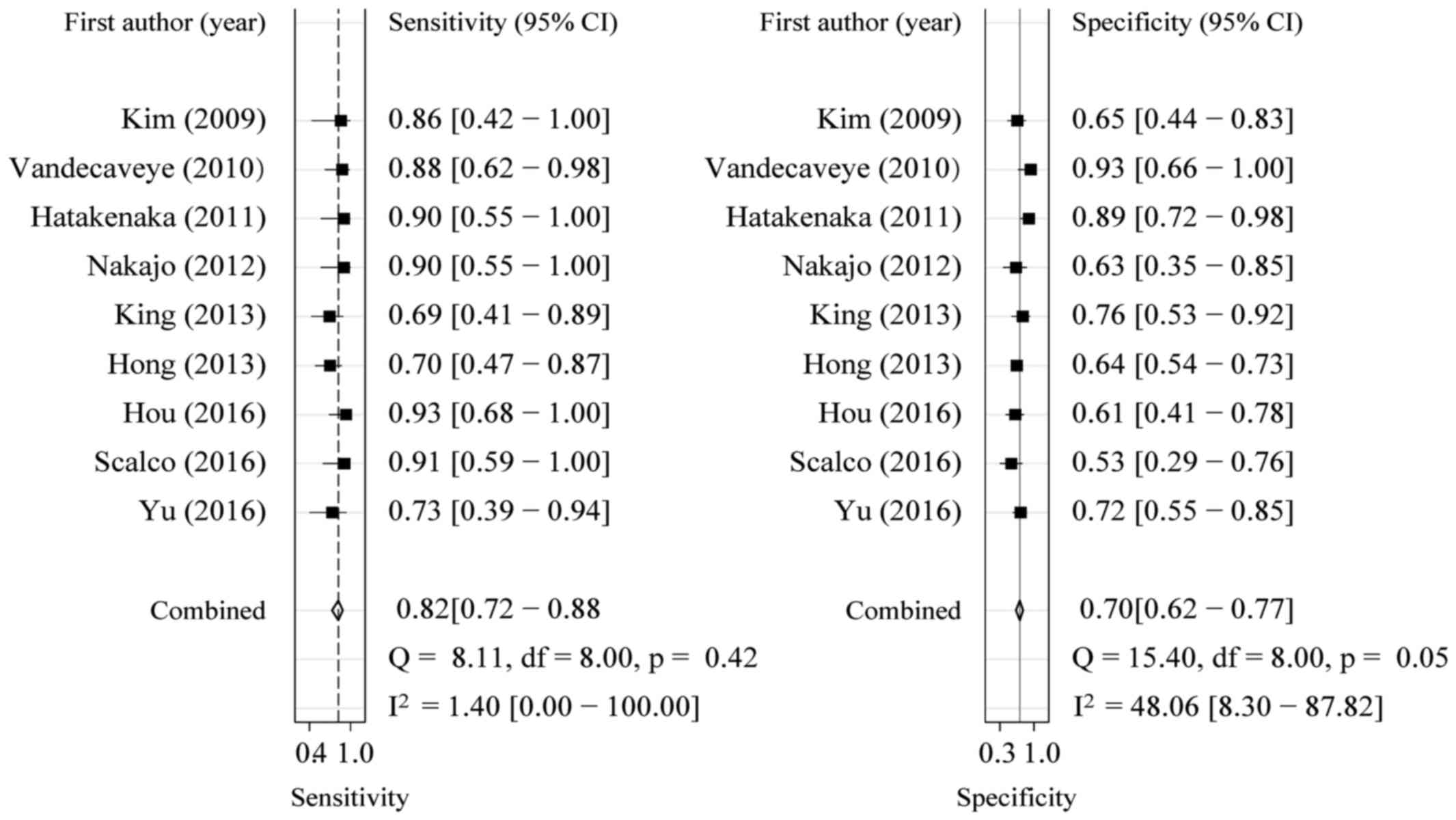
Meta-analysis
The sensitivity and specificity of DWI in the
prediction of locoregional failure of CRT in HNSCC patients ranged
from 0.69 to 0.95 and from 0.55 to 0.89, respectively, with pooled
estimates of 0.82 [95% confidence interval (CI): 0.72–0.88] and
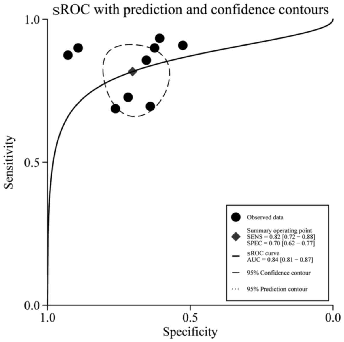
0.70 (95% CI: 0.62–0.77), respectively. The forest plots of
sensitivities and specificities are shown in Fig. 1. Using the sROC curve, the AUC was
0.84 (95% CI: 0.81–0.87; Fig. 2).
The positive LR, negative LR and the DOR of DWI were 2.7 (95% CI:
2.1–3.6), 0.26 (95% CI: 0.17–0.41) and 10.48 (95% CI: 5.35–20.53),
respectively. Among all 9 studies, no significant heterogeneity was
found in terms of pooled sensitivity (P=0.42, I2=1.4%),
specificity (P=0.05, I2=48.06%), or DOR (P=0.26,
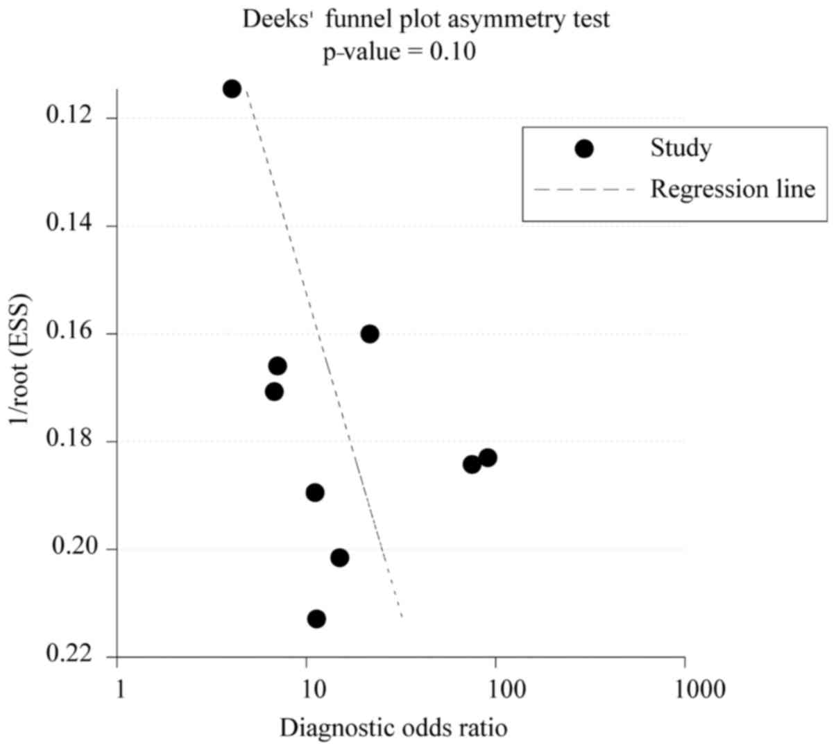
I2=20.6%). The Deeks' funnel plot asymmetry test
revealed no strong evidence for publication bias (P=0.10; Fig. 3).
Discussion
The present meta-analysis investigated the
feasibility of ADC as an imaging biomarker for the assessment of
locoregional failure of CRT in HNSCC patients. Accurate prediction
or monitoring of therapeutic efficacy before or during the early
stages of treatment is crucial for individual patients to modify
the ongoing treatment regimen in a timely manner. In addition,
patients with radioresistant tumors may be offered an escalated
radiation dose or alternative treatment options, such as early
surgical intervention.
To the best of our knowledge, this is the first
meta-analysis to determine the ability of DWI to predict local
failure of CRT in HNSCC patients. A pooled sensitivity of 0.82 (95%
CI: 0.72–0.88) and a specificity of 0.70 (95% CI: 0.62–0.77) were
calculated from a total of 421 patients in 9 studies who fulfilled
all the inclusion and exclusion criteria. Furthermore, according to
a widely accepted interpretation (17), a good predictive accuracy (AUC=0.84)
was observed between local failure and locally controlled
lesions.
A number of studies have investigated the role of
DWI in the prediction of response to therapeutic interventions. A
preclinical study by Hamstra et al (18) reported that a significant increase in
ADC was noted in an animal model of HNSCC 5 days after initiation
of CRT, which may be used as a marker to predict treatment
response. Kim et al (7) also
reported that patients with complete response to CRT had
significantly lower pretreatment ADC values than did patients with
partial response. In addition, the changes in ADC between baseline
and early intratreatment measurement may also demonstrate higher
test accuracy in separating favorable from unfavorable responders.
Contrary to Kim et al, King et al (19) reported that ADC values were more
effective after treatment, compared with before or during
treatment, in predicting response to CRT in HNSCC patients. All
these studies indicated that ADC may be used as an important
imaging biomarker for personalized treatment in HNSCC.
From a clinical perspective, optimized clinical
benefit may be achieved by adopting the most appropriate treatment
strategies or timely modification of a treatment plan on the basis
of an individual patient's response assessment. Therefore, ADC
values have been proposed to be of higher value as biomarkers
before treatment or in early intratreatment than they are in a
post-treatment setting. In our meta-analysis, only studies using
either pretreatment ADC (n=6) (7,8,10–12,15) or
percentage change in ADC values between baseline and early
intratreatment measurement (n=3) (13,14,16) were
included to predict locoregional failure of CRT in HNSCC patients,
with the optimum cutoff (threshold) values for ADC ranging from
0.86 to 1.11 for pretreatment and from 14 to 52.7% for percentage
change in ADC. According to the histopathological analysis
(20), high numbers of tumor-stromal
cells, which contribute to reducing the restriction of water
molecule motion in tumors, are significantly associated with high
pretreatment ADC values. Stromal cells play a crucial role in
supporting the growth of tumors by promoting tumor cell invasion,
protecting apoptosis, and then creating a barrier to systemic
therapy. This may partly explain the biological mechanisms
underlying the association between high pretreatment ADC values and
poor treatment response to CRT in HNSCC patients.
Currently, in addition to DWI, multiple imaging
modalities, including dynamic contrast-enhanced MRI (21), phosphorous MR spectroscopy (22), and fluorine-18-fluorodeoxyglucose
positron emission tomography with computed tomography (23) have been used to predict and monitor
treatment efficacy in HNSCC patients. The notable advantages of DWI
over these other imaging modalities are its simplicity and the fact
that the use of contrast agents is not required. Injection of
contrast agents may increase the cost and their use may not be
indicated in patients who have severe renal function impairment.
Therefore, it is more advantageous to use DWI to predict and
monitor the treatment response.
Several limitations should be acknowledged in this
meta-analysis. First, the number of included studies and samples
was moderate. This limitation may lead to an overestimation of
predictive accuracy (24). Second,
the reference standards regarding locoregional failure of CRT
included histopathological findings or clinical and imaging results
during the follow-up period, which may affect the predictive value
of DWI. Third, although the heterogeneity among the included
studies was not significant, it was present. Human papillomavirus
(HPV) status was not detailed in all the included studies. HPV
status is the primary etiological determinant of chemoradiotherapy
response, demonstrating a 25% improvement in 3-year overall
survival in large-scale cooperative group datasets (25). While not exclusively HPV-associated,
domestically, the vast majority of oropharyngeal cancers are
HPV-related (26). However, at
present, HPV status does not define a differential in terms of
standard of care treatment, which remains CRT for the majority of
head and neck cancers. Consequently, pooled analysis is appropriate
until results from cooperative group studies (such as NRG HN002)
(27) are mature enough to define
whether HPV status-based de-intensification is efficacious.
Finally, factors associated with the scanning protocol and analysis
method differed among the studies. To further establish DWI as a
routine clinical assessment tool, DWI scanning methods and analysis
protocols should be validated or standardized. Recently, the
Radiological Society of North America Quantitative Imaging
Biomarker Alliance have developed a standardized ADC measurement
phantom (28). The use of this
device to standardize ADC measurements across
facilities/devices/sequences is imperative for future DWI
implementation.
In conclusion, our meta-analysis suggests that DWI
is a promising imaging modality for predicting local failure of CRT
in HNSCC patients. However, considering the limitations of the
present study, the results must be interpreted with caution.
Larger-scale, prospective, randomized-controlled studies are
required to confirm the clinical value of DWI in predicting
treatment outcome in HNSCC.
Acknowledgements
The authors would like to thank Mrs. Tamara K. Locke
at the Department of Scientific Publications of MD Anderson Cancer
Center for providing editorial support in the preparation of this
manuscript.
References
|
1
|
Jemal A, Bray F, Center MM, Ferlay J, Ward
E and Forman D: Global cancer statistics. CA Cancer J Clin.
61:69–90. 2011. View Article : Google Scholar : PubMed/NCBI
|
|
2
|
King AD and Thoeny HC: Functional MRI for
the prediction of treatment response in head and neck squamous cell
carcinoma: Potential and limitations. Cancer Imaging. 16:232016.
View Article : Google Scholar : PubMed/NCBI
|
|
3
|
Wu LM, Zhu J, Hu J, Yin Y, Gu HY, Hua J,
Chen J and Xu JR: Is there a benefit in using magnetic resonance
imaging in the prediction of preoperative neoadjuvant therapy
response in locally advanced rectal cancer? Int J Colorectal Dis.
28:1225–1238. 2013. View Article : Google Scholar : PubMed/NCBI
|
|
4
|
Weis JA, Miga MI, Arlinghaus LR, Li X,
Abramson V, Chakravarthy AB, Pendyala P and Yankeelov TE:
Predicting the response of breast cancer to neoadjuvant therapy
using a mechanically coupled reaction-diffusion model. Cancer Res.
75:4697–4707. 2015. View Article : Google Scholar : PubMed/NCBI
|
|
5
|
Yoo RE, Choi SH, Kim TM, Lee SH, Park CK,
Park SH, Kim IH, Yun TJ, Kim JH and Sohn CH: Independent poor
prognostic factors for true progression after radiation therapy and
concomitant temozolomide in patients with glioblastoma:
Subependymal enhancement and low ADC value. AJNR Am J Neuroradiol.
36:1846–1852. 2015. View Article : Google Scholar : PubMed/NCBI
|
|
6
|
Padhani AR, Liu G, Koh DM, Chenevert TL,
Thoeny HC, Takahara T, Dzik-Jurasz A, Ross BD, Van Cauteren M,
Collins D, et al: Diffusion-weighted magnetic resonance imaging as
a cancer biomarker: Consensus and recommendations. Neoplasia.
11:102–125. 2009. View Article : Google Scholar : PubMed/NCBI
|
|
7
|
Kim S, Loevner L, Quon H, Sherman E,
Weinstein G, Kilger A and Poptani H: Diffusion-weighted magnetic
resonance imaging for predicting and detecting early response to
chemoradiation therapy of squamous cell carcinomas of the head and
neck. Clin Cancer Res. 15:986–994. 2009. View Article : Google Scholar : PubMed/NCBI
|
|
8
|
Hatakenaka M, Nakamura K, Yabuuchi H,
Shioyama Y, Matsuo Y, Ohnishi K, Sunami S, Kamitani T, Setoguchi T,
Yoshiura T, et al: Pretreatment apparent diffusion coefficient of
the primary lesion correlates with local failure in head-and-neck
cancer treated with chemoradiotherapy or radiotherapy. Int J Radiat
Oncol Biol Phys. 81:339–345. 2011. View Article : Google Scholar : PubMed/NCBI
|
|
9
|
Matoba M, Tuji H, Shimode Y, Toyoda I,
Kuginuki Y, Miwa K and Tonami H: Fractional change in apparent
diffusion coefficient as an imaging biomarker for predicting
treatment response in head and neck cancer treated with
chemoradiotherapy. AJNR Am J Neuroradiol. 35:379–385. 2014.
View Article : Google Scholar : PubMed/NCBI
|
|
10
|
Xiao-ping Y, Jing H, Fei-ping L, Yin H,
Qiang L, Lanlan W and Wei W: Intravoxel incoherent motion MRI for
predicting early response to induction chemotherapy and
chemoradiotherapy in patients with nasopharyngeal carcinoma. J Magn
Reson Imaging. 43:1179–1190. 2016. View Article : Google Scholar : PubMed/NCBI
|
|
11
|
Scalco E, Marzi S, Sanguineti G, Vidiri A
and Rizzo G: Characterization of cervical lymph-nodes using a
multi-parametric and multi-modal approach for an early prediction
of tumor response to chemo-radiotherapy. Phys Med. 32:1672–1680.
2016. View Article : Google Scholar : PubMed/NCBI
|
|
12
|
Hou J, Yu X, Hu Y, Li F, Xiang W, Wang L,
Wang H, Lu Q, Zhang Z and Zeng W: Value of intravoxel incoherent
motion and dynamic contrast-enhanced MRI for predicting the early
and short-term responses to chemoradiotherapy in nasopharyngeal
carcinoma. Medicine (Baltimore). 95:e43202016. View Article : Google Scholar : PubMed/NCBI
|
|
13
|
King AD, Chow KK, Yu KH, Mo FK, Yeung DK,
Yuan J, Bhatia KS, Vlantis AC and Ahuja AT: Head and neck squamous
cell carcinoma: Diagnostic performance of diffusion-weighted MR
imaging for the prediction of treatment response. Radiology.
266:531–538. 2013. View Article : Google Scholar : PubMed/NCBI
|
|
14
|
Hong J, Yao Y, Zhang Y, Tang T, Zhang H,
Bao D, Chen Y and Pan J: Value of magnetic resonance
diffusion-weighted imaging for the prediction of radiosensitivity
in nasopharyngeal carcinoma. Otolaryngol Head Neck Surg.
149:707–713. 2013. View Article : Google Scholar : PubMed/NCBI
|
|
15
|
Nakajo M, Nakajo M, Kajiya Y, Tani A,
Kamiyama T, Yonekura R, Fukukura Y, Matsuzaki T, Nishimoto K,
Nomoto M and Koriyama C: FDG PET/CT and diffusion-weighted imaging
of head and neck squamous cell carcinoma: Comparison of prognostic
significance between primary tumor standardized uptake value and
apparent diffusion coefficient. Clin Nucl Med. 37:475–480. 2012.
View Article : Google Scholar : PubMed/NCBI
|
|
16
|
Vandecaveye V, Dirix P, De Keyzer F, de
Beeck KO, Vander Poorten V, Roebben I, Nuyts S and Hermans R:
Predictive value of diffusion-weighted magnetic resonance imaging
during chemoradiotherapy for head and neck squamous cell carcinoma.
Eur Radiol. 20:1703–1714. 2010. View Article : Google Scholar : PubMed/NCBI
|
|
17
|
Jones CM and Athanasiou T: Summary
receiver operating characteristic curve analysis techniques in the
evaluation of diagnostic tests. Ann Thorac Surg. 79:16–20. 2005.
View Article : Google Scholar : PubMed/NCBI
|
|
18
|
Hamstra DA, Lee KC, Moffat BA, Chenevert
TL, Rehemtulla A and Ross BD: Diffusion magnetic resonance imaging:
An imaging treatment response biomarker to chemoradiotherapy in a
mouse model of squamous cell cancer of the head and neck. Transl
Oncol. 1:187–194. 2008. View Article : Google Scholar : PubMed/NCBI
|
|
19
|
King AD, Mo FK, Yu KH, Yeung DK, Zhou H,
Bhatia KS, Tse GM, Vlantis AC, Wong JK and Ahuja AT: Squamous cell
carcinoma of the head and neck: Diffusion-weighted MR imaging for
prediction and monitoring of treatment response. Eur Radiol.
20:2213–2220. 2010. View Article : Google Scholar : PubMed/NCBI
|
|
20
|
Driessen JP, Caldas-Magalhaes J, Janssen
LM, Pameijer FA, Kooij N, Terhaard CH, Grolman W and Philippens ME:
Diffusion-weighted MR imaging in laryngeal and hypopharyngeal
carcinoma: Association between apparent diffusion coefficient and
histologic findings. Radiology. 272:456–463. 2014. View Article : Google Scholar : PubMed/NCBI
|
|
21
|
Chawla S, Kim S, Dougherty L, Wang S,
Loevner LA, Quon H and Poptani H: Pretreatment diffusion-weighted
and dynamic contrast-enhanced MRI for prediction of local treatment
response in squamous cell carcinomas of the head and neck. AJR Am J
Roentgenol. 200:35–43. 2013. View Article : Google Scholar : PubMed/NCBI
|
|
22
|
King AD, Yeung DK, Yu KH, Mo FK, Bhatia
KS, Tse GM, Vlantis AC, Wong JK, Hu CW and Ahuja AT: Pretreatment
and early intratreatment prediction of clinicopathologic response
of head and neck cancer to chemoradiotherapy using 1H-MRS. J Magn
Reson Imaging. 32:199–203. 2010. View Article : Google Scholar : PubMed/NCBI
|
|
23
|
Wichmann G, Krüger A, Boehm A, Kolb M,
Hofer M, Fischer M, Müller S, Purz S, Stumpp P, Sabri O, et al:
Induction chemotherapy followed by radiotherapy for larynx
preservation in advanced laryngeal and hypopharyngeal cancer:
Outcome prediction after one cycle induction chemotherapy by a
score based on clinical evaluation, computed tomography-based
volumetry and (18)F-FDG-PET/CT. Eur J Cancer. 72:144–155. 2017.
View Article : Google Scholar : PubMed/NCBI
|
|
24
|
Brazzelli M, Sandercock PA, Chappell FM,
Celani MG, Righetti E, Arestis N, Wardlaw JM and Deeks JJ: Magnetic
resonance imaging versus computed tomography for detection of acute
vascular lesions in patients presenting with stroke symptoms.
Cochrane Database Syst Rev. 4:CD0074242009.
|
|
25
|
Ang KK, Harris J, Wheeler R, Weber R,
Rosenthal DI, Nguyen-Tân PF, Westra WH, Chung CH, Jordan RC, Lu C,
et al: Human papillomavirus and survival of patients with
oropharyngeal cancer. N Engl J Med. 363:24–35. 2010. View Article : Google Scholar : PubMed/NCBI
|
|
26
|
Adelstein DJ and Rodriguez CP: Human
papillomavirus: Changing paradigms in oropharyngeal cancer. Curr
Oncol Rep. 12:115–120. 2010. View Article : Google Scholar : PubMed/NCBI
|
|
27
|
Yom SS: NRG-HN002: A randomized phase ii
trial for patients with p16 positive, non-smoking associated,
locoregionally advanced oropharyngeal cancer. NCT02254278. 2014,
https://www.crcwm.org/Attachments/NRG%20HN002%20FastFacts.pdfJune
20–2016
|
|
28
|
Michael A, Boss TLC, Mark A, Rose Edward
F, Jackson, et al: QIBA PDF MRI technical committee: Activities in
diffusion MRI. 2014, https://qibawiki.rsna.org/images/b/bc/QIBA_PDF_DWI_Poster_2014_v1_0.pdfMay
24–2014
|