Introduction
Teratoma is a type of germ cell tumor, originating
from pluripotent cells with multi-directional potential of
differentiation into different types of somatic cells. The majority
of teratomas contain more than one embryonic germ cell layer,
mostly elements derived from ectoderm and least frequently from
endoderm; therefore, teratomas may contain skin, hair, teeth, brain
tissue, nerves, adipose tissue and cartilage, among others
(1). Teratoma presenting outside the
gonads is rare, while adrenal teratomas are even rarer. Teratomas
are divided into mature and immature, according to the degree of
tissue differentiation. Mature teratomas are benign, with a low
malignant potential, although they may develop malignant
transformation (2). Primary
retroperitoneal teratomas are uncommon in patients aged >30
years, with only 10.0% of reported cases being over that age
(3). The PubMed database was
searched for the term ‘adrenal teratoma’, and 239 articles
published between June 1952 and April 2017 were identified;
however, only 10 of those articles were considered relevant to the
present study. In total, these studies reported or described 18
cases of adult patients with primary adrenal teratomas (Table I), 7 cases of adrenal teratoma in
children aged <16 years (Table
II), and 8 cases of retroperitoneal teratoma of the adrenal
gland (Table III). The purpose of
this study was to review our experience with diagnosis and
treatment of primary adrenal teratoma, in order to determine the
clinical characteristics associated with this rare tumor.
 | Table I.Cases of adult primary adrenal
teratoma reported in the literature. |
Table I.
Cases of adult primary adrenal
teratoma reported in the literature.
| Case | Age (years) | Year | Sex | Presentation | Location | Size (cm) | Surgery | Follow-up
(months) | (Refs.) |
|---|
| 1 | 17 | 1987 | M | Pain | Left | 8.0 | NA | NA | (4) |
| 2 | 18 | 1999 | F | Back pain | Left | 11×8×7 | NA | NA | (5) |
| 3 | 17 | 1999 | M | Back pain for 2
months | Right | 7.5×6×3 | NA | NA | (5) |
| 4 | 37 | 1999 | F | Back pain for 1
year | Left | 10 | NA | NA | (5) |
| 5 | 57 | 2002 | F | Abdominal pain for 6
months | Left | 8.0×7.0×5.0 | NA | NA | (3) |
| 6 | 21 | 2004 | F | Increased abdominal
mass | Left | 38×30×30 | NA | NA | (6) |
| 7 | 61 | 2006 | F | Incidental
discovery | Left | 8.0 | Laparoscopic | 12 | (7) |
| 8 | 40 | 2010 | F | Pain | Right | 9×8×5 | Open | NA | (8) |
| 9 | 49 | 2015 | M | Incidental
discovery | Right | 6.0×7.0×11 |
Retroperitoneoscopic | 8 | (9) |
| 10 | 21 | 2015 | F | Incidental
discovery | Right | 8.5 |
Retroperitoneoscopic | 4–60 | (10) |
| 11 | 16 | 2015 | F | Incidental
discovery | Right | 9.0 |
Retroperitoneoscopic | 4–60 | (10) |
| 12 | 43 | 2015 | F | Incidental
discovery | Left | 4.9 |
Retroperitoneoscopic | 4–60 | (10) |
| 13 | 49 | 2015 | F | Incidental
discovery | Left | 5.3 |
Retroperitoneoscopic | 4–60 | (10) |
| 14 | 51 | 2015 | F | Incidental
discovery | Right | 2.4 |
Retroperitoneoscopic | 4–60 | (10) |
| 15 | 19 | 2015 | M | Flank pain for 9
months | Right | 8×6×4 | Open | 12 | (11) |
| 16 | 26 | 2017 | M | Flank pain | Right | 8.5×5.5×4.8 | Open | NA | (12) |
| 17 | 29 | 2017 | F | Incidental
discovery | Left | 2.5×2.1×0.5 | Laparoscopic | NA | (12) |
| 18 | 24 | 2017 | F | Abdominal pain | Left | 11.5×9.0×3 | Laparoscopic | NA | (12) |
| Table II.Pediatric cases of adrenal teratoma
reported in the literature. |
Table II.
Pediatric cases of adrenal teratoma
reported in the literature.
| Case | Age | Year | Sex | Presentation | Location | Size (cm) | Surgery | Follow-up
(months) | (Refs.) |
|---|
| 1 | 5 years | 1968 | M | Incidental
discovery | Right | 10×7.5×3.5 | Open | 6 | (13) |
| 2 | 8 years | 2006 | M | Lumbar pain after a
fall | Right | 8.0 | Laparoscopic | 3 | (7) |
| 3 | 45 days | 2009 | M | Incidental
discovery | Left | 5.5×4.5×3 | Open | 12 | (14) |
| 4 | 4 years | 2011 | F | Incidental
discovery | Left | 3.0 | Open | 13 | (15) |
| 5 | 8 years | 2011 | M | Abdominal pain | Right | 10×8×6 | Open | 6 | (16) |
| 6 | 3 months | 2013 | M | Abdominal
distension | Left | 14×10×8 | NA | NA | (17) |
| 7 | 3 months | 2017 | F | Abdominal
distention | Right | 10×10×8 | Open | NA | (18) |
| Table III.Cases of retroperitoneal adrenal
teratoma reported in the literature. |
Table III.
Cases of retroperitoneal adrenal
teratoma reported in the literature.
| Case | Age (years) | Year | Sex | Presentation | Location | Size (cm) | Surgery | Follow-up
(months) | (Refs.) |
|---|
| 1 | 33 | 2007 | M | Incidental
discovery | Left | 9×7×5 | Open | 18 | (19) |
| 2 | 37 | 2010 | M | Accidental
discovery | Left | 20×14×13 | Laparoscopic | 4 | (20) |
| 3 | 50 | 2011 | F | Back pain for 2
years | Left | 8×4×3.5 | Laparoscopic | 18 | (21) |
| 4 | 62 | 2011 | M | Incidental
discovery | Left | 4.7×3.0 | Laparoscopic | 48 | (22) |
| 5 | 22 | 2013 | M | Flank pain for
1month | Left | 9.0×9.0×10 | NA | 6 | (23) |
| 6 | 39 | 2014 | F | Dizziness for 6
months | Right | 22.5×17×7 | NA | 18 | (24) |
| 7 | 24 | 2016 | F | Pain and coughing
for 2 years | Left | 7.6×3×6.5 | NA | NA | (25) |
| 8 | 32 | 2016 | F | Incidental
discovery | Right | 5.1×3.6×3.4 |
Retroperitoneoscopic | 29 | (26) |
Case reports
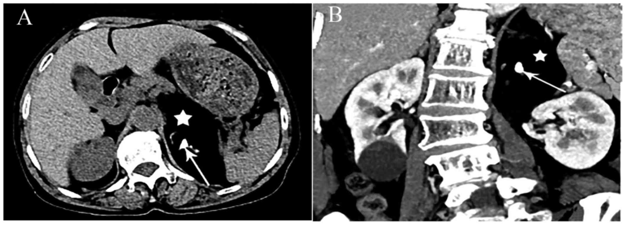
Case 1
A mass in the left abdomen developing for 3 months
was identified in a 69-year-old female patient who was admitted to
the hospital for further examination and treatment. No other
physical findings were observed. Computed tomography (CT)
examination after admission revealed a spindle-shaped mass of
heterogeneous density in the left adrenal gland, consisting mainly
of fat tissue; a small number of calcifications and soft tissue
density shadows were visible. The mass was sized ~9.3×4.9 cm
(Fig. 1). The results of complete
blood counts and routine biochemical assessment, including kidney
and liver function tests, revealed no major abnormalities. The test
results for serum cortisol, adrenocorticotropic hormone,
renin-angiotensin-aldosterone, plasma epinephrine, plasma
norepinephrine, plasma dopamine and urinary vanillylmandelic acid
were unremarkable. Therefore, the clinical diagnosis was left
adrenal mass. Subsequent retroperitoneal left adrenal lumpectomy
was successfully performed. After the tumor was completely removed,
gross postoperative examination revealed a grayish-yellow mass,
sized 10.0×6.0×4.0 cm, including visible gray cartilage tissue and
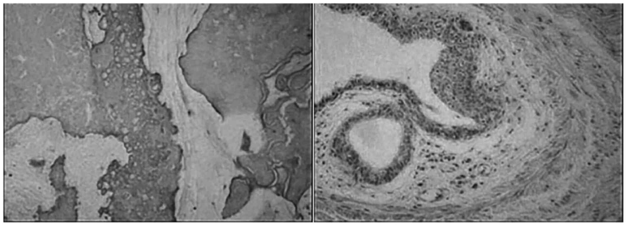
bone-like hard tissue. Hematoxylin and eosin (H&E) staining was
conducted as follows: The tissue was fixed using a 10% formalin
solution at 37°C for 24 h. Sections were cut to 6–8 µm thickness
then dried in an incubator at 45°C. Subsequently, sections were
stained with hematoxylin for 15 min (37°C), rinsed under running
water for 1 min then left standing in water for 5 min. Staining
with 0.5% eosin was also conducted for 3 min (37°C), followed by a
rinse under running water. Under examination with an optical
microscope, the tumor included adipose tissue, cartilage,
calcification, ossification and smooth muscle. Tubular structures
with stratified epithelium, with no obvious mitotic figures, were
regionally identified, and were surrounded by fiber and smooth
muscle. The tumor was infiltrated by inflammatory cells, and
included scattered tubular and vesicular structures lined by
ciliated columnar epithelium or flattened epithelium (Fig. 2). Based on these findings, the
pathological analysis of the specimens from the left adrenal gland
suggested the diagnosis of teratoma. The patient was stable, did
not complain of pain postoperatively, and exhibited no recurrence
at the 1-year follow-up.
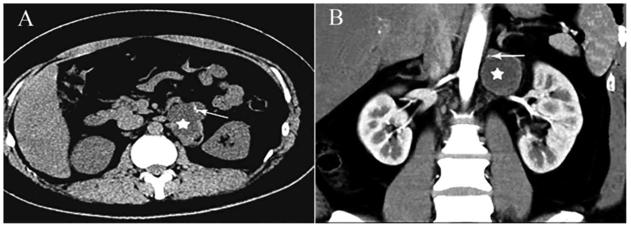
Case 2
An asymptomatic 29-year-old woman was admitted to
the hospital due to incidental discovery of a left adrenal mass 1
month prior. A CT examination after admission revealed an irregular
pelvic lesion, sized 6.0×4.0 cm, in the lower part of the left
adrenal gland (Fig. 3).
Calcification was identified at the edge of the tumor. Routine
blood examinations, such as liver and kidney function tests,
electrolytes and other routine tests, revealed no major
abnormalities. Plasma adrenal-associated hormone levels,
adrenocorticotropic hormone, renin activity, angiotensin,
aldosterone, cortisol, and urine vanillylmandelic acid levels were
within the normal range. Thus, the clinical diagnosis was left
adrenal gland mass. Subsequently, complete laparoscopic
retroperitoneal adrenalectomy was successfully performed.
Macroscopic examination of the sample revealed a grayish-red mass,
sized 2.5×2.1×0.5 cm, of mixed consistency. The capsule of the mass
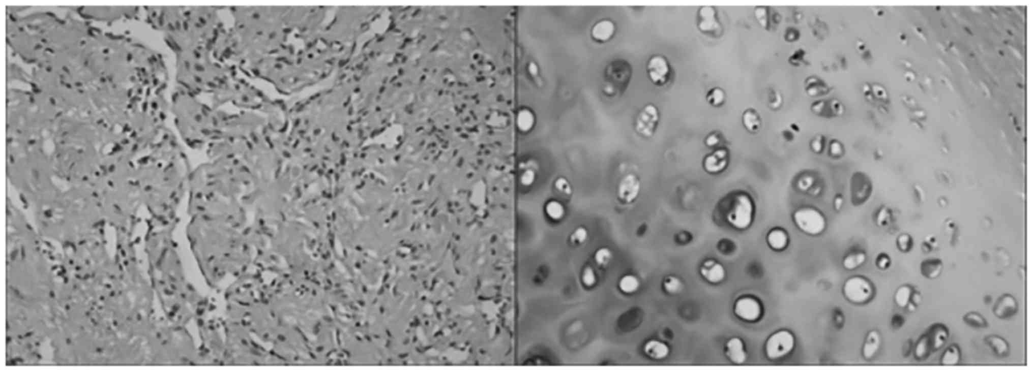
was gray and solid. H&E staining was conducted as
aforementioned. Microscopic examination revealed a tumor cyst wall
lined with squamous epithelium, and cartilage was also identified
(Fig. 4). Based on these findings,
the pathological analysis of the specimens from the left adrenal
gland suggested the diagnosis of mature teratoma. The patient was
stable postoperatively and no recurrence had occurred at the
12-month follow-up.
Results
A total of 18 cases of definitive adult primary
adrenal teratomain patients aged 17–61 years [mean ± standard
deviation (SD); 33.06±15.47 years] were identified in the
literature. Of the 18 patients, 13 were female (72.22%). A total of
8 patients (44.44%) visited a doctor after they incidentally found
an adrenal mass, and 7 patients (38.89%) visited a doctor due to
abdominal or back pain. Of the 18 patients, 9 (50%) experienced
pain, and teratoma was found in the left adrenal gland in 9
patients (50%). The median diameter of the tumor was 8.25 cm. The
majority of the patients consented to laparoscopic surgery between
2006 and 2017 (75%). Definitive follow-up data suggesting a good
prognosis were only reported for8 of the cases (44.44%).
Among patients aged <16 years, 7 pediatric cases
of adrenal teratoma were reported in the literature, of whom were 5
male (71.43%). A total of 3 cases (42.86%) were incidentally found
during examination, 2 cases (28.57%) visited a doctor due to pain,
and 2 cases (28.57%) visited a doctor due to abdominal distension.
The median tumor diameter was 10 cm. The oldest pediatric patient
was aged 8 years. Only 1 patient underwent laparoscopic tumor
resection, and there was no reported disease recurrence or death
during the follow-up period in 5 patients.
Due to the rarity of teratoma, there is no clear
clinical or pathological definition for this condition (3). It remains to be determined whether
primary adrenal teratoma should be considered as a pathological
entity similar to other retroperitoneal teratomas. According to its
definition, primary retroperitoneal teratoma is not specific to any
organ, and is most common in the left adrenal region. The present
research also identified 8 cases of adult adrenal gland
retroperitoneal teratomas (4 male and 4 female patients). These
patients were aged 24–62 years (mean ± SD; 37.38±13.27 years). Four
of the patients were admitted to the hospital due toan incidentally
discoveredadrenal mass, and in 6 cases (75%) the mass was located
in the left adrenal gland. The median tumor diameter was 8.5 cm.
The tumors were successfully removed laparoscopicallyin 4 cases
(50%). Complete follow-up data were available for 7 cases (87.5%),
and they all had a good prognosis. There was 1 reported death due
to hypertension (24).
Discussion
Adrenal mature teratomas are generally
non-functional, and the tumor exhibits latent growth. During the
early stages, the tumor causes no signs or symptoms. Later, as the
tumor increases in size, it may press on the surrounding organs,
which may cause abdominal discomfort and distension, lumbago, back
pain, nausea, vomiting, weight loss, urinary retention, intestinal
obstruction, even swelling of the lower extremity or genitals due
to lymphatic obstruction. There is no specificity regarding the
organs affected by this tumor or the symptoms. For example, when a
cystic teratoma ruptures, patients may suddenly experience
abdominal pain, peritoneal effusion or peritonitis (1), while other patients remain asymptomatic
(2).
B-scan ultrasound, CT and magnetic resonance imaging
(MRI) can clearly demonstrate the location, shap, and anatomical
associations of the tumor. Tumors on B-scan appear as a
heterogeneous group of strong light signals and contained a variety
of cystic/solid elements with uneven echo, reflecting the tumor
tissue composition. However, this qualitative diagnosis is not
sufficient. The appearance of teratoma on CT is quite
characteristic, and its diagnostic accuracy is superior to that
with B-ultrasound. CT images of mature teratoma reveal the presence
of calcification and adipose tissue, the growth site of the tumor
is clear, the capsule is continuous with the surrounding tissues
and organs, and the presence of infiltration and invasive growth
can be determined. This level of clarity is useful for providing
the basis for preoperative assessment and selection of the surgical
method (27). MRI can demonstrate
the internal characteristics of the tumor and the location of tumor
blood vessels, improve the resolution of soft tissue, clearly
delineate the border between the tumor capsule and the surrounding
adipose tissue, and determine the association between the teratoma
and major blood vessels and the presence of infiltration (1). Therefore, the results of preoperative
imaging and postoperative pathology are crucial for the final
diagnosis of teratoma.
Adrenal teratoma is usually treated by surgical
resection. The goal is complete removal of the tumor without
damaging adjacent tissues and organs, in order to relieve the
clinical symptoms and prevent malignant transformation (28). In addition, if the tumor can be
completely resected, a biopsy is not considered necessary.
Teratomas are not sensitive to radiotherapy or chemotherapy. The
surgical procedure is complicated, as adrenal mature teratoma is
often closely associated or wrapped around important blood vessels
with thin walls; this makes complete resection exceedingly
difficult. If necessary, the complicating blood vessels should be
severed and then ligated. In general, mature teratoma exhibits a
high degree of tissue differentiation (29). Complete resection is usually
curative, with only few reported cases of recurrence and metastasis
(30). A completely resected benign
teratoma may not require adjuvant chemotherapy, as it has been
reported that mature adrenal teratomas have been successfully
resected in patients as young as 8 and as old as61 years. Both
patients recovered well, with no recurrence (7).
At present, laparoscopic surgery remains the primary
choice for the treatment of adrenal tumors. Laparoscopic techniques
have been used in adrenal surgery since 1992, have developed
rapidly and are increasingly being used in urology (31). Due to the small incision and fast
recovery, laparoscopic surgery has become the preferred treatment
method for mature adrenal teratomas. Laparoscopic surgery is
currently the gold standard for the removal of adrenal lesions
(32).
Pediatric adrenal teratomas are uncommon,
representing~5% of all pediatric teratomas (12). Although pediatric cases of adrenal
teratomas have not been found in our literature review, primary
adrenal teratomas have been reported in neonates, infants and
children. Interestingly, primary adrenal teratoma has been reported
to have a maximum onset age of 8 years, with no reports in the
literature of such tumors in older children.
In conclusion, primary adrenal teratoma is a rare
tumor, which is more common in adults compared with children. Adult
primary adrenal teratoma occurs more often in men; however, among
pediatric cases, there were more female compared with male
patients. In both adults and children, adrenal primary adrenal
teratoma usually manifests as a large adrenal mass, preoperative
diagnosis is difficult, and diagnosis can only be confirmed by
histopathological examination following surgical removal of the
tumor. Laparoscopic surgery is the primary treatment choice for
adult adrenal teratoma, but children with primary adrenal teratoma
usually undergo open surgery. Based on the available follow-up
data, adults and children with primary adrenal teratoma have a good
prognosis with treatment. Due to the limited number of cases
reported in the literature, the current results are not
comprehensive. More studies and clinical cases of teratoma with
long-term follow-up data must be published and analyzed to confirm
these trends.
Acknowledgements
Not applicable.
Funding
The present study was supported by the National
Natural Science Foundation of China (grant no. 81101922), Science
and Technology Development Fund Project of Shenzhen (grant nos.
JCYJ20160429090753103 and JCYJ20170307111334308), the fund of
‘San-ming’ Project of Medicine in Shenzhen (grant no.
SZSM201612066), the Fund of Shenzhen Key Laboratory (grant no.
ZDSYS201504301045406) and the fund of Guangdong Key Medical
Subject.
Availability of data and materials
The analyzed data sets generated during the study
are available from the corresponding author on reasonable
request.
Authors' contributions
LZ and XP performed the experimental work, data
collection and interpretation. TH and YL participated in the design
and coordination of experimental work, and acquisition of data. WL,
YH, LN and SY participated in the study design, data collection,
analysis of data and preparation of the manuscript. YL conducted
the study design, the analysis and interpretation of data and
drafted the manuscript. All authors read and approved the final
manuscript.
Ethics approval and consent to
participate
Consent to participate was obtained from all
patients.
Consent for publication
Consent for publication of any associated data and
accompanying images was obtained from all patients.
Competing interests
The authors declare that they have no competing
interests.
References
|
1
|
Scott AL, Abbassi-Ghadi N, Archer CM,
Swamy R and Gupta S: Neuroendocrine carcinoma arising within a
retroperitoneal mature teratoma. Ann R Coll Surg Engl. 92:W5–W8.
2010. View Article : Google Scholar : PubMed/NCBI
|
|
2
|
Gatcombe HG, Assikis V, Kooby D and
Johnstone PA: Primary retroperitoneal teratomas: A review of the
literature. J Surg Oncol. 86:107–113. 2004. View Article : Google Scholar : PubMed/NCBI
|
|
3
|
Bedri S, Erfanian K, Schwaitzberg S and
Tischler AS: Mature cystic teratoma involving adrenal gland. Endocr
Pathol. 13:59–64. 2002. View Article : Google Scholar : PubMed/NCBI
|
|
4
|
McMillan A and Horwich A: Malignant
teratoma presenting with an adrenal mass. Clin Radiol. 38:327–328.
1987. View Article : Google Scholar : PubMed/NCBI
|
|
5
|
Lam KY and Lo CY: Teratoma in the region
of adrenal gland: A unique entity masquerading as lipomatous
adrenal tumor. Surgery. 126:90–94. 1999. View Article : Google Scholar : PubMed/NCBI
|
|
6
|
Polo JL, Villarejo PJ, Molina M, Yuste P,
Menéndez JM, Babé J and Puente S: Giant mature cystic teratoma of
the adrenal region. AJR Am J Roentgenol. 183:837–838. 2004.
View Article : Google Scholar : PubMed/NCBI
|
|
7
|
Castillo OA, Vitagliano G, Villeta M,
Arellano L and Santis O: Laparoscopic resection of adrenal
teratoma. JSLS. 10:522–524. 2006.PubMed/NCBI
|
|
8
|
Shrestha MK and Lalchan S: Adrenal gland
teratoma in a 40-year-old woman. Nepal Med Coll J. 12:201–202.
2010.PubMed/NCBI
|
|
9
|
Li H, Zhao T, Wei Q, Yuan H, Cao D, Shen
P, Liu L, Zeng H and Chen N: Laparoscopic resection of a huge
mature cystic teratoma of the right adrenal gland through
retroperitoneal approach: A case report and literature review.
World J Surg Oncol. 13:3182015. View Article : Google Scholar : PubMed/NCBI
|
|
10
|
Li S, Li H, Ji Z, Yan W and Zhang Y:
Primary adrenal teratoma: Clinical characteristics and
retroperitoneal laparoscopic resection in five adults. Oncol Lett.
10:2865–2870. 2015. View Article : Google Scholar : PubMed/NCBI
|
|
11
|
Nadeem M, Ather MH, Sulaiman MN and Pervez
S: ‘Looks Can Be Deceiving’: Adrenal Teratoma Causing Diagnostic
Difficulty. Case Rep Urol. 2015:2325912015.PubMed/NCBI
|
|
12
|
Kuo EJ, Sisk AE, Yang Z, Huang J, Yeh MW
and Livhits MJ: Adrenal Teratoma: A Case Series and Review of the
Literature. Endocr Pathol. 28:152–158. 2017. View Article : Google Scholar : PubMed/NCBI
|
|
13
|
Engel RM, Elkins RC and Fletcher BD:
Retroperitoneal teratoma. Review of the literature and presentation
of an unusual case. Cancer. 22:1068–1073. 1968. View Article : Google Scholar : PubMed/NCBI
|
|
14
|
Oguzkurt P, Ince E, Temiz A, Demir S,
Akabolat F and Hicsonmez A: Prenatal diagnosis of a mass in the
adrenal region that proved to be a teratoma. J Pediatr Hematol
Oncol. 31:350–351. 2009. View Article : Google Scholar : PubMed/NCBI
|
|
15
|
Li Y, Zhong Z and Zhao X: Primary mature
teratoma presenting as an adrenal tumor in a child. Urology.
78:689–691. 2011. View Article : Google Scholar : PubMed/NCBI
|
|
16
|
Ersoz S, Kucuk H, Mungan S, Turgutalp H,
Imamoglu M and Kosucu P: Neurocytoma arising in an adrenal gland
mature teratoma. Fetal Pediatr Pathol. 30:275–279. 2011. View Article : Google Scholar : PubMed/NCBI
|
|
17
|
Ciftci I, Cihan T, Koksal Y, Ugras S and
Erol C: Giant mature adrenal cystic teratoma in an infant. Acta
Inform Med. 21:140–141. 2013. View Article : Google Scholar : PubMed/NCBI
|
|
18
|
Garg A, Pollak-Christian E and
Unnikrishnan N: A Rare Adrenal Mass in a 3-Month-Old: A Case Report
and Literature Review. Case Rep Pediatr.
2017:45423212017.PubMed/NCBI
|
|
19
|
Rais-Bahrami S, Varkarakis IM, Lujan G and
Jarrett TW: Primary retroperitoneal teratoma presenting as an
adrenal tumor in an adult. Urology. 69(185): e181–e182. 2007.
|
|
20
|
Sato F, Mimata H and Mori K: Primary
retroperitoneal mature cystic teratoma presenting as an adrenal
tumor in an adult. Int J Urol. 17:8172010. View Article : Google Scholar : PubMed/NCBI
|
|
21
|
Chen JC, Khiyami A and McHenry CR:
Retroperitoneal cystic teratoma masquerading as an incidentally
discovered adrenal mass. Endocr Pract. 17:e130–e134. 2011.
View Article : Google Scholar : PubMed/NCBI
|
|
22
|
Giordano R, Giraudo G, Forno D, Bosco M,
Delsedime L, Morino M and Arvat E: A case of primary
retroperitoneal teratoma presenting as an adrenal incidentaloma. J
Endocrinol Invest. 34:645–646. 2011. View Article : Google Scholar : PubMed/NCBI
|
|
23
|
Bhatti A, Al-Hindi H, Azzam A, Amin T and
Abu-Zaid A: Mature (benign) cystic retroperitoneal teratoma
involving the left adrenal gland in a 22-year-old male: A case
report and literature review. Case Rep Oncol Med.
2013:6102802013.PubMed/NCBI
|
|
24
|
Tang DD, Zhang XS, Hao ZY, Zhou J and
Liang CZ: A giant primary retroperitoneal mature cystic teratoma in
right adrenal region in a 39-year-old female. Int J Clin Exp Med.
7:1611–1613. 2014.PubMed/NCBI
|
|
25
|
Bhatia V, Sharma S, Sood S, Mardi K and
Venkat B: Case 231: Retroperitoneal Adrenal Teratoma Presenting as
Trichoptysis. Radiology. 280:317–321. 2016. View Article : Google Scholar : PubMed/NCBI
|
|
26
|
Kataoka M, Fukushima H, Nakanishi Y,
Yokoyama M, Funata N, Motoi T, Tobisu KI and Koga F:
Retroperitoneal Teratoma in an Adult: A Potential Pitfall in the
Differential Diagnosis of Adrenal Myelolipoma. Case Rep Urol.
2016:51417692016.PubMed/NCBI
|
|
27
|
Wang LJ, Chu SH, Ng KF and Wong YC:
Adenocarcinomas arising from primary retroperitoneal mature
teratomas: CT and MR imaging. Eur Radiol. 12:1546–1549. 2002.
View Article : Google Scholar : PubMed/NCBI
|
|
28
|
Yoon SS, Tanabe KK and Warshaw AL: Adult
primary retroperitoneal teratoma. Surgery. 137:663–664. 2005.
View Article : Google Scholar : PubMed/NCBI
|
|
29
|
Jones NM and Kiely EM: Retroperitoneal
teratomas-potential for surgical misadventure. J Pediatr Surg.
43:184–186; discussion 187. 2008. View Article : Google Scholar : PubMed/NCBI
|
|
30
|
James J, Dhillon GS, Blewett CJ,
Halldorsson A and Cecalupo AJ: A large adrenal teratoma in a
neonate. Am Surg. 75:347–349. 2009.PubMed/NCBI
|
|
31
|
Gagner M, Lacroix A and Bolté E:
Laparoscopic adrenalectomy in Cushing's syndrome and
pheochromocytoma. N Engl J Med. 327:10331992. View Article : Google Scholar : PubMed/NCBI
|
|
32
|
Chuan-Yu S, Yat-Faat H, Wei-Hong D,
Yuan-Cheng G, Qing-Feng H, Ke X, Bin G and Guo-Wei X: Laparoscopic
adrenalectomy for adrenal tumors. Int J Endocrinol.
2014:2418542014. View Article : Google Scholar : PubMed/NCBI
|