Introduction
Gastrointestinal subepithelial lesions (SELs) are
often found incidentally on esophagogastroduodenoscopy (EGD)
(1). Gastrointestinal stromal tumors
(GISTs) may require treatment intervention, whereas benign lesions
such as leiomyomas and neurilemomas can often be followed up.
Therefore, the differential diagnosis of GISTs from benign lesions
is important in deciding treatment strategy (2,3).
Follow-up observation has previously been
recommended for GISTs <20 mm in diameter, but because of
possible occult malignancy, surgery for smaller lesions <20 mm
is now recommended in principle (4,5).
Diagnosis of a small GIST by conventional endoscopic biopsy and
imaging can be difficult. Thus, endoscopic ultrasound-guided fine
needle aspiration (EUS-FNA) to acquire tissue samples for
immunohistological diagnosis is essential (6).
EUS-FNA to evaluate SELs is useful (7), and although tissue collection from
small lesions <20 mm has been reported, the adequate sample
rates have been slightly low (8).
EUS-FNA with a 19G needle can be valuable for histologic diagnosis,
since an adequate tissue sample is likely to be obtained for
immunostaining (9,10). However, because a 19G needle is
stiffer than a 22G needle, the difficulty of adequately positioning
the scope, and in particular, the angulation of the needle relative
to the scope, makes lesion puncture more difficult (11). This led to design of a thin needle to
acquire adequate tissue, and in 2016, the Franseen needle was
developed to improve diagnostic performance (12).
This study evaluated diagnostic accuracy rates of
EUS-FNA for SELs and compared adequate sample rates between a 22G
conventional needle and a 22G Franseen needle. In particular,
utility for lesions <20 mm in diameter was compared.
Materials and methods
The present study was approved by the Ethical Review
Board at Saitama Medical University International Medical Center
(Saitama, Japan) and complied with the Declaration of Helsinki, as
revised in Brazil 2013. All patients provided written, informed
consent for EUS-FNA. This study included 57 consecutive lesions (61
sessions) of EUS-FNA using a 22G conventional needle or a 22G
Franseen needle to evaluate SELs at our medical center between July
2013 and October 2017. Patient data were retrospectively analyzed.
The primary endpoint of this study was to compare adequate sample
rates between a 22G conventional needle group and a 22G Franseen
needle group. The secondary endpoint was to compare other data,
including tumor size, procedure time, number of punctures, and
tumor site.
EUS-FNA procedures
EUS-FNA procedures were performed using a convex
linear-array echoendoscope (GF-UCT260; Olympus Optical Co Ltd,
Tokyo, Japan) paired with an ultrasound machine (EU-ME2 Premier
Plus; Olympus Optical Co Ltd). EUS was performed with the patient
under conscious sedation using intravenous midazolam and pethidine
hydrochloride. After excluding regional or collateral vasculature,
the mass was punctured. The stylet was removed, and continuous
suction was applied with a 20-ml syringe.
Then, 20–30 rapid strokes were made within the
lesion, suction was released, and the needle was removed. The
aspirated samples were smeared onto glass slides by inserting the
stylet and applying air pressure. The samples were examined
visually for white color and then fixed in formalin for histologic
examination.
Because on-site cytological examination was not
performed, EUS-FNA was repeated whenever the procedure could be
continued under our supervision with a cytology technician until
there was visual confirmation of an adequate sample for
histopathology and immunostaining.
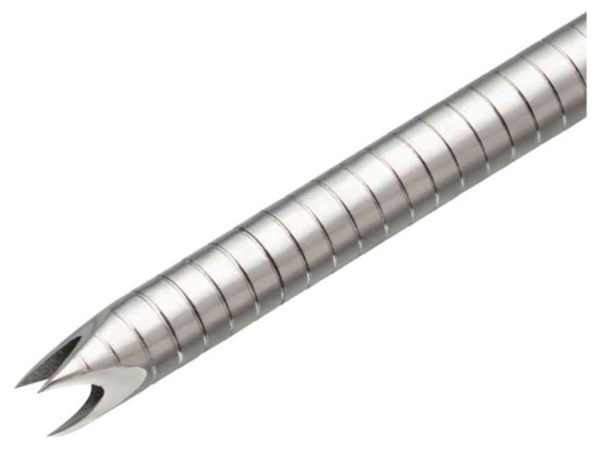
The puncture needles used were a 22G conventional
needle (C group) and a 22G Franseen needle (F group).
Expect® (Boston Scientific Japan, Tokyo, Japan) was used
in the C group, and Acquire® (Boston Scientific Japan)
was used in the F group (Fig. 1).
The C needle was primarily used in patients between July 2013 and
December 2016, and the F needle was primarily used in patients
thereafter. EUS-FNA procedures were performed by five endoscopists.
Three endoscopists were trainees, and two were experienced in
performing EUS-FNA. The three trainee endoscopists had sufficient
experience, having conducted more than 1,000 regular EGDs, 500
colonoscopies, and 20 EUS procedures. They had also attended 20
EUS-FNA procedures performed by EUS-FNA experts as assistants. The
two expert endoscopists had performed regular EGD, colonoscopy, and
EUS procedures. They had performed more than 50 EUS-FNA procedures
before the beginning of this study.
Histological evaluation
EUS-FNA specimens were fixed in 10% formalin and
embedded in paraffin. Sections were stained with hematoxylin and
eosin (Η&Ε) for histologic examination, and immunostaining was
performed. These were examined by two pathology technicians and two
pathologists. Only the histologic diagnosis was analyzed in this
study. Immunohistochemical (IHC) staining was subsequently
performed. A diagnosis of GIST was made by positive c-kit staining,
with or without positive CD34 and DOG-1 IHC staining. Leiomyoma was
diagnosed by positive desmin staining, and schwannoma was diagnosed
by positive S-100 staining. The results of IHC were described as
positive or negative. Positive IHC staining was defined as staining
of >50% of the tumor cells. Negative IHC staining was defined as
either focal positivity or staining of <50% of the tumor
cells.
Study definitions
Adequate samples were defined as samples sufficient
for immunohistological examination. The final diagnosis was based
on postoperative histopathology of lesions that were surgically
resectable. When a diagnosis could not be made on the first
examination, EUS-FNA was performed in the second session if the
patient agreed.
Statistical analysis
Categorical variables are expressed as absolute
(n) and relative (%) frequencies, and they were compared
using Fisher's exact test. For comparisons of continuous data, a
2-sample t-test was used if a normal distribution was likely, and
the Mann-Whitney test was used if normality could not be
demonstrated. P<0.05 was considered to indicate a statistically
significant difference. Statistical calculations were performed
using SAS 9.4 and SAS JMP 12.2.0 for Windows (SAS Institute Inc,
Cary, NC, USA).
Results
Median age, tumor size, number of punctures, and
procedure time were 67.0 years, 23.9 mm, 3 times, and 25.0 min,
respectively (Table I). Among the 57
lesions, the most common FNA diagnosis was GIST in 32 patients,
followed by leiomyoma in 9 patients. In 10 patients, there was
insufficient material for an FNA diagnosis (Table II). Among the 10 lesions without a
definitive diagnosis, 5 were <20 mm in size.
 | Table I.Clinical features of patients and
outcomes of endoscopic ultrasound-guided fine needle
aspiration. |
Table I.
Clinical features of patients and
outcomes of endoscopic ultrasound-guided fine needle
aspiration.
| Variable | All | C group | F group | P-value |
|---|
| Sex, male/female | 37/24 | 27/17 | 10/7 | >0.99 |
| Age (years), median
(IQR) | 67.0 (58.0–74.5) | 67.0 (55.0–74.8) | 72.0 (58.5–74.5) | 0.71 |
| Tumor size (mm),
median (IQR) | 23.9 (17.3–33.0) | 23.9 (16.4–30.8) | 26.7 (19.7–40.2) | 0.47 |
| Number of punctures,
median (IQR) | 3 (3–4) | 3 (3–4) | 3 (3–4) | 0.55 |
| Procedure time (min),
median (IQR) | 25.0 (18.5–32.0) | 25.5 (18.0–32.0) | 23.0 (19.0–32.0) | 0.96 |
| Table II.Endoscopic ultrasound-guided fine
needle aspiration diagnoses. |
Table II.
Endoscopic ultrasound-guided fine
needle aspiration diagnoses.
| EUS-FNA
diagnosis | No. of lesions
(%) |
|---|
| GIST | 56.1 (32) |
| Schwannoma | 3.5
(2) |
| Leiomyoma | 15.8 (9) |
| Ectopic pancreas | 3.5 (2) |
| Lymph node
metastasis | 1.8 (1) |
| Lipoma | 1.8 (1) |
| Insufficient
material | 17.5
(10) |
Consent for surgery was obtained from 26 patients
who had a preoperative FNA diagnosis of GIST or other suspected
malignancy. The postoperative diagnosis in these patients was GIST
in 22, schwannoma in 2, lymph node metastases from esophageal
cancer in 1, and other in 1 patient. None of the patients had any
EUS-FNA-related complications.
The diagnostic accuracy rate of EUS-FNA for GIST was
95.5% (Table III). The overall
adequate sample rate was 80.3%; the rates by lesion site were
esophagus 100% (5/5), stomach 78.2% (43/55), and rectum 100%
(1/1).
| Table III.Final diagnoses. |
Table III.
Final diagnoses.
| Final diagnosis | Accuracy of EUS-FNA,
% (n of total) |
|---|
| GIST | 95.5
(21/22) |
| Other tumors | 50 (2/4) |
| Overall | 88.5 (23/26) |
A comparison of patient characteristics between the
C group and the F group showed no significant differences in age,
sex, tumor size, or tumor site (Table
I). The overall adequate sample rates in the C group and the F
group were 75.0% (33/44) and 94.1% (16/17), respectively. Although
the difference was not significant, the adequate sample rate tended
to be higher in the F group. It was possible to collect specimens
in both groups even for tumors located on the greater curvature
side in the stomach (Table IV).
| Table IV.Comparison of endoscopic
ultrasound-guided fine needle aspiration outcomes between the
Franseen needle group and the conventional needle group. |
Table IV.
Comparison of endoscopic
ultrasound-guided fine needle aspiration outcomes between the
Franseen needle group and the conventional needle group.
| Obtaining an adequate
specimen, n (%) | All (%) | C group (%) | F group (%) | P-value |
|---|
| Location |
|
Esophagus | 5/5 (100) | 5/5 (100) | – | >0.99 |
|
Stomach | 43/55 (78.2) | 28/39 (71.8) | 15/16 (93.8) | 0.15 |
|
Greater curvature
side | 19/22 (86.4) | 11/14 (78.6) | 8/8 (100) | 0.27 |
|
Lesser curvature
side | 24/33 (72.7) | 17/25 (68.0) | 7/8 (87.5) | 0.39 |
|
Rectum | 1/1 (100) | – | 1/1 (100) | >0.99 |
|
≥20 mm | 35/41 (85.4) | 24/29 (82.8) | 11/12 (91.7) | 0.65 |
|
<20 mm | 14/20 (70.0) | 9/15 (60) | 5/5 (100) | 0.26 |
| Overall | 49/61 (80.3) | 33/44 (75.0) | 16/17 (94.1) | 0.15 |
The adequate sample rates were also compared based
on tumor size. For lesions ≥20 mm, the adequate sample rates were
82.8% (24/29) in the C group and 91.7% (11/12) in the F group, 8.9
percentage points higher in the F group. However, for lesions
<20 mm, the adequate sample rates were 60% (9/15) in the C group
and 100% (5/5) in the F group, 40 percentage points higher in the F
group (P=0.65, 0.26). Thus, the adequate sample rate was markedly
higher in the F group for lesions <20 mm in diameter (Table IV). Although there were a few cases
of examination, even trainee endoscopists could collect adequate
samples (Table V).
| Table V.Comparison of endoscopic
ultrasound-guided fine needle aspiration outcomes using a Franseen
needle between the trainee and expert groups. |
Table V.
Comparison of endoscopic
ultrasound-guided fine needle aspiration outcomes using a Franseen
needle between the trainee and expert groups.
| Obtaining an adequate
specimen, n (%) | All (%) | Trainee (%) | Expert (%) | P-value |
|---|
| Location |
|
Esophagus | – | – | – | – |
|
Stomach | 15/16 (93.8) | 4/4 (100) | 11/12 (91.7) | >0.99 |
|
Greater curvature
side | 8/8 (100) | 2/2 (100) | 6/6 (100) | >0.99 |
|
Lesser curvature
side | 7/8 (87.5) | 2/2 (100) | 5/6 (83.3) | >0.99 |
|
Rectum | 1/1 (100) | – | 1/1 (100) | >0.99 |
|
≥20 mm | 11/12 (91.7) | 3/3 (100) | 8/9 (88.9) | >0.99 |
|
<20 mm | 5/5 (100) | 1/1 (100) | 4/4 (100) | >0.99 |
| Overall | 16/17 (94.1) | 4/4 (100) | 12/13 (92.3) | >0.99 |
Discussion
EUS-FNA is a useful, minimally invasive procedure
for tissue acquisition from SELs, with reported diagnostic accuracy
rates of 52–92% (13–18). GISTs ≥20 mm in diameter are
associated with a high rate of metastases, and previous guidelines
recommended surgery only for lesions ≥20 mm (19). However, a more recent study of 43
surgical cases found that, even for tumors <20 mm, 23% were at
intermediate risk for possible metastases based on modified
Fletcher criteria (8). Liver
metastases after surgery for GISTs <20 mm have also been
reported (20).
Furthermore, with the increase in minimally invasive
procedures such as laparoscopic and endoscopic cooperative surgery
(LECS) (21), primary surgical
resection is now being recommended for resectable GISTs without
metastases regardless of size (22).
Therefore, the need for diagnosis and treatment of smaller SELs is
increasingly being recognized. This calls for improved diagnostic
performance of EUS-FNA, but the sensitivity for lesions <20 mm
has been insufficient (8).
Novel needles, including those with a side hole
(23) and fork-tip (14), have recently been developed for use
in EUS-FNA. Despite the increase in needle options, no specific
needle has been uniformly adopted. The needle sizes mainly used are
19G, 22G, and 25G, and although a 19G needle may provide sufficient
tissue, strong puncture resistance can make using a 19G needle very
difficult, especially in smaller lesions. Therefore, a 22G needle
is more commonly used for EUS-FNA.
The 22G Franseen needle used in the present study
provided a high adequate sample rate and was particularly useful
for lesions <20 mm. The Franseen needle has 3 tips to puncture
and grip tissue and 3 cutting planes to cut tissue for sample
acquisition. This enables acquisition of an adequate tissue sample
(12).
In our view, EUS-FNA is indicated for SELs larger
than 10 mm. Using the current EUS-FNA system including needles, it
is difficult to puncture SELs smaller than 10 mm (8), and metastatic lesions have not been
reported in a small tumor.
This study was a retrospective analysis of a
single-center experience with EUS-FNA to evaluate gastrointestinal
SELs at our medical center, including a comparison of utility
between a conventional needle and Franseen needle to evaluate
smaller SELs.
The overall adequate sample rate for FNA diagnosis
of SELs at our medical center was 80.3%, which is similar to
previously reported studies. In patients in whom sample acquisition
was difficult, factors besides needle selection and lesion size,
including differences in lesion site and differences in disease
type, may have had an influence. Therefore, further investigation
is needed. In an analysis limited to cases in which a final
diagnosis was possible, the diagnostic accuracy rate for GIST at
our medical center was 95.5%. Although these are good results, in
many cases of a final diagnosis of an SEL, surgery was not
performed, and during follow-up observation, there were no changes
in imaging findings. Thus, assessment based on the clinical course
in these patients was difficult.
This study examined the utility of a 22G-Franseen
needle in small SELs, for which it is increasingly being recognized
that early diagnosis and treatment are important.
Comparison between the Franseen needle group and the
conventional needle group showed no marked differences in patient
age, sex, tumor size, or number of punctures. Although there was no
significant difference in the F group, the findings suggest that
the Franseen needle may be useful to improve adequate sample rates,
particularly, for small lesions <20 mm in size. Although the
possibility that puncture performance of the Franseen needle might
be inferior to the C needle was considered due to the shape of the
tip of the needle, the present results showed no significant
difference. The reason for this may be that when we puncture, the
up angle is used to fix the lesion firmly and to take a quick
puncture.
Because this study was retrospective, some
limitations must be considered when interpreting the results. Some
influence of needle selection on each examination day cannot be
excluded. In addition, all data were retrospectively collected from
a single center. A prospective study with many cases will be
necessary. However, the utility of the Franseen needle has already
been reported (12), so the present
results are consistent for SELs. In particular, the utility of the
Franseen needle tended to be higher for small lesions <20 mm in
the present study. In conclusion, the current findings suggest that
a Franseen needle may improve the adequate sample rate and
diagnostic accuracy rate of EUS-FNA in small SELs.
Acknowledgements
Not applicable.
Funding
No funding was received.
Availability of data and materials
All data generated or analyzed during this study are
included in this published article.
Authors' contributions
AF designed the study and drafted the article. AF,
SR, MK, YT and TK performed all of the endoscopic ultrasound-guided
fine needle aspiration procedures, and RA performed the statistical
analysis. KM, MM, and KN critically revised the article for
important intellectual content. SR supervised the study and gave
final approval of the version to be published. The final version of
the manuscript was read and approved by all of the authors.
Ethics approval and consent to
participate
The present study was approved by the Ethical Review
Board at Saitama Medical University International Medical Center
(Saitama, Japan) and complied with the Declaration of Helsinki, as
revised in Brazil 2013. All patients provided written informed
consent.
Patient consent for publications
Not applicable.
Competing interests
The authors declare that they have no competing
interests.
References
|
1
|
Hedenbro JL, Ekelund M and Wetterberg P:
Endoscopic diagnosis of submucosal gastric lesions. The results
after routine endoscopy. Surg Endosc. 5:20–23. 1991. View Article : Google Scholar : PubMed/NCBI
|
|
2
|
Ando N, Goto H, Niwa Y, Hirooka Y, Ohmiya
N, Nagasaka T and Hayakawa T: The diagnosis of GI stromal tumors
with EUS-guided fine needle aspiration with immunohistochemical
analysis. Gastrointest Endosc. 55:37–43. 2002. View Article : Google Scholar : PubMed/NCBI
|
|
3
|
Miettinen M and Lasota J: Gastrointestinal
stromal tumors-definition, clinical, histological,
immunohistochemical and molecular genetic features and differential
diagnosis. Virchows Arch. 438:1–12. 2001. View Article : Google Scholar : PubMed/NCBI
|
|
4
|
Blay JY, Bonvalot S, Casali P, Choi H,
Debiec-Richter M, Dei Tos AP, Emile JF, Gronchi A, Hogendoorn PC,
Joensuu H, et al: Consensus meeting for the management of
gastrointestinal stromal tumors. Report of the GIST consensus
conference of 20–21 March 2004, under the auspices of ESMO. Ann
Oncol. 16:566–578. 2005. View Article : Google Scholar : PubMed/NCBI
|
|
5
|
Nishida T, Hirota S, Yanagisawa A, Sugino
Y, Minami M, Yamamura Y, Otani Y, Shimada Y, Takahashi F and Kubota
T: GIST Guideline Subcommittee: Clinical practice guidelines for
gastrointestinal stromal tumor (GIST) in Japan: English version.
Int J Clin Oncol. 13:416–430. 2008. View Article : Google Scholar : PubMed/NCBI
|
|
6
|
Okasha HH, Naguib M, El Nady M, Ezzat R,
Al-Gemeie E, Al-Nabawy W, Aref W, Abdel-Moaty A, Essam K and Hamdy
A: Role of endoscopic ultrasound and endoscopic-ultrasound-guided
fine-needle aspiration in endoscopic biopsy negative
gastrointestinal lesions. Endosc Ultrasound. 6:156–161. 2017.
View Article : Google Scholar : PubMed/NCBI
|
|
7
|
Mekky MA, Yamao K, Sawaki A, Mizuno N,
Hara K, Nafeh MA, Osman AM, Koshikawa T, Yatabe Y and Bhatia V:
Diagnostic utility of EUS-guided FNA in patients with gastric
submucosal tumors. Gastrointest Endosc. 71:913–919. 2010.
View Article : Google Scholar : PubMed/NCBI
|
|
8
|
Akahoshi K, Oya M, Koga T, Koga H,
Motomura Y, Kubokawa M, Gibo J and Nakamura K: Clinical usefulness
of endoscopic ultrasound-guided fine needle aspiration for gastric
subepithelial lesions smaller than 2 cm. J Gastrointestin Liver
Dis. 23:405–412. 2014.PubMed/NCBI
|
|
9
|
Iwashita T, Yasuda I, Doi S, Ando N,
Nakashima M, Adachi S, Hirose Y, Mukai T, Iwata K, Tomita E, et al:
Use of samples from endoscopic ultrasound-guided 19-gauge
fine-needle aspiration in diagnosis of autoimmune pancreatitis.
Clin Gastroenterol Hepatol. 10:316–322. 2012. View Article : Google Scholar : PubMed/NCBI
|
|
10
|
Yasuda I, Goto N, Tsurumi H, Nakashima M,
Doi S, Iwashita T, Kanemura N, Kasahara S, Adachi S, Hara T, et al:
Endoscopic ultrasound-guided fine needle aspiration biopsy for
diagnosis of lymphoproliferative disorders: Feasibility of
immunohistological, flow cytometric and cytogenetic assessments. Am
J Gastroenterol. 107:397–404. 2012. View Article : Google Scholar : PubMed/NCBI
|
|
11
|
Song TJ, Kim JH, Lee SS, Eum JB, Moon SH,
Park DY, Seo DW, Lee SK, Jang SJ, Yun SC and Kim MH: The
prospective randomized, controlled trial of endoscopic
ultrasound-guided fine-needle aspiration using 22G and 19G
aspiration needles for solid pancreatic or peripancreatic masses.
Am J Gastroenterol. 105:1739–1745. 2010. View Article : Google Scholar : PubMed/NCBI
|
|
12
|
Bang JY, Hebert-Magee S, Hasan MK,
Navaneethan U, Hawes R and Varadarajulu S: Endoscopic
ultrasonography-guided biopsy using a Franseen needle design:
Initial assessment. Dig Endosc. 29:338–346. 2017. View Article : Google Scholar : PubMed/NCBI
|
|
13
|
Akahoshi K, Sumida Y, Matsui N, Oya M,
Akinaga R, Kubokawa M, Motomura Y, Honda K, Watanabe M and Nagaie
T: Preoperative diagnosis of gastrointestinal stromal tumor by
endoscopic ultrasound-guided fine needle aspiration. World J
Gastroenterol. 13:2077–2082. 2007. View Article : Google Scholar : PubMed/NCBI
|
|
14
|
El Chafic AH, Loren D, Siddiqui A, Mounzer
R, Cosgrove N and Kowalski T: Comparison of FNA and fine-needle
biopsy for EUS-guided sampling of suspected GI stromal tumors.
Gastrointest Endosc. 86:510–515. 2017. View Article : Google Scholar : PubMed/NCBI
|
|
15
|
Hoda KM, Rodriguez SA and Faigel DO:
EUS-guided sampling of suspected GI stromal tumors. Gastrointest
Endosc. 69:1218–1223. 2009. View Article : Google Scholar : PubMed/NCBI
|
|
16
|
Kim GH, Cho YK, Kim EY, Kim HK, Cho JW,
Lee TH and Moon JS: Korean EUS Study Group: Comparison of 22-gauge
aspiration needle with 22-gauge biopsy needle in endoscopic
ultrasonography-guided subepithelial tumor sampling. Scand J
Gastroenterol. 49:347–354. 2014. View Article : Google Scholar : PubMed/NCBI
|
|
17
|
Philipper M, Hollerbach S, Gabbert HE,
Heikaus S, Böcking A, Pomjanski N, Neuhaus H, Frieling T and
Schumacher B: Prospective comparison of endoscopic
ultrasound-guided fine-needle aspiration and surgical histology in
upper gastrointestinal submucosal tumors. Endoscopy. 42:300–305.
2010. View Article : Google Scholar : PubMed/NCBI
|
|
18
|
Watson RR, Binmoeller KF, Hamerski CM,
Shergill AK, Shaw RE, Jaffee IM, Stewart L and Shah JN: Yield and
performance characteristics of endoscopic ultrasound-guided fine
needle aspiration for diagnosing upper GI tract stromal tumors. Dig
Dis Sci. 56:1757–1762. 2011. View Article : Google Scholar : PubMed/NCBI
|
|
19
|
Miettinen M, Sobin LH and Lasota J:
Gastrointestinal stromal tumors of the stomach: A
clinicopathologic, immunohistochemical and molecular genetic study
of 1765 cases with long-term follow-up. Am J Surg Pathol. 29:52–68.
2005. View Article : Google Scholar : PubMed/NCBI
|
|
20
|
Aso A, Ihara E, Kubo H, Osoegawa T, Oono
T, Nakamura K, Ito T, Kakeji Y, Mikako O, Yamamoto H, et al:
Gastric gastrointestinal stromal tumor smaller than 20 mm with
liver metastasis. Clin J Gastroenterol. 6:29–32. 2013. View Article : Google Scholar : PubMed/NCBI
|
|
21
|
Hiki N, Yamamoto Y, Fukunaga T, Yamaguchi
T, Nunobe S, Tokunaga M, Miki A, Ohyama S and Seto Y: Laparoscopic
and endoscopic cooperative surgery for gastrointestinal stromal
tumor dissection. Surg Endosc. 22:1729–1735. 2008. View Article : Google Scholar : PubMed/NCBI
|
|
22
|
ESMO/European Sarcoma Network Working
Group: Gastrointestinal stromal tumors: ESMO clinical practice
guidelines for diagnosis, treatment and follow-up. Ann Oncol. 23
Suppl 7:vii49–vii55. 2012.PubMed/NCBI
|
|
23
|
Inoue T, Okumura F, Mizushima T, Nishie H,
Iwasaki H, Anbe K, Ozeki T, Kachi K, Fukusada S, Suzuki Y and Sano
H: Assessment of factors affecting the usefulness and diagnostic
yield of core biopsy needles with a side hole in endoscopic
ultrasound-guided fine-needle aspiration. Gut Liver. 10:51–57.
2016. View
Article : Google Scholar : PubMed/NCBI
|