Spandidos Publications Logo
  • About
    • About Spandidos
    • Aims and Scopes
    • Abstracting and Indexing
    • Editorial Policies
    • Reprints and Permissions
    • Job Opportunities
    • Terms and Conditions
    • Contact
  • Journals
    • All Journals
    • Oncology Letters
      • Oncology Letters
      • Information for Authors
      • Editorial Policies
      • Editorial Board
      • Aims and Scope
      • Abstracting and Indexing
      • Bibliographic Information
      • Archive
    • International Journal of Oncology
      • International Journal of Oncology
      • Information for Authors
      • Editorial Policies
      • Editorial Board
      • Aims and Scope
      • Abstracting and Indexing
      • Bibliographic Information
      • Archive
    • Molecular and Clinical Oncology
      • Molecular and Clinical Oncology
      • Information for Authors
      • Editorial Policies
      • Editorial Board
      • Aims and Scope
      • Abstracting and Indexing
      • Bibliographic Information
      • Archive
    • Experimental and Therapeutic Medicine
      • Experimental and Therapeutic Medicine
      • Information for Authors
      • Editorial Policies
      • Editorial Board
      • Aims and Scope
      • Abstracting and Indexing
      • Bibliographic Information
      • Archive
    • International Journal of Molecular Medicine
      • International Journal of Molecular Medicine
      • Information for Authors
      • Editorial Policies
      • Editorial Board
      • Aims and Scope
      • Abstracting and Indexing
      • Bibliographic Information
      • Archive
    • Biomedical Reports
      • Biomedical Reports
      • Information for Authors
      • Editorial Policies
      • Editorial Board
      • Aims and Scope
      • Abstracting and Indexing
      • Bibliographic Information
      • Archive
    • Oncology Reports
      • Oncology Reports
      • Information for Authors
      • Editorial Policies
      • Editorial Board
      • Aims and Scope
      • Abstracting and Indexing
      • Bibliographic Information
      • Archive
    • Molecular Medicine Reports
      • Molecular Medicine Reports
      • Information for Authors
      • Editorial Policies
      • Editorial Board
      • Aims and Scope
      • Abstracting and Indexing
      • Bibliographic Information
      • Archive
    • World Academy of Sciences Journal
      • World Academy of Sciences Journal
      • Information for Authors
      • Editorial Policies
      • Editorial Board
      • Aims and Scope
      • Abstracting and Indexing
      • Bibliographic Information
      • Archive
    • International Journal of Functional Nutrition
      • International Journal of Functional Nutrition
      • Information for Authors
      • Editorial Policies
      • Editorial Board
      • Aims and Scope
      • Abstracting and Indexing
      • Bibliographic Information
      • Archive
    • International Journal of Epigenetics
      • International Journal of Epigenetics
      • Information for Authors
      • Editorial Policies
      • Editorial Board
      • Aims and Scope
      • Abstracting and Indexing
      • Bibliographic Information
      • Archive
    • Medicine International
      • Medicine International
      • Information for Authors
      • Editorial Policies
      • Editorial Board
      • Aims and Scope
      • Abstracting and Indexing
      • Bibliographic Information
      • Archive
  • Articles
  • Information
    • Information for Authors
    • Information for Reviewers
    • Information for Librarians
    • Information for Advertisers
    • Conferences
  • Language Editing
Spandidos Publications Logo
  • About
    • About Spandidos
    • Aims and Scopes
    • Abstracting and Indexing
    • Editorial Policies
    • Reprints and Permissions
    • Job Opportunities
    • Terms and Conditions
    • Contact
  • Journals
    • All Journals
    • Biomedical Reports
      • Information for Authors
      • Editorial Policies
      • Editorial Board
      • Aims and Scope
      • Abstracting and Indexing
      • Bibliographic Information
      • Archive
    • Experimental and Therapeutic Medicine
      • Information for Authors
      • Editorial Policies
      • Editorial Board
      • Aims and Scope
      • Abstracting and Indexing
      • Bibliographic Information
      • Archive
    • International Journal of Epigenetics
      • Information for Authors
      • Editorial Policies
      • Editorial Board
      • Aims and Scope
      • Abstracting and Indexing
      • Bibliographic Information
      • Archive
    • International Journal of Functional Nutrition
      • Information for Authors
      • Editorial Policies
      • Editorial Board
      • Aims and Scope
      • Abstracting and Indexing
      • Bibliographic Information
      • Archive
    • International Journal of Molecular Medicine
      • Information for Authors
      • Editorial Policies
      • Editorial Board
      • Aims and Scope
      • Abstracting and Indexing
      • Bibliographic Information
      • Archive
    • International Journal of Oncology
      • Information for Authors
      • Editorial Policies
      • Editorial Board
      • Aims and Scope
      • Abstracting and Indexing
      • Bibliographic Information
      • Archive
    • Medicine International
      • Information for Authors
      • Editorial Policies
      • Editorial Board
      • Aims and Scope
      • Abstracting and Indexing
      • Bibliographic Information
      • Archive
    • Molecular and Clinical Oncology
      • Information for Authors
      • Editorial Policies
      • Editorial Board
      • Aims and Scope
      • Abstracting and Indexing
      • Bibliographic Information
      • Archive
    • Molecular Medicine Reports
      • Information for Authors
      • Editorial Policies
      • Editorial Board
      • Aims and Scope
      • Abstracting and Indexing
      • Bibliographic Information
      • Archive
    • Oncology Letters
      • Information for Authors
      • Editorial Policies
      • Editorial Board
      • Aims and Scope
      • Abstracting and Indexing
      • Bibliographic Information
      • Archive
    • Oncology Reports
      • Information for Authors
      • Editorial Policies
      • Editorial Board
      • Aims and Scope
      • Abstracting and Indexing
      • Bibliographic Information
      • Archive
    • World Academy of Sciences Journal
      • Information for Authors
      • Editorial Policies
      • Editorial Board
      • Aims and Scope
      • Abstracting and Indexing
      • Bibliographic Information
      • Archive
  • Articles
  • Information
    • For Authors
    • For Reviewers
    • For Librarians
    • For Advertisers
    • Conferences
  • Language Editing
Login Register Submit
  • This site uses cookies
  • You can change your cookie settings at any time by following the instructions in our Cookie Policy. To find out more, you may read our Privacy Policy.

    I agree
Search articles by DOI, keyword, author or affiliation
Search
Advanced Search
presentation
Molecular and Clinical Oncology
Join Editorial Board Propose a Special Issue
Print ISSN: 2049-9450 Online ISSN: 2049-9469
Journal Cover
May-2019 Volume 10 Issue 5

Full Size Image

Sign up for eToc alerts
Recommend to Library

Journals

International Journal of Molecular Medicine

International Journal of Molecular Medicine

International Journal of Molecular Medicine is an international journal devoted to molecular mechanisms of human disease.

International Journal of Oncology

International Journal of Oncology

International Journal of Oncology is an international journal devoted to oncology research and cancer treatment.

Molecular Medicine Reports

Molecular Medicine Reports

Covers molecular medicine topics such as pharmacology, pathology, genetics, neuroscience, infectious diseases, molecular cardiology, and molecular surgery.

Oncology Reports

Oncology Reports

Oncology Reports is an international journal devoted to fundamental and applied research in Oncology.

Experimental and Therapeutic Medicine

Experimental and Therapeutic Medicine

Experimental and Therapeutic Medicine is an international journal devoted to laboratory and clinical medicine.

Oncology Letters

Oncology Letters

Oncology Letters is an international journal devoted to Experimental and Clinical Oncology.

Biomedical Reports

Biomedical Reports

Explores a wide range of biological and medical fields, including pharmacology, genetics, microbiology, neuroscience, and molecular cardiology.

Molecular and Clinical Oncology

Molecular and Clinical Oncology

International journal addressing all aspects of oncology research, from tumorigenesis and oncogenes to chemotherapy and metastasis.

World Academy of Sciences Journal

World Academy of Sciences Journal

Multidisciplinary open-access journal spanning biochemistry, genetics, neuroscience, environmental health, and synthetic biology.

International Journal of Functional Nutrition

International Journal of Functional Nutrition

Open-access journal combining biochemistry, pharmacology, immunology, and genetics to advance health through functional nutrition.

International Journal of Epigenetics

International Journal of Epigenetics

Publishes open-access research on using epigenetics to advance understanding and treatment of human disease.

Medicine International

Medicine International

An International Open Access Journal Devoted to General Medicine.

Journal Cover
May-2019 Volume 10 Issue 5

Full Size Image

Sign up for eToc alerts
Recommend to Library

  • Article
  • Citations
    • Cite This Article
    • Download Citation
    • Create Citation Alert
    • Remove Citation Alert
    • Cited By
  • Similar Articles
    • Related Articles (in Spandidos Publications)
    • Similar Articles (Google Scholar)
    • Similar Articles (PubMed)
  • Download PDF
  • Download XML
  • View XML
Case Report Open Access

Rectotumoral fistula formation occurring more than 5 years after carbon ion radiotherapy for sacral chordoma: A case report

  • Authors:
    • Yuichiro Ukon
    • Hidetatsu Outani
    • Shigenori Nagata
    • Eiichi Konishi
    • Reiko Imai
    • Yusuke Demizu
    • Tomoaki Okimoto
    • Norifumi Naka
    • Nobuhito Araki
  • View Affiliations / Copyright

    Affiliations: Department of Orthopaedic Surgery, Osaka University Graduate School of Medicine, Suita, Osaka 565-0871, Japan, Department of Diagnostic Pathology and Cytology, Osaka International Cancer Institute, Osaka, Osaka 541-8567, Japan, Department of Surgical Pathology, Kyoto Prefectural University of Medicine, Kamigyo-ku, Kyoto 602-8566, Japan, Research Center Hospital for Charged Particle Therapy, National Institute of Radiological Sciences, Chiba, Chiba 263-8555, Japan, Department of Radiation Oncology, Hyogo Ion Beam Medical Center Kobe Proton Center, Kobe, Hyogo 650-0047, Japan, Department of Radiology, Hyogo Ion Beam Medical Center, Tatsuno, Hyogo 679-5165, Japan, Musculoskeletal Oncology Service, Osaka International Cancer Institute, Osaka, Osaka 541-8567, Japan
    Copyright: © Ukon et al. This is an open access article distributed under the terms of Creative Commons Attribution License.
  • Pages: 487-491
    |
    Published online on: March 5, 2019
       https://doi.org/10.3892/mco.2019.1821
  • Expand metrics +
Metrics: Total Views: 0 (Spandidos Publications: | PMC Statistics: )
Metrics: Total PDF Downloads: 0 (Spandidos Publications: | PMC Statistics: )
Cited By (CrossRef): 0 citations Loading Articles...

This article is mentioned in:


Abstract

Chordoma is a rare tumor that originates from the notochord. Half of chordomas involve the sacral region. Surgery is considered to be the standard treatment for sacral chordoma. However, carbon ion radiotherapy (CIRT) has recently emerged as a promising treatment for unresectable sacral chordoma. Little is known about the long-term complications of CIRT. We present two cases of rectotumoral fistula formation that occurred >5 years after CIRT for sacral chordoma. We considered two possible explanations for fistula formation: radiation enterocolitis after CIRT might cause formation of the fistula long-term, and tumor regrowth might compress the rectum and cause fistula formation. A biopsy in Case 1 showed that regrowth tumor was post-CIRT. It is important to be aware of the possibility of rectal complications after CIRT, and if found, resection of the rectum should be considered. This is a first report of rectotumoral fistula formation that occurred >5 years after CIRT for sacral chordoma.

Introduction

Chordoma is a rare tumor that originates from the notochord, with an incidence of <0.1/100,000 (1). Half of chordomas involve the sacral region, followed by the skull base (35%) and the vertebral column (15%) (2). Surgery is still considered to be the standard cure management for sacral chordoma, whereas radiotherapy has been used for radical treatment in inoperable patients. However, carbon ion radiotherapy (CIRT) has recently emerged as a promising treatment for unresectable sacral chordoma in Japan (3). Heavy ions, such as carbon ions, have less lateral scattering than that of photons, which leads to further improvement in the dose distribution to the target area (4). A distinguishing feature of carbon ions versus photons is the release of low-level radiation along their travel paths except for the maximum energy at the end of their range (Bragg peak) (4). Thus, CIRT can selectively irradiate tumors by sparing the surrounding normal tissues (4,5). Furthermore, the relative biological effectiveness (RBE) in the tumor region is higher for heavy ions than for photons (5). Previous reports have demonstrated that patients who received marginal or intralesional excision of sacral chordoma often have worse prognoses, and adequate margins are only achieved in roughly 50% of cases. In addition, even for widely resected sacral chordoma, the 5-year local control rate has been found to be 80% (6–10). On the other hand, Imai et al reported a 5-year local control rate of sacral chordoma treated by CIRT of 88%, although >80% of tumors in the study were located higher than the S2 level (3). Additionally, CIRT has fewer short-term complications than does surgical resection for sacral chordoma (3,11). However, when sacral chordoma is close to the intestine, spacers are often placed between the tumor and the intestine to reduce the risk of gastrointestinal complications (4). In Japan, CIRT has increasingly been used for treatment of sacral chordoma. Here we present two cases of rectotumoral fistula formation that occurred >5 years after CIRT for sacral chordoma. We obtained written informed consent from the two patients.

Case report

Case 1

A 73-year-old man was referred to our outpatient clinic for sacral tumor detected on magnetic resonance imaging (MRI). The tumor was pathologically confirmed by biopsy as a chordoma. Since the tumor invaded the upper level of the sacrum (S1-5) and resection of the tumor would cause severe impairments, the patient decided to receive CIRT instead of surgery.

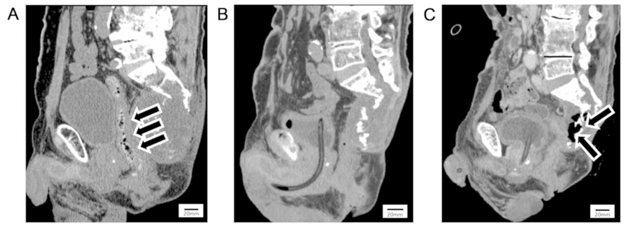
Since the tumor was contiguous with the rectum (Fig. 1A), we tried to place a spacer between the tumor and rectum to prevent radiation enterocolitis. However, we could not place the spacer because of adhesion of the tumor to the surroundings, so we performed a colostomy before CIRT. We performed CIRT at a total dose of 70.4 Gy (RBE) in 16 fractions. Colostomy closure was not performed because of a neurogenic rectal disturbance that occurred before CIRT. The tumor shrank markedly and partially ossified 6 years after CIRT, as shown by computed tomography (CT) (Fig. 1B) and MRI. Seven years after CIRT, the patient visited a general practitioner because of a high-grade fever and worsening sacral pain. A colonoscopy revealed rectotumoral fistula formation. After antibiotic treatment failed to reduce the high fever, he was referred to our outpatient clinic. Contrast-enhanced CT showed a 
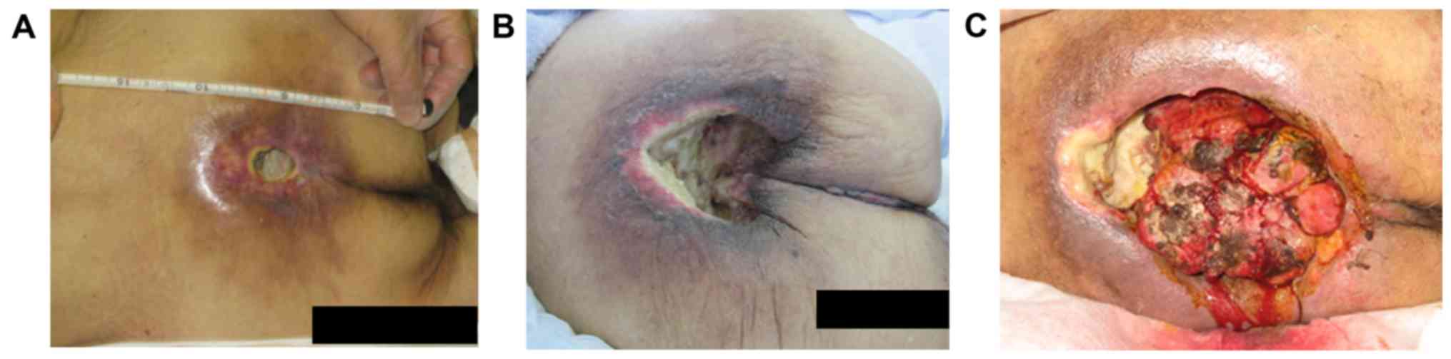
rectotumoral fistula and emphysema inside the tumor (Fig. 1C). Physical examination showed skin flare that was centrally ulcerated around the sacrum (Fig. 2A), and a tumor was observed in the dimple of ulceration. A blood test showed that his white blood cell (WBC) count and C-reactive protein (CRP) were elevated: WBC, 1.12×10⁴/µl; CRP, 13.1 mg/dl. He was diagnosed as having sepsis from the rectotumoral fistula with tumor recurrence and received continuous intravenous antibiotic treatment. Resection of the fistula and residual rectum with anal closure and necrotic sacral bone excision were performed to control the infection 1 
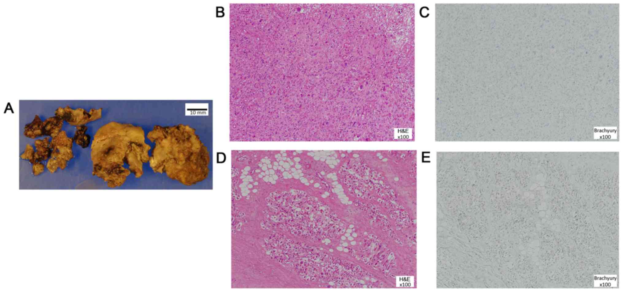
month after admission. During the procedures, a tumor biopsy was performed. However, after the operation, the ulceration enlarged and the tumor kept growing (Fig. 2B). Pathological examination revealed that most components appeared as undifferentiated pleomorphic sarcoma and some parts still resembled a chordoma, however, brachyury staining, which is diagnostic marker for chordoma, was negative. The tumor showed no identifiable line of differentiation based on the immunohistochemical negativity for pan-cytokeratin, epithelial membrane antigen (EMA), desmin, α-smooth muscle actin (α-SMA), S100, and CD34; all of which were negative in the morphologically chordoma-like areas as well. The specimen was considered as either a post-CIRT sarcoma or a recurrent dedifferentiated chordoma (Fig. 3). Despite trans-arterial embolization (TAE) and chemical debridement using Mohs paste, the tumor kept growing and bleeding. Finally, he died 9 years after CIRT because of bleeding from the tumor (Fig. 2C).

Figure 1.

Sagittal computed tomographic images in Case 1. (A) Image prior to CIRT: The tumor is close to the rectum (arrows). (B) Image 6 years following CIRT: The tumor has shrunk markedly and is partially ossified. (C) Image 7 years following CIRT: A rectotumoral fistula has formed and emphysema can be seen inside the tumor (arrows). CIRT, carbon ion radiotherapy.

Figure 2.

Images of the hip in Case 1. (A) Image of the patient at time of admission to the hospital: Skin flare is centrally ulcerated around the sacrum. (B) Image following several surgeries: Large ulceration without wound healing. (C) Image several days prior to mortality: Larger ulceration prior and a tumor inside the dimple with continuous bleeding are presented.

Figure 3.

Biopsy of Case 1. (A) Tumors biopsied in the dimple. Pathological examination of high grade sarcoma lesion by (B) hematoxylin and eosin (H&E) and (C) brachyry staining. Pathological examination of chordoma-like appearance lesion by (D) H&E and (E) brachyry staining. Magnification, ×100.

Case 2

A 55-year-old man was referred to our outpatient clinic for an expanding tumor, which was previously diagnosed as a benign tumor by biopsy when he received surgery for rectal cancer 7 years ago. We performed a biopsy again, and the specimen was pathologically confirmed as chordoma. Surgical resection had a risk of inducing severe neurological deficit because the tumor invaded the upper level of the sacrum (S1-5). Thus, he rejected the surgery and accepted CIRT instead. The total dose was 70.4 GyE in 16 
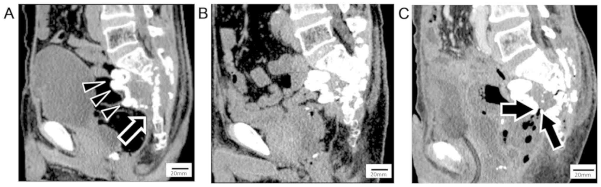
fractions. A 15×20-cm polytetrafluoroethylene spacer was placed between the rectum and tumor before CIRT to prevent radiation enterocolitis. The tumor in front of the coccyx could not be covered because of adhesion of the rectal surroundings caused by previous surgery (Fig. 4A). The tumor had shrunk markedly and become partially ossified 8 years after CIRT (Fig. 4B). However, follow-up MRI and CT revealed tumor regrowth and lung metastasis 10 years after CIRT. Surgery had not been performed because the tumor had invaded the pelvic structures, and radiotherapy could not be delivered because of the previous CIRT. He visited our hospital because of a high fever 1 month after diagnosis of the tumor regrowth. A blood test revealed high WBC count (1.12×10⁴/µl) and CRP (20.0 mg/dl), and a blood culture was positive for proteus vulgaris. Contrast-enhanced CT revealed that the rectotumoral fistula at the site of the spacer was uncovered and emphysema was inside the tumor (Fig. 4C). He was diagnosed as having sepsis from the rectotumoral fistula that was difficult to remove and received intravenous antibiotic treatment. He died of uncontrolled sepsis 7 months after admission despite continuous antibiotic treatment.

Figure 4.

Sagittal computed tomographic images in Case 2. (A) Image prior to CIRT: The placed spacer placed shown (triangles); however, the tumor in front of the coccyx is uncovered (arrows). (B) Image 8 years following CIRT: The tumor has shrunk markedly and become partially ossified. (C) Image 10 years following CIRT: A rectotumoral fistula has formed at the site uncovered by the spacer (arrows) and emphysema may be seen inside the tumor. CIRT, carbon ion radiotherapy.

Discussion

There are few reports about complications of CIRT for sacral chordoma. Neuropathic pain (15–16%) and skin toxicity (5–22%) have been reported (3,11). Imai et al reported 188 cases analysis of CIRT for unresectable sacral chordoma. In that study, the median follow-up period was 62 month (6.8–147.5) and 70% of patients were followed for over 5 years or until death. The 5-year local control rates were 77.2% (3,12). The local recurrence rate is not high. Among the locally recurrent cases there are no previous report about fistula formation. Thus, it is quite rare complication, and to our best knowledge, this is the first report of rectotumoral fistula formation after CIRT for sacral chordoma.

We considered two possible explanations for the fistula formation: First, radiation enterocolitis after CIRT might have caused formation of the fistula over a long period of time (13). Wallner et al reported that 8 (0.3%) of 2464 patients developed a radiation-related fistula after prostate brachytherapy (13). The average volume of the rectum receiving 100% of the prescription dose was moderately higher for the fistula patients than for the non-fistula patients. Time to fistulation ranged from 23 to 66 months (median, 25 months) (13). Thus, irradiation of the gastrointestinal tract around the tumor has a risk for fistula formation. Although both of our patients were operated on for spacer protection, coverage of the rectum was not achieved, which led to fistulation. Therefore, it is important to be aware that when sufficient protection during CIRT cannot be provided, careful follow-up to monitor potential fistulation is necessary. Second, tumor regrowth can compress and invade the rectum and enforce fistula formation. Imai et al reported that the local recurrence rate for 5 years was 22.8% (12). However, the long-term recurrence rate remains unknown. In our cases, the patients experienced tumor regrowth 7 years (Case 1) and 10 years (Case 2) after CIRT and increased rapidly. Late tumor regrowth might affect the long latency of fistula formation.

We confirmed dedifferentiated morphology similar to undifferentiated pleomorphic sarcoma by biopsy 7 years after CIRT in Case 1. Huvos et al proposed the following criteria for diagnosis of post-radiation sarcoma: i) The patient received irradiation, ii) the neoplasm occurred in the radiation field, iii) a latent period of some years had elapsed, and iv) there was histological or roentgenographic evidence for the pre-existing osseous condition, if present, in addition to microscopic proof of a sarcoma (14). The risk of post-radiation sarcoma has been found to be 0.06% at a mean latency of 15 years (3–50 years) (15). It is difficult to determine whether our cases involved dedifferentiated chordomas or post-CIRT sarcomas. However, according to Huvos' criteria, we concluded that it was post-CIRT sarcoma.

The limitation of our study is little pathological evidences to show sarcoma formation from chorodoma. In Case 1, unfortunately, the pathology preparation slide before CIRT was lost. However, needle biopsy under CT guide was performed. The pathological report was slow growing chordoma. We confirmed the diagnosis as chordoma with the result of this needle biopsy and the typical radiographic feature of MRI and CT. The radiographic feature represent no evidence of high grade sarcoma before CIRT. After CIRT, some parts of tumor still resembled a chorodma. However, brachyury staining was negative. In Case 2, we also could not find the pathological preparation slide nor paraffin block of initial open biopsy specimen in case 2. However, this case was irradiated in National Institute of Radiological Sciences in Chiba, where all cases were pathologically and radiographically reviewed by the board members. This case was also centrally reviewed and diagnosed as typical chordoma. We did not perform biopsy for recurrent tumor because of the sepsis and malnourishment.

It is important to prevent fistula formation at the tumor because treatments of the wound within the radiation area of CIRT are very difficult. Unfortunately, when a tumor is found that shows regrowth, resection of the tumor including the residual rectum should be considered.

In conclusion, we experienced two cases of rectotumoral fistula formation that occurred >5 years after CIRT. It is important to be aware of the possibility of rectal complications and to investigate rectal conditions after CIRT during long-term follow-up. If rectal complications are revealed, resection of the rectum should be considered. Additionally, tumor regrowth can worsen rectal complications.

Acknowledgements

Not applicable.

Funding

No funding was received.

Availability of data and materials

The datasets generated and/or analyzed during the current study are not publicly available to maintain the privacy of the patients but are available from the corresponding author on reasonable request.

Authors' contributions

YU collected the patient data and was a principal contributor to the writing of the manuscript. NA contributed to the design and conception of the present study, and corrected the manuscript. HO additionally contributed to the design and conception of the present study, and reviewed the manuscript. SN and EK performed the histological examination of chordoma specimens. RI treated the patient by carbon ion radiotherapy in Case 2 and contributed to data collection. YD and TO treated the patient by carbon ion radiotherapy in Case 1 and contributed to data collection. NN contributed to the data collection. All authors contributed to the interpretation, discussion and critical review of the manuscript. All authors read and approved the final manuscript.

Ethics approval and consent to participate

The present study was approved by the Osaka International Cancer Institutional Review Board (Osaka, Japan).

Patient consent for publication

The patients provided informed consent regarding medical information and the publication of the present study.

Competing interests

The authors declare that they have no competing interests.

References

1 

Yu E, Koffer PP, DiPetrillo TA and Kinsella TJ: Incidence, treatment, and survival patterns for sacral chordoma in the United States, 1974–2011. Front Oncol. 6:2032016. View Article : Google Scholar : PubMed/NCBI

2 

Chugh R, Tawbi H, Lucas DR, Biermann JS, Schuetze SM and Baker LH: Chordoma: The nonsarcoma primary bone tumor. Oncologist. 12:1344–1350. 2007. View Article : Google Scholar : PubMed/NCBI

3 

Imai R, Kamada T, Sugahara S, Tsuji H and Tsujii H: Carbon ion radiotherapy for sacral chordoma. Br J Radiol. 84:S48–S54. 2011. View Article : Google Scholar : PubMed/NCBI

4 

Lorenzo C, Andrea P, Barbara V, Denis P, Rosaria FM, Piero F, Viviana V, Alberto I, Mario C, Brugnatelli S, et al: Surgical spacer placement prior carbon ion radiotherapy (CIRT): An effective feasible strategy to improve the treatment for sacral chordoma. World J Surg Oncol. 14:2112016. View Article : Google Scholar : PubMed/NCBI

5 

Kamada T, Tsujii H, Blakely EA, Debus J, De Neve W, Durante M, Jäkel O, Mayer R, Orecchia R, Pötter R, et al: Carbon ion radiotherapy in Japan: An assessment of 20 years of clinical experience. Lancet Oncol. 16:e93–e100. 2015. View Article : Google Scholar : PubMed/NCBI

6 

Varga PP, Szövérfi Z, Fisher CG, Boriani S, Gokaslan ZL, Dekutoski MB, Chou D, Quraishi NA, Reynolds JJ, Luzzati A, et al: Surgical treatment of sacral chordoma: Prognostic variables for local recurrence and overall survival. Eur Spine J. 24:1092–1101. 2015. View Article : Google Scholar : PubMed/NCBI

7 

Radaelli S, Stacchiotti S, Ruggieri P, Donati D, Casali PG, Palmerini E, Collini P, Gambarotti M, Porcu L, Boriani S, et al: Sacral chordoma: Long-term outcome of a large series of patients surgically treated at two reference centers. Spine. 41:1049–1057. 2016. View Article : Google Scholar : PubMed/NCBI

8 

Yang Y, Niu X, Li Y, Liu W and Xu H: Recurrence and survival factors analysis of 171 cases of sacral chordoma in a single institute. Eur Spine J. 26:1910–1916. 2017. View Article : Google Scholar : PubMed/NCBI

9 

Stacchiotti S and Sommer J; Chordoma Global Consensus Group, : Building a global consensus approach to chordoma: A position paper from the medical and patient community. Lancet Oncol. 16:e71–e83. 2015. View Article : Google Scholar : PubMed/NCBI

10 

Asavamongkolkul A and Waikakul S: Wide resection of sacral chordoma via a posterior approach. Int Orthop. 36:607–612. 2012. View Article : Google Scholar : PubMed/NCBI

11 

Demizu Y, Jin D, Sulaiman NS, Nagano F, Terashima K, Tokumaru S, Akagi T, Fujii O, Daimon T, Sasaki R, et al: Particle therapy using protons or carbon ions for unresectable or incompletely resected bone and soft tissue sarcomas of the pelvis. Int J Radiat Oncol Biol Phys. 98:367–374. 2017. View Article : Google Scholar : PubMed/NCBI

12 

Imai R, Kamada T, Araki N, Abe S, Iwamoto Y, Ozaki T, Kanehira C, Kaya M, Takahashi K, Chuman H, et al Working Group for Bone and Soft Tissue Sarcomas, : Carbon ion radiation therapy for unresectable sacral chordoma: An analysis of 188 cases. Int J Radiat Oncol Biol Phys. 95:322–327. 2016. View Article : Google Scholar : PubMed/NCBI

13 

Wallner K, Sutlief S, Bergsagel C and Merrick GS: Severe rectal complications after prostate brachytherapy. Radiother Oncol. 114:272–275. 2015. View Article : Google Scholar : PubMed/NCBI

14 

Huvos AG, Woodard HQ, Cahan WG, Higinbotham NL, Stewart FW, Butler A and Bretsky SS: Postradiation osteogenic sarcoma of bone and soft tissues. A clinicopathologic study of 66 patients. Cancer. 55:1244–1255. 1985. View Article : Google Scholar : PubMed/NCBI

15 

Mavrogenis AF, Pala E, Guerra G and Ruggieri P: Post-radiation sarcomas. Clinical outcome of 52 Patients. J Surg Oncol. 105:570–576. 2012. View Article : Google Scholar : PubMed/NCBI

Related Articles

  • Abstract
  • View
  • Download
  • Twitter
Copy and paste a formatted citation
Spandidos Publications style
Ukon Y, Outani H, Nagata S, Konishi E, Imai R, Demizu Y, Okimoto T, Naka N and Araki N: Rectotumoral fistula formation occurring more than 5 years after carbon ion radiotherapy for sacral chordoma: A case report. Mol Clin Oncol 10: 487-491, 2019.
APA
Ukon, Y., Outani, H., Nagata, S., Konishi, E., Imai, R., Demizu, Y. ... Araki, N. (2019). Rectotumoral fistula formation occurring more than 5 years after carbon ion radiotherapy for sacral chordoma: A case report. Molecular and Clinical Oncology, 10, 487-491. https://doi.org/10.3892/mco.2019.1821
MLA
Ukon, Y., Outani, H., Nagata, S., Konishi, E., Imai, R., Demizu, Y., Okimoto, T., Naka, N., Araki, N."Rectotumoral fistula formation occurring more than 5 years after carbon ion radiotherapy for sacral chordoma: A case report". Molecular and Clinical Oncology 10.5 (2019): 487-491.
Chicago
Ukon, Y., Outani, H., Nagata, S., Konishi, E., Imai, R., Demizu, Y., Okimoto, T., Naka, N., Araki, N."Rectotumoral fistula formation occurring more than 5 years after carbon ion radiotherapy for sacral chordoma: A case report". Molecular and Clinical Oncology 10, no. 5 (2019): 487-491. https://doi.org/10.3892/mco.2019.1821
Copy and paste a formatted citation
x
Spandidos Publications style
Ukon Y, Outani H, Nagata S, Konishi E, Imai R, Demizu Y, Okimoto T, Naka N and Araki N: Rectotumoral fistula formation occurring more than 5 years after carbon ion radiotherapy for sacral chordoma: A case report. Mol Clin Oncol 10: 487-491, 2019.
APA
Ukon, Y., Outani, H., Nagata, S., Konishi, E., Imai, R., Demizu, Y. ... Araki, N. (2019). Rectotumoral fistula formation occurring more than 5 years after carbon ion radiotherapy for sacral chordoma: A case report. Molecular and Clinical Oncology, 10, 487-491. https://doi.org/10.3892/mco.2019.1821
MLA
Ukon, Y., Outani, H., Nagata, S., Konishi, E., Imai, R., Demizu, Y., Okimoto, T., Naka, N., Araki, N."Rectotumoral fistula formation occurring more than 5 years after carbon ion radiotherapy for sacral chordoma: A case report". Molecular and Clinical Oncology 10.5 (2019): 487-491.
Chicago
Ukon, Y., Outani, H., Nagata, S., Konishi, E., Imai, R., Demizu, Y., Okimoto, T., Naka, N., Araki, N."Rectotumoral fistula formation occurring more than 5 years after carbon ion radiotherapy for sacral chordoma: A case report". Molecular and Clinical Oncology 10, no. 5 (2019): 487-491. https://doi.org/10.3892/mco.2019.1821
Follow us
  • Twitter
  • LinkedIn
  • Facebook
About
  • Spandidos Publications
  • Careers
  • Cookie Policy
  • Privacy Policy
How can we help?
  • Help
  • Live Chat
  • Contact
  • Email to our Support Team