Introduction
Since the first report by Brunn in 1939 as a primary
myofibroblastic tumor of the lung (1), an inflammatory myofibroblastic tumor
(IMT) has been acknowledged as a distinctive, though rare,
intermediate soft tissue tumor that commonly in the orbit and lung
(2,3). Although previously believed to be
proliferative lesions deriving from submucosal stroma, of low or
indeterminate malignant potential (4), IMT is now considered to be different
from pseudotumor for the distinct histological and molecular
features, specifically characteristic cellular spindle cell
proliferation alongside mutations in the anaplastic lymphoma kinase
(ALK)-1 gene loci (5), and there
has been an increasing incidence to be found in the pelvic cavity,
head and neck, trunk, retroperitoneum, abdomen and limbs (6).
An IMT diagnosis is characterized by the lymphocytic
infiltrate and spindle myoepithelial cell proliferation (7). Coffin et al (8) described the IMT with three different
histological patterns, which may be present simultaneously within a
single specimen. Based in fact that IMT is considered as a low
malignant potential neoplasm, surgical resection is the first choice
of treatment, and several studies proved that successful resolution
with radiotherapy, chemotherapy, steroids, or even non-steroidal
inflammatory drugs (2,4,9).
According to a previous study, ~50% of tumours present with ALK
overexpression, which is considered to identify the characteristics
and provide the opportunity for targeted therapy (10). Although the exact incidence remains
unclear, it has been reported that the recurrence rate and distant
metastasis rate of IMT are low (recurrence: 2% in pulmonary, 25% in
extrapulmonary sites; metastasis: less than 5%) (3).
Since the first study of a thirty-two-year-old woman
of bladder IMT in 1980(11), the
bladder has become one of the common sites of the tumor involving
urinary system, and more than 200 cases of bladder IMT were
reported mostly in the ophthalmologic and pathologic literature.
Due to the lack of specificity clinical symptoms and laboratory
results, a preoperative imaging-based examination is very important
for determining bladder IMT and selecting appropriate ways to
manage conditions of patients requiring emergency treatment
(including bladder cancer and lymphoma) (6). However, to the best of our knowledge,
there are seldom studies systematically analyzing the CT features
of bladder IMT in the radiology literature. The purpose of the
present study was to describe the characteristic CT findings of 9
patients with bladder IMT confirmed by pathology.
Materials and methods
Patients
The study was exempt from the Institutional Review
Board of the first Hospital of Zhengzhou University. The written
informed consent was waived by the Institutional Review Board
because this is a retrospective study. Between 2011 and 2021, a
review of medical records based on the pathology records and the
PACS system of our institution revealed patients with bladder IMT
proved on pathologic examination, and the requirement for written
informed consent was waived in this retrospective study.
CT evaluation
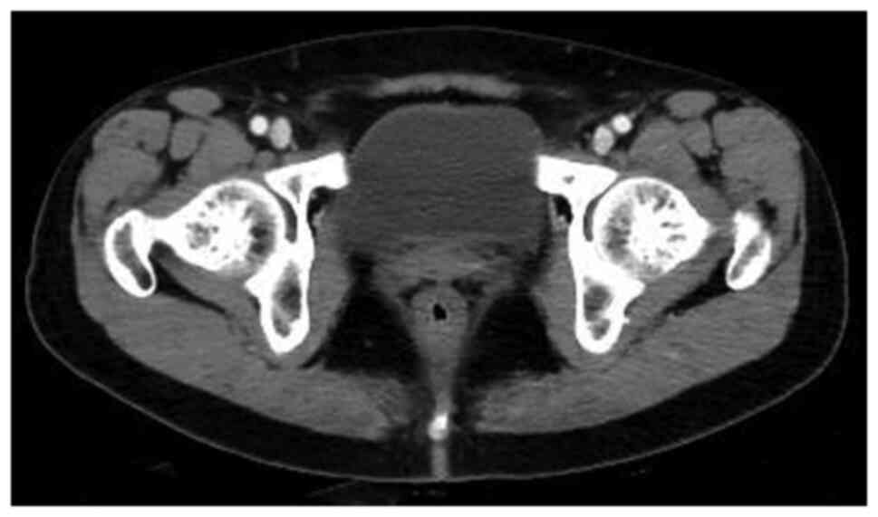
The pelvic CT scans (Discovery CT750HD; GE
Healthcare) were typically obtained after the bladder had a
sensation of urine (Fig. 1) and
intravenous administration of non-ionic iohexol (iopromide, 370
mg/ml, GE Medical Systems, 1.5 ml/kg, and 5 ml/s) by a dual-head
pump injector (Medrad, Inc.; Bayer AG), with a section thickness of
5 mm and a pitch of 1.5.
Image analyses
All CT images were reviewed blindly by two abdominal
radiologists with 3 and 6 years of experience at AW4.6 workstation
(GE Healthcare). The following CT features of the bladder IMT were
assessed: i) Diameter of the lesion, ii) location of the lesion
(posterior wall; superior wall; front wall; left wall; right wall),
iii) contour of the lesion (polypoid; cauliflower-like; limited
thick-walled), iv) growth pattern of the lesion (endophytic;
exophytic; mixed), v) margin of the lesion (smooth; lobulated), vi)
boundary of the lesion (clear; ill-defined), vii) density of the
lesion on non-enhancement CT scan (low density; iso-density;
slightly high density), viii) presence of non-enhancement
low-density area within the lesion (homogeneous; heterogeneous),
ix) type of enhancement of the lesion (ring-shaped; heterogeneous)
and x) degree of enhancement of the lesion (significant; moderate;
mild). The lesion showed a symmetrical change in the center of the
lesion after enhancement: taking the base of the lesion as the
tangent position, and making the median line perpendicular to the
base of the lesion, it could be observed that the lesion showed a
symmetrical change. In addition, the clinical data (including age,
sex and symptoms) were also recorded by the review of medical
records.
Results
Patient characteristics
The nine patients (four men and five women) who were
evaluated were 7-75 years old (mean age, 40 years). The most common
presenting clinical symptoms were gross hematuria (9/9), followed
by frequent urination (4/9) and painful urination (4/9). Relevant
history included appendicitis resection (1/9) and nephrotic
syndrome (1/9). Of the 9 patients, 6 (67%) had irregular blood
clots in the bladder. The mean Ki67 value of the presence of an
irregular blood clot in the bladder was 18% and that of no blood
clot was 12% (Tables I and
II).
 | Table ISummary of CT findings for nine
patients with IMT of bladder. |
Table I
Summary of CT findings for nine
patients with IMT of bladder.
| Patient | Sex | Age, years | Clinical
Symptom(s) | Diameter
(mm2) | Location | Number | Contour | Growth pattern | Margin | Boundary | Tumor Uniformity | Type of
enhancement | Ki67 (%) |
|---|
| 1 | Female | 63 | Gross hematuria,
frequent urination, painful urination | 48x51 | Posterior wall | One | Polypoid | Exophytic | Lobulated | Ill-defined | Ring-shaped | Persistent | 5 |
| 2 | Female | 60 | Gross hematuria,
painful urination, irregular blood clot | 33x34 | Superior wall | One | Cauliflower-
like | Endophytic | Smooth | Clear | Ring-shaped | Persistent | 5 |
| 3 | Female | 35 | Gross hematuria, | 33x34 | Superior wall | One | Polypoid | Endophytic | Smooth | Clear | Ring-shaped | Persistent | 20 |
| 4 | Male | 28 | Gross hematuria,
frequent urination, painful urination, irregular blood clot | 34x48 | Superior wall | One | Polypoid | Exophytic | Smooth | Clear | Ring-shaped | Persistent | 30 |
| 5 | Female | 14 | Gross hematuria,
irregular blood clot | 22x19 | Superior wall | One | Polypoid | Endophytic | Smooth | Clear | Ring-shaped | Persistent | 20 |
| 6 | Male | 35 | Gross hematuria,
frequent urination, irregular blood clot | 44x47 | Front wall | One | Limited thick-
walled | Mixed | Smooth | Clear | Heterogeneous | Persistent | 30 |
| 7 | Female | 7 | Gross hematuria,
painful urination, irregular blood clot | 29x32 | Left wall | One | Polypoid | Endophytic | Smooth | Clear | Ring-shaped | Persistent | 20 |
| 8 | Male | 75 | Gross hematuria,
frequent urination, irregular blood clot | 35x48 | Posterior wall | One | Limited thick-
walled | Mixed | Smooth | Clear | Heterogeneous | Persistent | 15 |
| 9 | Male | 47 | Gross
hematuria | 13x21 | Superior wall | Multiple | Limited thick-
walled | Mixed | Smooth | Clear | Heterogeneous | Persistent | 10 |
| Table IISummary of clinical manifestations
for nine patients with IMT of bladder. |
Table II
Summary of clinical manifestations
for nine patients with IMT of bladder.
| Patient | Sex | Age, years |
Immunohistochemistry | Operation | Other
treatment | Follow-up time | Follow-up
method | Prognosis |
|---|
| 1 | Female | 63 | CK+,
Vimentin+, SMA+, CD34 (Vascular+),
LCA (Stove+) | Cystoscopy | | 1 month | Ultrasound | No recurrence |
| 2 | Female | 60 | Desmin
(Individual+), ALK+ | Transurethral
resection of bladder tumor | | 1 month | Ultrasound | No recurrence |
| 3 | Female | 35 | CK+,
EMA+, ALK+, SMA (Stove+), CD30
(Minority+) | Partial
cystectomy | | 3 months | Bladder
endoscopy | No recurrence |
| 4 | Male | 28 | CK+,
EMA+, ALK+, Vimentin+, SMA
(Stove+), CD68 (Scattered+) | Partial cystectomy
and pelvic lymph node dissection | | 7 months | CT, ultrasound,
Magnetic resonance imaging | No recurrence |
| 5 | Female | 14 | AE1/AE3
(Weak+), CD34 (Vascular+), ALK+,
SMA+, Desmin (Stove+) | Transurethral
resection of bladder tumor | Bladder perfusion
therapy | 2 months | Bladder
endoscopy | No recurrence |
| 6 | Male | 35 | SMA
(Stove+), Desmin (Stove+), ALK
(Stove+) | Transurethral
resection of bladder tumor plus partial cystectomy | | 19 months | Bladder endoscopy,
CT, ultrasound | No recurrence |
| 7 | Female | 7 | ALK+,
AE1/AE3 (Partial+), SMA (Partial+), Desmin
(Partial+) | Bladder tumor
excision | | 2 months | Ultrasound | No recurrence |
| 8 | Male | 75 | CK7+,
CK20 (Epithelial+), SMA+, ERG
(Partial+), GATA-3+ | Rod-shaped
prostatic dilatation plus electrotomy of bladder neck and
mouth | | 1 month | Magnetic resonance
imaging | No recurrence |
| 9 | Male | 47 |
AE1/AE3+, CD117
(Stove+), Vimentin+, CD99+, P53
(60%) | Laparoscopic
radical cystectomy and in situ cystectomy | | 4 months | CT | No recurrence |
CT findings
The CT findings showed that 8 (89%) patients had one
tumor and 1 (11%) patient had multiple tumors. The bladder IMT size
ranged from 1.3x2.1 to 4.8x5.1 cm2. Tumors occurred in
the posterior wall in 2 (22%) patients, 5 (30%) patients had tumors
occurred in the superior wall, 1 (11%) patient had tumors occurred
in the front wall, and 1 (11%) patient had tumor occurred in the
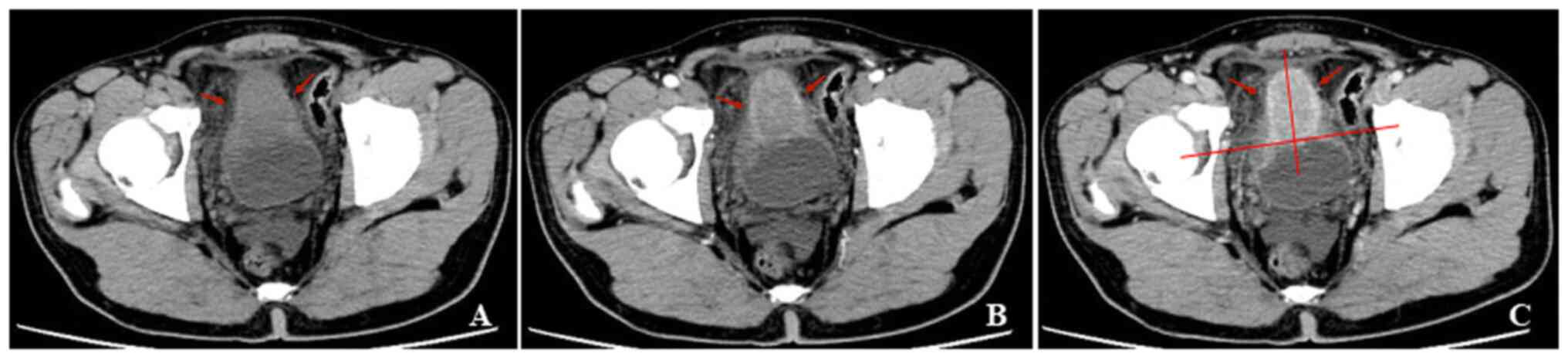
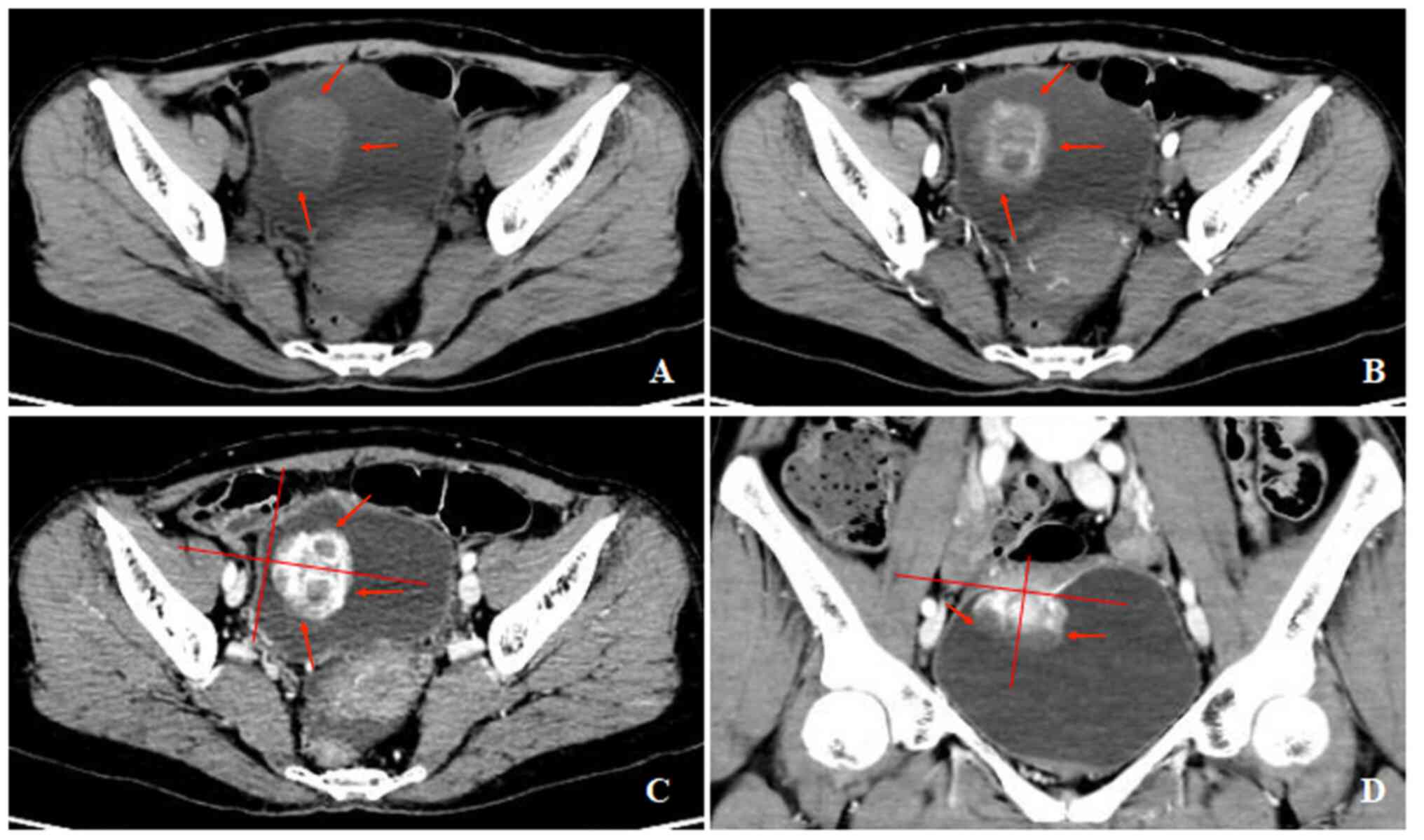
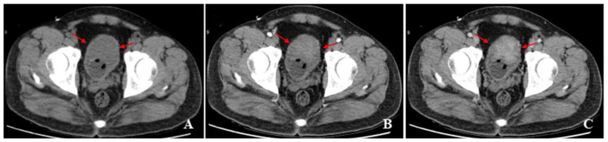
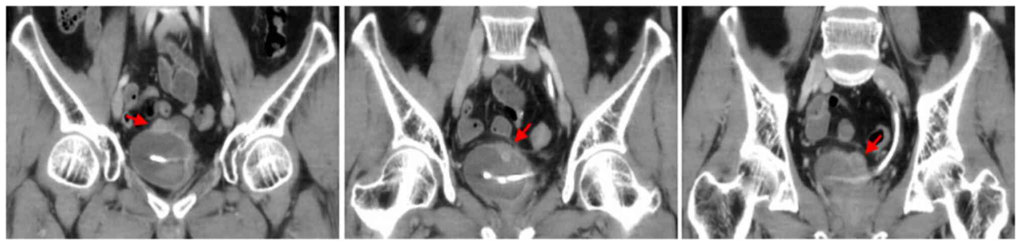
left wall (Fig. 2, Fig. 3, Fig.
4 and Fig. 5). An endophytic
growth pattern (Fig. 3) was
identified in 4 (44%) patients, an exophytic growth pattern
(Fig. 2) was observed in 2 (22%)
patients, and a mixed growth pattern (Fig. 5) was revealed in 3 (33%) patients.
The tumor manifests morphologically as either polypoid (n=5), or
cauliflower-like (n=1) soft-tissue mass with a wide base in the
cavity, or a limited thick-walled (n=3) in the bladder. The tumor
margins were smooth (n=8) or lobulated (n=1) and the tumor
boundaries were either clear (n=7) or ill-defined (n=2).
The unenhanced CT examination of the lesions
revealed either low density (n=4), iso-density (n=3), or slightly
high density (n=1), and density was either homogeneous (n=3) or
heterogeneous (n=6). The enhanced CT examination of the lesions
showed either ring-shaped (n=3) or heterogeneous (n=6), and the
degree of enhancement was either significant (n=6), or moderate
(n=3). The enhancement pattern was persistent (n=9). In addition,
all polypoid and cauliflower-like soft-tissue masses showed a
symmetrical change in the center of the lesion after enhancement on
the CT image.
Discussion
IMT is a rare tumor with intermediate biological
potential. It is characterized by irregular proliferation of
spindle cells in a mucoid to the collagenous stroma, inflammatory
infiltration mainly composed of plasma cells and lymphocytes, and
occasional mixing of eosinophils and neutrophils (12). Since the first description of IMT
in 1939, the understanding of its biological and clinical
characteristics has undergone significant changes (9). Based on morphological and
histopathological features, IMT used to be described as fibrous
histiocytoma, plasma cell granuloma, inflammatory pseudotumor,
inflammatory fibroid polyp, fibroxanthoma, xanthoma and
xanthogranuloma (9). At present,
in the fourth edition of the World Health Organization
Classification of Soft Tissue and Bone Tumors, IMT is classified as
an intermediate tumor lesion (invasive, occasionally metastatic)
(10). Microscopic examination
showed that IMT is a tumor with myxoid/vascular type, compact
spindle cell type, and hypocellular fibrous type, which could
easily lead to misdiagnosis, including inflammatory malignant
fibrous histiocytoma, leiomyosarcoma, gastrointestinal stromal
tumor rich in inflammatory cells and solitary fibrous tumors
(13). The diagnosis of IMT can be
consolidated by immunohistochemical analysis (13). IMT tumor cells characteristically
stain positive for ALK-1 in 87.5% cases, smooth muscle actin (SMA)
in 90% cases, pan-cytokeratin focally in >50% cases, and desmin
in ~50% cases (14). In addition,
a young woman diagnosed as having IMT of the urinary bladder showed
the expression of CD10 in a recently published study (14). The pathogenesis of the tumor
remains unknown, and it may be related to infections,
immunosuppression, chemotherapy, radiotherapy, local trauma and
autoimmune disorders (15). In
addition, several diseases, including Crohn's disease, congenital
neutropenia, gastrointestinal stromal tumor, and pregnancy have
been related to the development of IMT (16). Previous studies have reported the
correlation between ALK-1 gene expression and local recurrence,
distant metastasis, and overall prognosis of IMT patients (17-20).
Moreover, in certain studies (8,21,22),
the bladder IMT was associated with the Epstein-Barr virus,
bacteria such as Campylobacter Equi, the human herpes virus
HHV8, Campylobacter jejuni, Escherichia coli, trauma,
radio- and steroid therapy. Previous studies reported that a
history of inflammation in the bladder, previous instrumentation,
radiation, or a history of surgery was a potential cause of IMT
(6). One patient in the present
study had a history of appendicitis resection, and one patient had
a history of nephrotic syndrome; however, it could not be confirmed
whether the surgery was considered to be a causative factor of IMT,
because the lesion occurred in the other parts of the pelvis.
There have been certain case reports of bladder IMT,
but the shortcomings are obvious. As this tumor is a rare tumor, it
is easy to be misdiagnosed and missed. At present, there is a lack
of literature studies on the systematic analysis of imaging
manifestations of bladder IMT, leading to a lack of understanding
among clinicians. Therefore, the clinical imaging data of 9
patients were systematically analyzed and combined with previous
literature studies, some new insights were found, aiming to improve
our understanding of bladder IMT.
The bladder IMT is uncommon accounting for less than
1% of all bladder tumors (23).
The distribution of 182 IMT of the bladder was 51.7% in the female
patients, and 48.3% in the male patients, with a mean age of
38.9±16.6 years, according to Teoh et al (24). In a previous study by Harik et
al (25), the age of onset of
bladder IMT ranged from 7 to 77 years (average 47 years), and the
incidence rate of male patients was 3.2 times that of female
patients. According to a previous study (6), the average age of onset of bladder
IMT was 53 years old, and the incidence rate of male patients was
slightly higher than that of female patients. In the current study,
the age of patients ranged from 7 to 75 years (average, 40 years),
and the proportion of women preferring men to women was 4:5. Due to
the limited number of cases, this demographic difference in the
study may be caused by selection bias.
The most common clinical symptoms of patients with
bladder IMT are anemia and gross hematuria, accompanied by pain
occasionally during urination (6,26).
In addition, there was one case report by Harik et al
(25), which had no clinical
symptoms. Due to the rarity of this tumor, it is not always
possible to predict the presence of IMT preoperatively based solely
on clinical and radiological findings (24).
CT is a common examination method for bladder
space-occupying lesions, and it has been reported in the literature
on the detection, diagnosis and differentiation of bladder lesions
as well as the judgment of curative effect. Since bladder IMT is a
rare tumor, if some typical imaging diagnostic information can be
obtained before treatment, it will be helpful to develop treatment
methods and improve the prognosis of patients. Therefore, it is
very necessary to systematically analyze the imaging findings of
bladder IMT.
Due to its rarity, the CT-based analysis of the
bladder IMT remains largely unknown. Lack of understanding often
brings difficulties to the preoperative diagnosis and treatment of
such tumors. Therefore, it was decided to systematically review the
bladder IMT and focus on image-based analysis. Usually, the tumor
is identified as a polypoid or cauliflower-like soft-tissue mass
with a broad base on the CT image. Occasionally, narrow-base
polypoid lesions, nodules, or limited thick-walled bladders have
been reported (6,27,28).
In the present study, tumors of 5 patients (30%) were located in
the superior wall, and 2 (22%) patients' tumors were located in the
posterior wall, which was more similar to the distribution reported
by Liang et al (6). In
addition, it was found that 8 of the included patients (89%) had
well-defined boundaries, and in the aforementioned study (6), more than five-six of patients also
had a well-defined tumor in the bladder. Thus, well-defined tumors
were considered to be a feature of the bladder IMT on CT imaging.
In the present study, there was a patient with a tumor invasion in
the perivesical soft tissue of the bladder. According to the
results of the histopathological examination, it was confirmed that
it is caused by inflammatory changes. Liang et al (6) proposed that the relationship between
the number of lesion and biological behavior needed further
investigation. In the current study, there were eight patients with
one lesion, and one patient with multiple lesions. The present data
showed that there was no significant relationship between one
lesion and bladder IMT. Liang et al (6) stated that endophytic tumors are more
common in bladder IMT. In addition, there have been previous
studies that early-stage bladder IMT can appear as a limited
thick-walled bladder (27,28). Similarly, the present series of
studies revealed that endophytic tumors were more common than
exophytic tumors; 44% of the cases were endophytic, 11% exophytic
and 22% mixed growth pattern.
Although blood clot is a non-specific sign of any
bladder tumor and can be observed in other diseases such as
hemorrhagic cystitis, the present study aimed to analyze the
relationship between blood clots in the bladder and Ki67 value of
the bladder IMT. Of the 9 patients, 6 (67%) had irregular blood
clots in the bladder and 9 (100%) had gross hematuria. Therefore,
the presence of irregular blood clots in the bladder was considered
to be helpful in the diagnosis of bladder IMT (6). The mean Ki67 value of an irregular
blood clot of the bladder was 18 and that of no blood clot was 12.
It was found that a large Ki67 value appeared to be related to the
irregular blood clot.
To expound the relationship between Ki67 value and
the prognosis of patients with bladder IMT is of great value for
improving the prognosis of patients. However, due to the limited
sample size included in the present study and the time of
follow-up, the prognosis assessment was limited. This is a
limitation to the current study. As a result, sample size will be
further accumulated to analyze the relationship between Ki67 value
and the prognosis of patients with bladder IMT in future
studies.
Although it is not a pathological diagnosis, it has
been reported that ring enhancement is the most significant feature
of bladder IMT, because more tumor cells gather at the edge of the
lesion, accompanied by inflammatory cell infiltration and a
relatively rich blood supply (6).
Similarly, all 9 lesions in the present study showed homogeneous or
heterogeneous enhancement, and six of the nine lesions also showed
ring enhancement on CT imaging. It is interesting to note that all
polypoid and cauliflower-like soft-tissue mass exhibited a
symmetrical change in the center of the lesion after enhancement on
the CT image, which had not been reported before. It is considered
that symmetrical enhancement may be the clue of the bladder IMT
diagnosis. In the present study, all 9 lesions were examined with
CT-enhanced examination, intense enhancement at arterial phase
scanning, followed by progressive enhancement at intravenous phase
scanning. The possible reason is the presence of a contrast agent
in the interstitial space in the center of the lesion (6).
The bladder IMT is often difficult to diagnose
correctly before surgery, leading to unnecessary radical bladder
resection. The analysis may be due to: i) The clinical symptoms and
signs are not characteristic, ii) Clinicians and radiologists have
no or insufficient knowledge of it and iii) Preoperative cystoscopy
is often due to insufficient materials, resulting in a low rate of
diagnosis.
Surgical treatment of the bladder IMT is preferred,
and the appropriate surgical method should be determined after a
comprehensive examination and full evaluation before surgery. Based
on the presence of inflammatory cell infiltration in the
pathological tissue of the bladder IMT, anti-infection therapy can
play a role in reducing the tumor, so perioperative
anti-inflammatory measures should be strengthened. The selection of
surgical methods should be comprehensively evaluated according to
the size of the tumor and the extent of bladder invasion. Although
transurethral resection of bladder tumor has the advantages of less
trauma and faster recovery, a multicenter study (24) showed that the rate of second
surgery after surgery was 21%, markedly higher than the rate of 2%
in patients who underwent partial cystectomy. Total cystotomy
should be carefully selected considering the patient's condition,
tumor size and invasion degree. Chemotherapy after bladder IMT is
controversial. Among the 9 patients admitted to our hospital, 1
patient with preoperative biopsy tendency to bladder malignant
tumor received bladder perfusion therapy, and the remaining 8
patients did not receive bladder perfusion therapy after the
operation. It remains to be verified whether bladder perfusion
could reduce the recurrence rate. It has been reported that
prednisone, COX-2 inhibitors and other anti-inflammatory drugs have
a favorable effect on bladder IMT in children before surgery
(29).
The prognosis of bladder IMT was favorable. The nine
patients in the current group were reviewed after surgery for
bladder endoscopy, CT, ultrasound and magnetic resonance
examination. The follow-up time was 1-19 months. No recurrence or
distant metastasis was observed in all patients. It has been
reported that the recurrence rate and distant metastasis rate of
IMT are 2-25% (3). Although most
bladder IMTs are benign tumors and still have malignant potential,
it is recommended that patients should be reexamined by cystoscopy
every 3 months after surgery for 2 years. Then, ultrasound and CT
have performed annually.
In conclusion, the bladder IMT had certain
characteristic CT findings that differ from other bladder lesions.
The present study of cases suggested that there may be a certain
relationship between Ki67 value and radiology feathers of the
bladder IMT, but the number of cases is limited, and further study
is needed for in-depth analysis.
Acknowledgements
Not applicable.
Funding
Funding: The present study was supported by the Outstanding
Youth Project in Henan for Young and Middle-aged Health and Health
Technology Innovation (grant no. YXKC2020053).
Availability of data and materials
The datasets used and/or analyzed during the current
study are available from the corresponding author on reasonable
request.
Authors' contributions
PL primarily wrote the manuscript. JG and PL
critically reviewed the paper and revised it. JG and PL confirm the
authenticity of all the raw data. PL and XR performed the database
search and literature review. PL, BZ and XR analyzed the data. All
authors read and approved the final manuscript.
Ethics approval and consent to
participate
The study was exempt from the Institutional Review
Board of the first Hospital of Zhengzhou University (Zhengzhou,
China). Written informed consent was waived by the Institutional
Review Board since this is a retrospective study.
Patient consent for publication
Not applicable.
Competing interests
The authors declare that they have no competing
interests.
References
|
1
|
Brunn H: Two interesting benign lung
tumours of contradictory histopathology: Remarks on the necessity
for maintaining the chest tumour registry. J Thorac Surg.
9:119–131. 1939.
|
|
2
|
Song D, Jiao W, Gao Z, Liu N, Zhang S,
Zong Y, Fang Z and Fan Y: Inflammatory myofibroblastic tumor of
urinary bladder with severe hematuria: A Case report and literature
review. Medicine (Baltimore). 98(e13987)2019.PubMed/NCBI View Article : Google Scholar
|
|
3
|
Chen B, Li S, Fang X, Xu H, Yu J, Liu L
and Wei Q: Inflammatory myofibroblastic tumor of the urinary system
on computed tomography at a high-volume institution in China. Urol
Int. 104:960–967. 2020.PubMed/NCBI View Article : Google Scholar
|
|
4
|
Xu H, He B, Tu X, Bao Y, Yang L, Zhuo H
and Wei Q: Minimally invasive surgery for inflammatory
myofibroblastic tumor of the urinary bladder: Three case reports.
Medicine (Baltimore). 97(e13474)2018.PubMed/NCBI View Article : Google Scholar
|
|
5
|
Cresser S, Nzenza T, Tripathy S and Hall
R: Case series: Inflammatory myofibroblastic bladder tumor in
regional Australia. Int J Surg Case Rep. 82(105898)2021.PubMed/NCBI View Article : Google Scholar
|
|
6
|
Liang W, Zhou X, Xu S and Lin S: CT
manifestations of inflammatory myofibroblastic tumors (Inflammatory
Pseudotumors) of the urinary system. AJR Am J Roentgenol.
206:1149–1155. 2016.PubMed/NCBI View Article : Google Scholar
|
|
7
|
Gass J, Beaghler M and Kwon M:
Inflammatory myofibroblastic tumor of the urinary bladder: A case
report. J Endourol Case Rep. 5:31–33. 2019.PubMed/NCBI View Article : Google Scholar
|
|
8
|
Coffin CM, Watterson J, Priest JR and
Dehner LP: Extrapulmonary inflammatory myofibroblastic tumor
(inflammatory pseudotumor). A clinicopathologic and
immunohistochemical study of 84 cases. Am J Surg Pathol.
19:859–872. 1995.PubMed/NCBI View Article : Google Scholar
|
|
9
|
Saleem MI, Ben-Hamida MA, Barrett AM, Bunn
SK, Huntley L, Wood KM and Yelbuz TM: Lower abdominal inflammatory
myofibroblastic tumor-an unusual presentation-a case report and
brief literature review. Eur J Pediatr. 166:679–683.
2007.PubMed/NCBI View Article : Google Scholar
|
|
10
|
Zeng X, Huang H, Li J, Peng J and Zhang J:
The clinical and radiological characteristics of inflammatory
myofibroblastic tumor occurring at unusual sites. Biomed Res Int.
2018(5679634)2018.PubMed/NCBI View Article : Google Scholar
|
|
11
|
Roth JA: Reactive pseudosarcomatous
response in urinary bladder. Urology. 16:635–637. 1980.PubMed/NCBI View Article : Google Scholar
|
|
12
|
Gupta A, Sharma S, Mittal A, Barwad A and
Rastogi S: Recurrent infantile inflammatory myofibroblastic tumor
of mesentery-Case report and review of imaging findings. Radiol
Case Rep. 16:504–510. 2020.PubMed/NCBI View Article : Google Scholar
|
|
13
|
Cerier E, Beal EW and Dillhoff ME:
Inflammatory myofibroblastic tumour: An unusual presentation
including small bowel obstruction and palpable abdominal mass. BMJ
Case Rep. 2018(bcr2018224549)2018.PubMed/NCBI View Article : Google Scholar
|
|
14
|
Patne SC, Katiyar R, Chaudhary D and
Trivedi S: Dysuria and fever in a young woman diagnosed as having
inflammatory myofibroblastic tumour of the urinary bladder. BMJ
Case Rep. 2016(bcr2015214059)2016.PubMed/NCBI View Article : Google Scholar
|
|
15
|
Huang YH, Zhong DJ, Tang J, Han JJ, Yu JD,
Wang J and Wan YL: Inflammatory myofibroblastic tumor of the liver
following renal transplantation. Ren Fail. 34:789–791.
2012.PubMed/NCBI View Article : Google Scholar
|
|
16
|
Chen CB, Chou CT, Hsueh C, Lee KW and Chen
YL: Hepatic inflammatory pseudotumor mimicking hepatocellular
carcinoma. J Chin Med Assoc. 76:299–301. 2013.PubMed/NCBI View Article : Google Scholar
|
|
17
|
Choi AH, Bohn OL, Beddow TD and McHenry
CR: Inflammatory myofibroblastic tumor of the small bowel
mesentery: An unusual cause of abdominal pain and uveitis. J
Gastrointest Surg. 15:584–588. 2011.PubMed/NCBI View Article : Google Scholar
|
|
18
|
Tsuzuki T, Magi-Galluzzi C and Epstein JI:
ALK-1 expression in inflammatory myofibroblastic tumor of the
urinary bladder. Am J Surg Pathol. 28:1609–1614. 2004.PubMed/NCBI View Article : Google Scholar
|
|
19
|
Telugu RB, Prabhu AJ, Kalappurayil NB,
Mathai J, Gnanamuthu BR and Manipadam MT: Clinicopathological study
of 18 cases of inflammatory myofibroblastic tumors with reference
to ALK-1 Expression: 5-Year experience in a tertiary care center. J
Pathol Transl Med. 51:255–263. 2021.PubMed/NCBI View Article : Google Scholar
|
|
20
|
Yagnik VD: ALK 1 negative inflammatory
myofibroblastic tumor of the ileum: A rare cause of ileocecal
intussusception. Surg J (N Y). 6:e101–e104. 2020.PubMed/NCBI View Article : Google Scholar
|
|
21
|
Dobrosz Z, Ryś J, Paleń P, Właszczuk P and
Ciepiela M: Inflammatory myofibroblastic tumor of the bladder-an
unexpected case coexisting with an ovarian teratoma. Diagn Pathol.
9(138)2014.PubMed/NCBI View Article : Google Scholar
|
|
22
|
Coffin CM, Humphrey PA and Dehner LP:
Extrapulmonary inflammatory myofibroblastic tumor: A clinical and
pathological survey. Semin Diagn Pathol. 15:85–101. 1998.PubMed/NCBI
|
|
23
|
Rotenberry C, Dowd K, Russell D, DeRiese
W, Teeple S and Cammack T: Robot-assisted partial cystectomy for
treatment of inflammatory myofibroblastic tumor of the bladder.
Urol Case Rep. 11:25–27. 2017.PubMed/NCBI View Article : Google Scholar
|
|
24
|
Teoh JY, Chan NH, Cheung HY, Hou SS and Ng
CF: Inflammatory myofibroblastic tumors of the urinary bladder: A
systematic review. Urology. 84:503–508. 2014.PubMed/NCBI View Article : Google Scholar
|
|
25
|
Harik LR, Merino C, Coindre JM, Amin MB,
Pedeutour F and Weiss SW: Pseudosarcomatous myofibroblastic
proliferations of the bladder: A clinicopathologic study of 42
cases. Am J Surg Pathol. 30:787–794. 2006.PubMed/NCBI View Article : Google Scholar
|
|
26
|
Cheng L, Foster SR, MacLennan GT,
Lopez-Beltran A, Zhang S and Montironi R: Inflammatory
myofibroblastic tumors of the genitourinary tract-single entity or
continuum? J Urol. 180:1235–1240. 2008.PubMed/NCBI View Article : Google Scholar
|
|
27
|
Wong-You-Cheong JJ, Woodward PJ, Manning
MA and Sesterhenn IA: From the Archives of the AFIP: Neoplasms of
the urinary bladder: Radiologic-pathologic correlation.
Radiographics. 26:553–580. 2006.PubMed/NCBI View Article : Google Scholar
|
|
28
|
Vikram R, Sandler CM and Ng CS: Imaging
and staging of transitional cell carcinoma: Part 1, lower urinary
tract. AJR Am J Roentgenol. 192:1481–1487. 2009.PubMed/NCBI View Article : Google Scholar
|
|
29
|
Berger A, Kim C, Hagstrom N and Ferrer F:
Successful preoperative treatment of pediatric bladder inflammatory
myofibroblastic tumor with anti-inflammatory therapy. Urology.
70:372.e13–5. 2007.PubMed/NCBI View Article : Google Scholar
|