Spandidos Publications Logo
  • About
    • About Spandidos
    • Aims and Scopes
    • Abstracting and Indexing
    • Editorial Policies
    • Reprints and Permissions
    • Job Opportunities
    • Terms and Conditions
    • Contact
  • Journals
    • All Journals
    • Oncology Letters
      • Oncology Letters
      • Information for Authors
      • Editorial Policies
      • Editorial Board
      • Aims and Scope
      • Abstracting and Indexing
      • Bibliographic Information
      • Archive
    • International Journal of Oncology
      • International Journal of Oncology
      • Information for Authors
      • Editorial Policies
      • Editorial Board
      • Aims and Scope
      • Abstracting and Indexing
      • Bibliographic Information
      • Archive
    • Molecular and Clinical Oncology
      • Molecular and Clinical Oncology
      • Information for Authors
      • Editorial Policies
      • Editorial Board
      • Aims and Scope
      • Abstracting and Indexing
      • Bibliographic Information
      • Archive
    • Experimental and Therapeutic Medicine
      • Experimental and Therapeutic Medicine
      • Information for Authors
      • Editorial Policies
      • Editorial Board
      • Aims and Scope
      • Abstracting and Indexing
      • Bibliographic Information
      • Archive
    • International Journal of Molecular Medicine
      • International Journal of Molecular Medicine
      • Information for Authors
      • Editorial Policies
      • Editorial Board
      • Aims and Scope
      • Abstracting and Indexing
      • Bibliographic Information
      • Archive
    • Biomedical Reports
      • Biomedical Reports
      • Information for Authors
      • Editorial Policies
      • Editorial Board
      • Aims and Scope
      • Abstracting and Indexing
      • Bibliographic Information
      • Archive
    • Oncology Reports
      • Oncology Reports
      • Information for Authors
      • Editorial Policies
      • Editorial Board
      • Aims and Scope
      • Abstracting and Indexing
      • Bibliographic Information
      • Archive
    • Molecular Medicine Reports
      • Molecular Medicine Reports
      • Information for Authors
      • Editorial Policies
      • Editorial Board
      • Aims and Scope
      • Abstracting and Indexing
      • Bibliographic Information
      • Archive
    • World Academy of Sciences Journal
      • World Academy of Sciences Journal
      • Information for Authors
      • Editorial Policies
      • Editorial Board
      • Aims and Scope
      • Abstracting and Indexing
      • Bibliographic Information
      • Archive
    • International Journal of Functional Nutrition
      • International Journal of Functional Nutrition
      • Information for Authors
      • Editorial Policies
      • Editorial Board
      • Aims and Scope
      • Abstracting and Indexing
      • Bibliographic Information
      • Archive
    • International Journal of Epigenetics
      • International Journal of Epigenetics
      • Information for Authors
      • Editorial Policies
      • Editorial Board
      • Aims and Scope
      • Abstracting and Indexing
      • Bibliographic Information
      • Archive
    • Medicine International
      • Medicine International
      • Information for Authors
      • Editorial Policies
      • Editorial Board
      • Aims and Scope
      • Abstracting and Indexing
      • Bibliographic Information
      • Archive
  • Articles
  • Information
    • Information for Authors
    • Information for Reviewers
    • Information for Librarians
    • Information for Advertisers
    • Conferences
  • Language Editing
Spandidos Publications Logo
  • About
    • About Spandidos
    • Aims and Scopes
    • Abstracting and Indexing
    • Editorial Policies
    • Reprints and Permissions
    • Job Opportunities
    • Terms and Conditions
    • Contact
  • Journals
    • All Journals
    • Biomedical Reports
      • Information for Authors
      • Editorial Policies
      • Editorial Board
      • Aims and Scope
      • Abstracting and Indexing
      • Bibliographic Information
      • Archive
    • Experimental and Therapeutic Medicine
      • Information for Authors
      • Editorial Policies
      • Editorial Board
      • Aims and Scope
      • Abstracting and Indexing
      • Bibliographic Information
      • Archive
    • International Journal of Epigenetics
      • Information for Authors
      • Editorial Policies
      • Editorial Board
      • Aims and Scope
      • Abstracting and Indexing
      • Bibliographic Information
      • Archive
    • International Journal of Functional Nutrition
      • Information for Authors
      • Editorial Policies
      • Editorial Board
      • Aims and Scope
      • Abstracting and Indexing
      • Bibliographic Information
      • Archive
    • International Journal of Molecular Medicine
      • Information for Authors
      • Editorial Policies
      • Editorial Board
      • Aims and Scope
      • Abstracting and Indexing
      • Bibliographic Information
      • Archive
    • International Journal of Oncology
      • Information for Authors
      • Editorial Policies
      • Editorial Board
      • Aims and Scope
      • Abstracting and Indexing
      • Bibliographic Information
      • Archive
    • Medicine International
      • Information for Authors
      • Editorial Policies
      • Editorial Board
      • Aims and Scope
      • Abstracting and Indexing
      • Bibliographic Information
      • Archive
    • Molecular and Clinical Oncology
      • Information for Authors
      • Editorial Policies
      • Editorial Board
      • Aims and Scope
      • Abstracting and Indexing
      • Bibliographic Information
      • Archive
    • Molecular Medicine Reports
      • Information for Authors
      • Editorial Policies
      • Editorial Board
      • Aims and Scope
      • Abstracting and Indexing
      • Bibliographic Information
      • Archive
    • Oncology Letters
      • Information for Authors
      • Editorial Policies
      • Editorial Board
      • Aims and Scope
      • Abstracting and Indexing
      • Bibliographic Information
      • Archive
    • Oncology Reports
      • Information for Authors
      • Editorial Policies
      • Editorial Board
      • Aims and Scope
      • Abstracting and Indexing
      • Bibliographic Information
      • Archive
    • World Academy of Sciences Journal
      • Information for Authors
      • Editorial Policies
      • Editorial Board
      • Aims and Scope
      • Abstracting and Indexing
      • Bibliographic Information
      • Archive
  • Articles
  • Information
    • For Authors
    • For Reviewers
    • For Librarians
    • For Advertisers
    • Conferences
  • Language Editing
Login Register Submit
  • This site uses cookies
  • You can change your cookie settings at any time by following the instructions in our Cookie Policy. To find out more, you may read our Privacy Policy.

    I agree
Search articles by DOI, keyword, author or affiliation
Search
Advanced Search
presentation
Molecular and Clinical Oncology
Join Editorial Board Propose a Special Issue
Print ISSN: 2049-9450 Online ISSN: 2049-9469
Journal Cover
November-2023 Volume 19 Issue 5

Full Size Image

Sign up for eToc alerts
Recommend to Library

Journals

International Journal of Molecular Medicine

International Journal of Molecular Medicine

International Journal of Molecular Medicine is an international journal devoted to molecular mechanisms of human disease.

International Journal of Oncology

International Journal of Oncology

International Journal of Oncology is an international journal devoted to oncology research and cancer treatment.

Molecular Medicine Reports

Molecular Medicine Reports

Covers molecular medicine topics such as pharmacology, pathology, genetics, neuroscience, infectious diseases, molecular cardiology, and molecular surgery.

Oncology Reports

Oncology Reports

Oncology Reports is an international journal devoted to fundamental and applied research in Oncology.

Experimental and Therapeutic Medicine

Experimental and Therapeutic Medicine

Experimental and Therapeutic Medicine is an international journal devoted to laboratory and clinical medicine.

Oncology Letters

Oncology Letters

Oncology Letters is an international journal devoted to Experimental and Clinical Oncology.

Biomedical Reports

Biomedical Reports

Explores a wide range of biological and medical fields, including pharmacology, genetics, microbiology, neuroscience, and molecular cardiology.

Molecular and Clinical Oncology

Molecular and Clinical Oncology

International journal addressing all aspects of oncology research, from tumorigenesis and oncogenes to chemotherapy and metastasis.

World Academy of Sciences Journal

World Academy of Sciences Journal

Multidisciplinary open-access journal spanning biochemistry, genetics, neuroscience, environmental health, and synthetic biology.

International Journal of Functional Nutrition

International Journal of Functional Nutrition

Open-access journal combining biochemistry, pharmacology, immunology, and genetics to advance health through functional nutrition.

International Journal of Epigenetics

International Journal of Epigenetics

Publishes open-access research on using epigenetics to advance understanding and treatment of human disease.

Medicine International

Medicine International

An International Open Access Journal Devoted to General Medicine.

Journal Cover
November-2023 Volume 19 Issue 5

Full Size Image

Sign up for eToc alerts
Recommend to Library

  • Article
  • Citations
    • Cite This Article
    • Download Citation
    • Create Citation Alert
    • Remove Citation Alert
    • Cited By
  • Similar Articles
    • Related Articles (in Spandidos Publications)
    • Similar Articles (Google Scholar)
    • Similar Articles (PubMed)
  • Download PDF
  • Download XML
  • View XML

  • Supplementary Files
    • Supplementary_Data.pdf
Case Report

A rare intrahepatic splenosis mimicking hepatocellular carcinoma: A case report

  • Authors:
    • Issei Umeda
    • Hideo Tomihara
    • Sakae Maeda
    • Akihiro Kitagawa
    • Kana Miyamoto
    • Ryohei Kawabata
    • Kazuhiro Nishikawa
    • Shingo Noura
    • Yumiko Yasuhara
    • Atsushi Miyamoto
  • View Affiliations / Copyright

    Affiliations: Department of Gastroenterological Surgery, Sakai City Medical Center, Sakai, Osaka 593‑8304, Japan, Department of Pathology and Laboratory, Sakai City Medical Center, Sakai, Osaka 593‑8304, Japan
  • Article Number: 91
    |
    Published online on: September 27, 2023
       https://doi.org/10.3892/mco.2023.2687
  • Expand metrics +
Metrics: Total Views: 0 (Spandidos Publications: | PMC Statistics: )
Metrics: Total PDF Downloads: 0 (Spandidos Publications: | PMC Statistics: )
Cited By (CrossRef): 0 citations Loading Articles...

This article is mentioned in:


Abstract

Intrahepatic splenosis (IHS) is a rare disease that is considered to result from heterotopic autotransplantation or implantation of splenic tissue after splenic trauma or surgery. A 46‑year‑old man with a treatment history of a left lateral liver segmentectomy and splenectomy for a road traffic injury 30 years earlier presented to Sakai City Medical Center (Sakai, Japan) with acute abdominal pain in November 2019. Physical examination showed no significant signs, and serum data were normal. Computed tomography revealed a hypodense mass measuring 2.5x1.7 cm in segment 7 of the liver. Gadoxetic acid‑enhanced magnetic resonance imaging showed early enhancement in the arterial phase and washout in the delayed phase. Therefore, laparoscopic surgery was performed with a preoperative diagnosis of hepatocellular carcinoma. Pathological examination of the tumor showed IHS. The postoperative course was uneventful, and the patient developed no new abnormal region in the liver during 2 years of follow‑up. The present study presented a case of IHS assumed to be hepatocellular carcinoma. IHS should be considered as a differential diagnosis of a liver mass detected years after splenic trauma or surgery, even in cases with imaging patterns suggesting malignancy.

Introduction

Splenosis is a rare benign condition occurring after trauma, splenectomy, or other procedures involving splenic tissue. It occurs when splenic sinus cells are transplanted directly into different compartments of the abdominal cavity or into the thorax (1). Distant transplantation of splenic tissue can occur by hematogenous spread to different organs, such as the liver, skin, or brain (2). Intrahepatic splenosis (IHS) is a condition in which splenic tissue is embedded within the liver parenchyma, and it was first described in the literature in 1939(3). It is usually detected incidentally during a physical examination or on imaging, some cases presented with diarrhea, pain, or bowel obstruction. The average interval between the initial trauma and detection is 16 years (range: 5 months to 32 years) (4), and cases of up to 60 years interval have been reported (5). Radiological findings are usually non-specific (6-10). Typical findings are hypodense areas on abdominal ultrasonography and non-contrast CT. Following contrast administration, IHS is hyperdense in the arterial phase, and hypodense in the delayed phase (3). On MRI, IHS is described as areas of homogeneous hypo-intensity in T1WI and hyperintensity in T2WI (10). Clinically, it is sometimes preoperatively diagnosed as a malignant tumor and surgical treatment should be inevitable. We report our experience with a rare case of IHS in which the imaging pattern was suggestive of hepatocellular carcinoma (HCC).

Case report

A 46-year-old man with a previous treatment history of a left lateral segmentectomy of the liver and splenectomy for a road traffic injury 30 years earlier presented to our emergency department with acute abdominal pain. The patient had no weight loss and no history of hepatitis or heavy drinking. The routine blood tests, including liver and renal function, were normal, and the serological biomarkers of hepatitis B and C virus infections were negative. Serum tumor marker levels for alpha-fetoprotein, CEA, CA19-9, and CA-125 were normal.

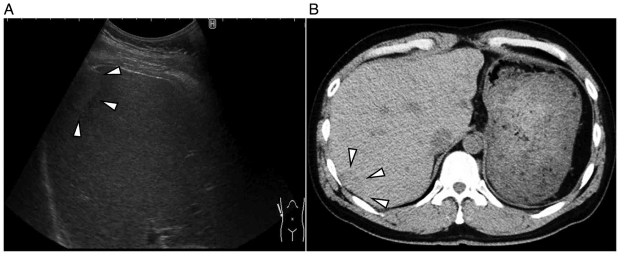
Abdominal ultrasonography (US) revealed a 2.4x1.4 cm isoechoic lesion with a hypoechoic zone at the margins in segment 7 of the right lobe of the liver (Fig. 1A). Abdominal plain-computed tomography (CT) revealed a 
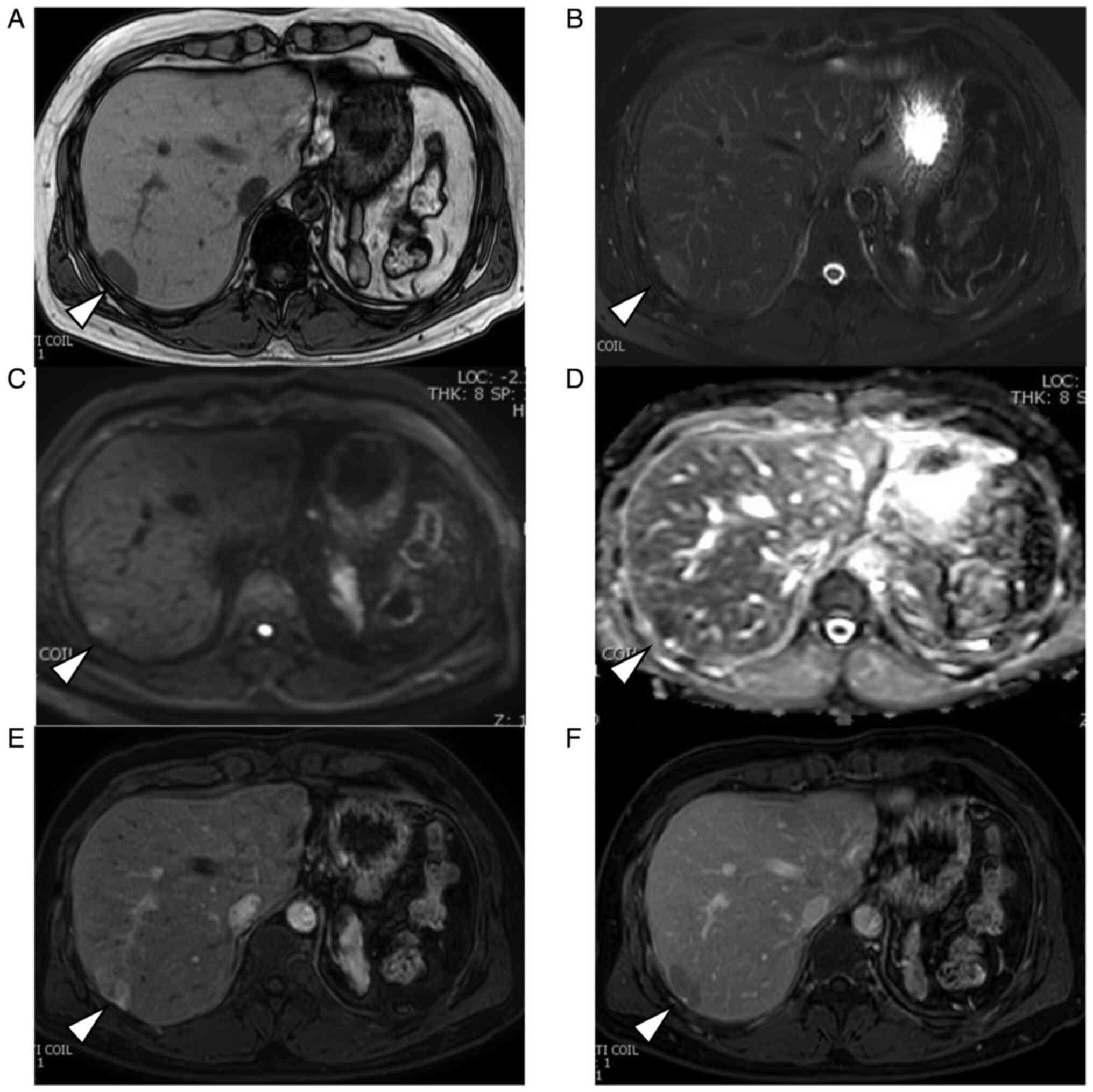
hypodense mass measuring 2.5x1.7 cm (Fig. 1B). Abdominal magnetic resonance imaging (MRI) revealed a homogeneously hypointense mass in T1-weighted images and hyperintensity in T2-weighted images (Fig. 2A and B). The mass showed a heterogeneous hyperintensity in diffusion-weighted images and signal reduction in apparent diffusion coefficient. (Fig. 2C and D). After injecting gadoxetic acid, the lesion appeared strongly heterogeneous and hyperintense during the early phase and relatively hypointense during delayed phase that is ‘washout pattern’ (Fig. 2E and F). An indication of a pseudocapsule was also seen.

Figure 1

Abdominal ultrasonography and abdominal plain-computed tomography imaging of the IHS. (A) An abdominal ultrasonography revealed a 2.4x1.4 cm isoechoic lesion (arrows) with a hypoechoic zone at the margins in segment 7 of the liver. (B) An abdominal plane-computed tomography scan revealed a hypodense mass (arrows) measuring 2.5x1.7 cm in segment 7 of the liver.

Figure 2

Magnetic resonance imaging of the IHS (arrows). The IHS was detected with (A) a homogeneous hypointensity in T1-weighted images, (B) slight hyperintensity in T2-weighted images. (C) Lesion showed a heterogeneous hyperintensity in diffusion-weighted images and (D) signal reduction in apparent diffusion coefficient. After the injection of gadoxetic acid, (E) the lesion appeared heterogeneously hyperintense during the early phase and (F) relatively hypointense during the delayed phase. HIS, intrahepatic splenosis.

Radiographic features suggested a differential diagnosis of HCC, hepatic hemangioma, and hepatocellular adenoma. Surgical intervention was proposed, and the patient decided to 
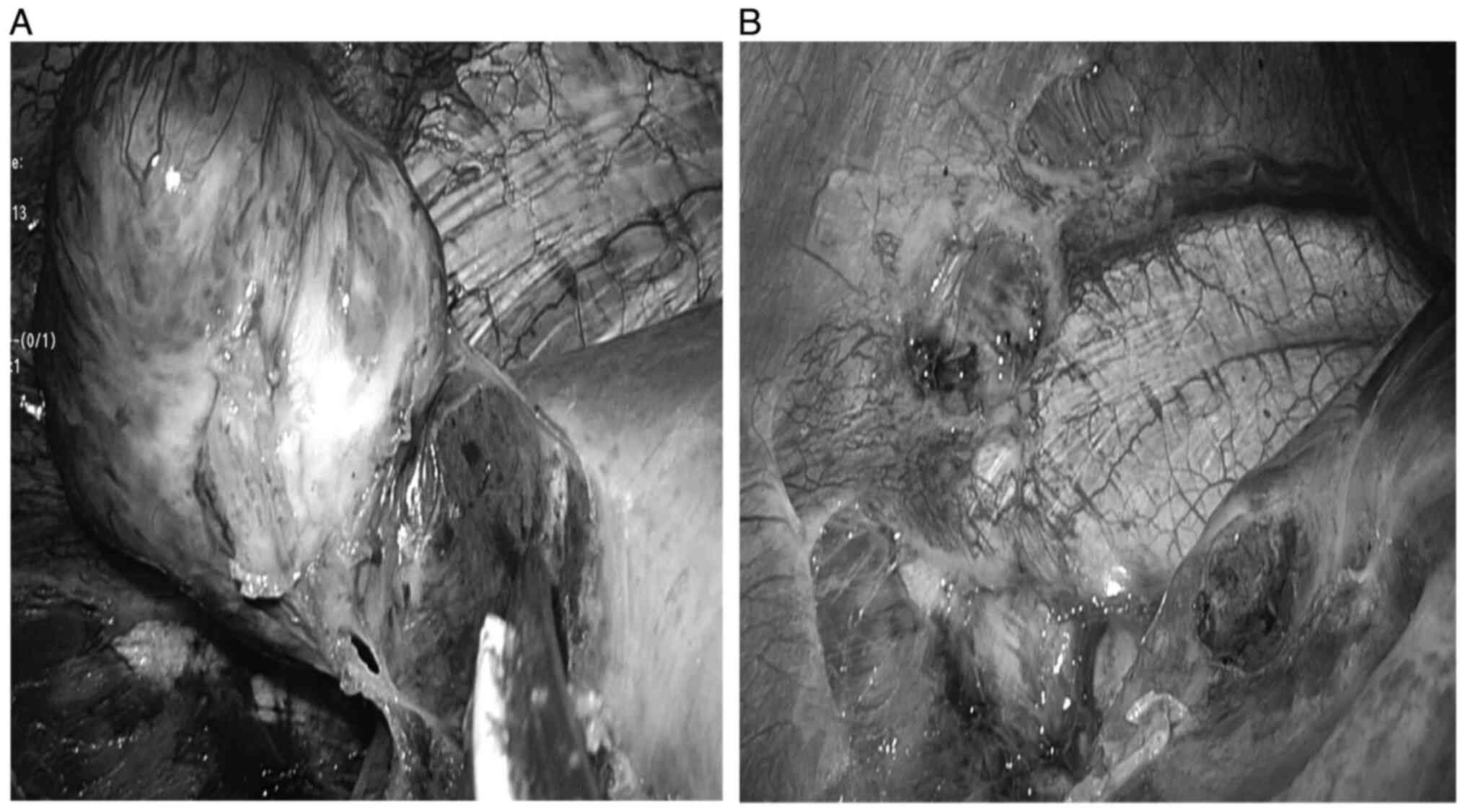
undergo a laparoscopic surgery. During surgery, the tumor was found in segment 7 of the right lobe (Fig. 3A). The tumor's feeding artery and drainage vessel were found to be from the inferior phrenic artery and vein (Fig. 3B). Liver tumor was successfully 
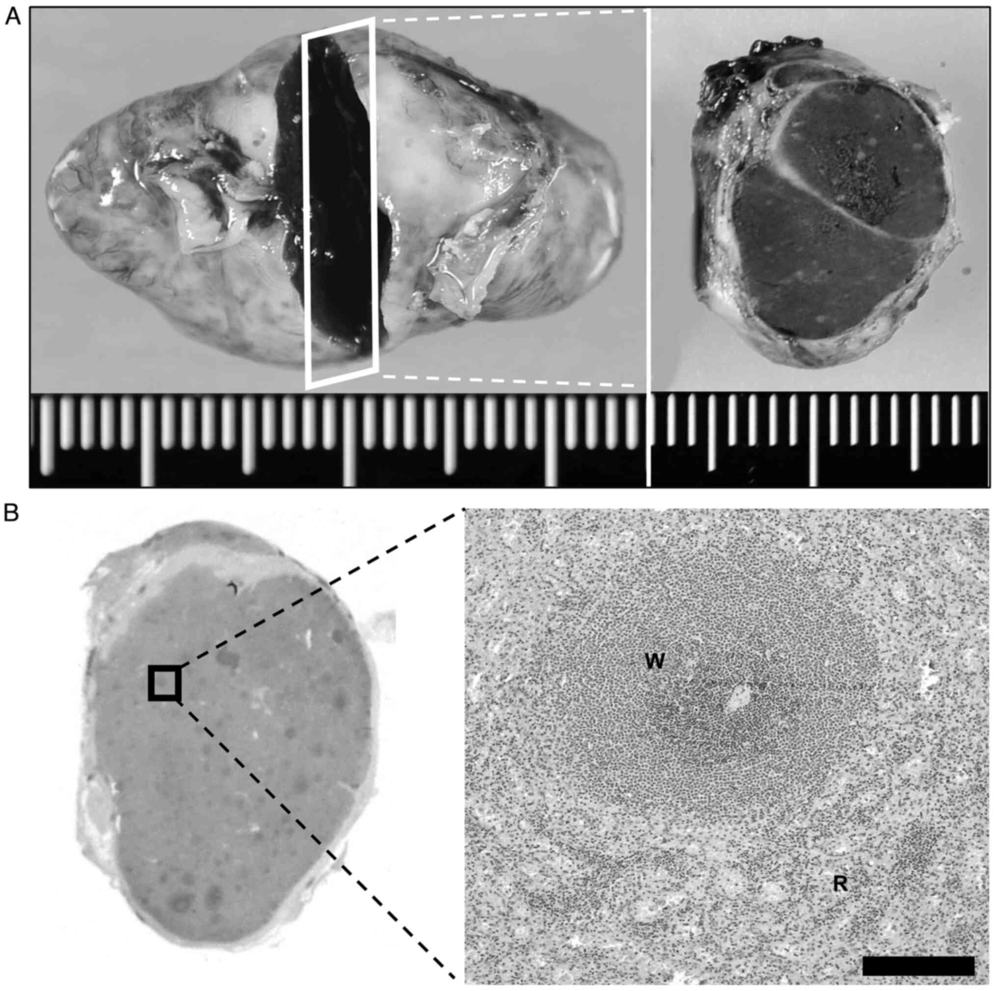
resected laparoscopically. Then, histopathologic examination of the resected specimen revealed that the tumor was totally consisted of splenic tissue surrounded by a fibrous capsule (Fig. 4A and B). The postoperative diagnosis was confirmed as IHS. The patient recovered uneventfully and was discharged on the fourth postoperative day. The patient visited outpatient care for 2 years of postoperative follow-up without any trouble.

Figure 3

During the surgery. (A) Tumor was buried in the liver and adherent to the diaphragm. It had a capsule and was soft texture. (B) Drainage vessels of the tumor were recognized from the inferior phrenic artery and vein.

Figure 4

Histopathologic findings. (A) Resected specimen was dark red and 2.7 cm in size. (B) Hematoxylin and eosin staining (magnification, x20). The resected specimen was composed of white and red pulp surrounded by a fibrous capsule (W: white pulp, R: red pulp). Scale bar, 200 µM.

Discussion

Splenosis is usually caused by heterotopic autotransplantation or implantation of splenic tissue after elective splenectomy or traumatic splenic rupture. Approximately 70% of patients with IHS are reported to have a history of splenic rupture or surgery (11). IHS is rare because splenosis usually occurs in the mesentery, omentum, or peritoneum in the left upper abdomen (12).

After a literature search, using the search term ‘intrahepatic splenosis’ on the PubMed, we identified and reviewed 52 cases. Of these, 14 cases were excluded because of lacking clinical details, and the remaining 38 cases were included in our review (Table SI). There were 31 (81.5%) male and 7 (18.4%) female patients, with a mean age of 47.9 (±13) years. There was a previous history of abdominal trauma in 89% of the patients, and 97% had a history of splenectomy, which is consistent with a previous report (12). Seventy-three percent of patients were asymptomatic on admission. The mean tumor size on imaging was 3.7 (±1.2) cm. Fifty-eight percent of the patients underwent surgery, including hepatectomy, with the preoperative diagnosis of HCC or liver metastases (3,7,10,13).

The radiographic appearance of IHS generally varies. Typical findings are hypodense areas on non-contrast CT. Following contrast administration, the lesions are hyperdense in the arterial phase, iso-dense in the portal venous phase, and hypodense in the delayed phase (3,14). On MRI, IHS is usually described as areas of homogeneous hypo-intensity in T1WI and hyperintensity in T2WI (10). Following contrast administration, IHS shows a heterogeneous enhancement in the arterial phase, which becomes homogeneous in the later phases (8,9,15). In the delayed phase, the signal intensity of IHS may be lower than that of the liver parenchyma (10). The imaging features are similar to the signal and enhancement patterns of the spleen, usually described as geographic or zebra patterns of enhancement. In the previous studies, 61.5% of patients had typical radiological features. In our case, the enhancement pattern of the tumor in the enhanced MRI showed early enhancement in the arterial phase and washout in the delayed phase that was similar to the radiographic features of moderately differentiated HCC.

There are currently reported to be two dominant mechanisms of IHS occurring. One hypothesis is that an invagination or an exophytic growth of splenic tissue directly seeded into the liver capsule. With this mechanism, the most frequent site of IHS is the area surrounded by the left lobe and the diaphragm because it can be easily seeded with splenic tissue during splenectomy (14,16-18). The other is that hematogenous spread due to the entry of an erythrocyte progenitor cell into the portal venous system. In the latter pattern, the margin between IHS and liver parenchyma is usually unclear (2,19,20). In our case, tumor happened to metastasize in the right lobe near the inferior phrenic vessels without seeding at the most frequent site, and it was extrahepatically fed by these vessels. The tumor showed early enhancement in the arterial phase and washout in the delayed phase that was characteristic imaging pattern for HCC, although IHS shows a heterogeneous enhancement in the arterial phase, and homogeneous in the later phases (8,9,15). In our case, IHS was extrahepatically fed, so that the radiological findings were typical for HCC. It was difficult to make a correct diagnosis of IHS preoperatively. Surgical procedure was successfully performed.

We reported a rare case of IHS mimicking HCC. With a history of traumatic rupture of the spleen and splenectomy, even when the tumor has typical radiological patterns for HCC, IHS might be a different diagnosis.

Supplementary Material

Previous reports of patients with intrahepatic splenosis.

Acknowledgements

Not applicable.

Funding

Funding: No funding was received.

Availability of data and materials

The datasets used and/or analyzed during the current study are available from the corresponding author on reasonable request.

Authors' contributions

IU, HT, KM, AK, SM, RK, KN, SN, YY and AM participated in the diagnosis and treatment of the patient and wrote the first draft of this manuscript. All authors have read and approved the final manuscript.

Ethics approval and consent to participate

Not applicable.

Patient consent for publication

Written informed consent was obtained from the patient to publish this case report and any accompanying images.

Competing interests

The authors declare that they have no competing interests.

References

1 

Gandhi D, Sharma P, Garg G, Songmen S, Solanki S and Singh T: Intrahepatic splenosis demonstrated by diffusion weighted MRI with histologic confirmation. Radiol Case Rep. 15:602–606. 2020.PubMed/NCBI View Article : Google Scholar

2 

Menth M, Herrmann K, Haug A, Raziorrouh B, Zachoval R, Jung CM and Otto C: Intra-hepatic splenosis as an unexpected cause of a focal liver lesion in a patient with hepatitis C and liver cirrhosis: A case report. Cases J. 2(8335)2009.PubMed/NCBI View Article : Google Scholar

3 

Toh WS, Chan KS, Ding CSL, Tan CH and Shelat VG: Intrahepatic splenosis: A world review. Clin Exp Hepatol. 6:185–198. 2020.PubMed/NCBI View Article : Google Scholar

4 

Rickert CH, Maasjosthusmann U, Probst-Cousin S, August C and Gullotta F: A unique case of cerebral spleen. Am J Surg Pathol. 22:894–896. 1998.PubMed/NCBI View Article : Google Scholar

5 

Baldari G, Sindoni A, Belletti A, Baldari S and Ruffini L: (99m)Tc disphosphonate uptake due to splenosis. Incidental finding 60 years after splenectomy. Clin Nucl Med. 40:533–535. 2015.PubMed/NCBI View Article : Google Scholar

6 

De Vuysere S, Van Steenbergen W, Aerts R, Van Hauwaert H, Van Beckevoort D and Van Hoe L: Intrahepatic splenosis: Imaging features. Abdom Imaging. 25:187–189. 2000.PubMed/NCBI View Article : Google Scholar

7 

Yeh ML, Wang LY, Huang CI, Hsieh MY, Lin ZY, Chuang WL, Chang WT, Wu CC and Chen CY: Abdominal splenosis mimicking hepatic tumor: A case report. Kaohsiung J Med Sci. 24:602–606. 2008.PubMed/NCBI View Article : Google Scholar

8 

Lin WC, Lee RC, Chiang JH, Wei CJ, Chu LS, Liu RS and Chang CY: MR features of abdominal splenosis. AJR Am J Roentgenol. 180:493–496. 2003.PubMed/NCBI View Article : Google Scholar

9 

Inchingolo R, Peddu P and Karani J: Hepatic splenosis presenting as arterialised liver lesion in a patient with NASH. Eur Rev Med Pharmacol Sci. 17:2853–2856. 2013.PubMed/NCBI

10 

Sato N, Abe T, Suzuki N, Waragai M, Teranishi Y, Takano Y, Sato A, Azami A and Gotoh M: Intrahepatic splenosis in a chronic hepatitis C patient with no history of splenic trauma mimicking hepatocellular carcinoma. Am J Case Rep. 15:416–420. 2014.PubMed/NCBI View Article : Google Scholar

11 

Case records of the Massachusetts general hospital. Case 29-1995: A 65-year-old man with mediastinal Hodgkin's disease and a pelvic mass. N Engl J Med. 333:784–791. 1995.PubMed/NCBI View Article : Google Scholar

12 

Mescoli C, Castoro C, Sergio A, Ruol A, Farinati F and Rugge M: Hepatic spleen nodules (HSN). Scand J Gastroenterol. 45:628–632. 2010.PubMed/NCBI View Article : Google Scholar

13 

Kang KC, Cho GS, Chung GA, Kang GH, Kim YJ, Lee MS, Kim HK and Park SJ: Intrahepatic splenosis mimicking liver metastasis in a patient with gastric cancer. J Gastric Cancer. 11:64–68. 2011.PubMed/NCBI View Article : Google Scholar

14 

Tsitouridis I, Michaelides M, Sotiriadis C and Arvaniti M: CT and MRI of intraperitoneal splenosis. Diagn Interv Radiol. 16:145–149. 2010.PubMed/NCBI View Article : Google Scholar

15 

Pekkafali Z, Karsli AF, Silit E, Başekim CC, Narin Y, Mutlu H and Kizilkaya E: Intrahepatic splenosis: A case report. Eur Radiol. 12:S62–S65. 2002.PubMed/NCBI View Article : Google Scholar

16 

Liu K, Liang Y, Liang X, Yu H, Wang Y and Cai X: Laparoscopic resection of isolated hepatic splenosis mimicking liver tumors: Case report with a literature review. Surg Laparosc Endosc Percutan Tech. 22:e307–e311. 2012.PubMed/NCBI View Article : Google Scholar

17 

Teles GNS, Monteiro PEZ and Raphe R: Intrahepatic splenosis mimicking hepatic neoplasia. Int J Surg Case Rep. 44:47–50. 2018.PubMed/NCBI View Article : Google Scholar

18 

Choi GH, Ju MK, Kim JY, Kang CM, Kim KS, Choi JS, Han KH, Park MS, Park YN, Lee WJ and Kim BR: Hepatic splenosis preoperatively diagnosed as hepatocellular carcinoma in a patient with chronic hepatitis B: A case report. J Korean Med Sci. 23:336–341. 2008.PubMed/NCBI View Article : Google Scholar

19 

Seguchi S, Yue F, Asanuma K and Sasaki K: Experimental splenosis in the liver and lung spread through the vasculature. Cell Tissue Res. 360:287–296. 2015.PubMed/NCBI View Article : Google Scholar

20 

Kwok CM, Chen YT, Lin HT, Su CH, Liu YS and Chiu YC: Portal vein entrance of splenic erythrocytic progenitor cells and local hypoxia of liver, two events cause intrahepatic splenosis. Med Hypotheses. 67:1330–1332. 2006.PubMed/NCBI View Article : Google Scholar

Related Articles

  • Abstract
  • View
  • Download
  • Twitter
Copy and paste a formatted citation
Spandidos Publications style
Umeda I, Tomihara H, Maeda S, Kitagawa A, Miyamoto K, Kawabata R, Nishikawa K, Noura S, Yasuhara Y, Miyamoto A, Miyamoto A, et al: A rare intrahepatic splenosis mimicking hepatocellular carcinoma: A case report. Mol Clin Oncol 19: 91, 2023.
APA
Umeda, I., Tomihara, H., Maeda, S., Kitagawa, A., Miyamoto, K., Kawabata, R. ... Miyamoto, A. (2023). A rare intrahepatic splenosis mimicking hepatocellular carcinoma: A case report. Molecular and Clinical Oncology, 19, 91. https://doi.org/10.3892/mco.2023.2687
MLA
Umeda, I., Tomihara, H., Maeda, S., Kitagawa, A., Miyamoto, K., Kawabata, R., Nishikawa, K., Noura, S., Yasuhara, Y., Miyamoto, A."A rare intrahepatic splenosis mimicking hepatocellular carcinoma: A case report". Molecular and Clinical Oncology 19.5 (2023): 91.
Chicago
Umeda, I., Tomihara, H., Maeda, S., Kitagawa, A., Miyamoto, K., Kawabata, R., Nishikawa, K., Noura, S., Yasuhara, Y., Miyamoto, A."A rare intrahepatic splenosis mimicking hepatocellular carcinoma: A case report". Molecular and Clinical Oncology 19, no. 5 (2023): 91. https://doi.org/10.3892/mco.2023.2687
Copy and paste a formatted citation
x
Spandidos Publications style
Umeda I, Tomihara H, Maeda S, Kitagawa A, Miyamoto K, Kawabata R, Nishikawa K, Noura S, Yasuhara Y, Miyamoto A, Miyamoto A, et al: A rare intrahepatic splenosis mimicking hepatocellular carcinoma: A case report. Mol Clin Oncol 19: 91, 2023.
APA
Umeda, I., Tomihara, H., Maeda, S., Kitagawa, A., Miyamoto, K., Kawabata, R. ... Miyamoto, A. (2023). A rare intrahepatic splenosis mimicking hepatocellular carcinoma: A case report. Molecular and Clinical Oncology, 19, 91. https://doi.org/10.3892/mco.2023.2687
MLA
Umeda, I., Tomihara, H., Maeda, S., Kitagawa, A., Miyamoto, K., Kawabata, R., Nishikawa, K., Noura, S., Yasuhara, Y., Miyamoto, A."A rare intrahepatic splenosis mimicking hepatocellular carcinoma: A case report". Molecular and Clinical Oncology 19.5 (2023): 91.
Chicago
Umeda, I., Tomihara, H., Maeda, S., Kitagawa, A., Miyamoto, K., Kawabata, R., Nishikawa, K., Noura, S., Yasuhara, Y., Miyamoto, A."A rare intrahepatic splenosis mimicking hepatocellular carcinoma: A case report". Molecular and Clinical Oncology 19, no. 5 (2023): 91. https://doi.org/10.3892/mco.2023.2687
Follow us
  • Twitter
  • LinkedIn
  • Facebook
About
  • Spandidos Publications
  • Careers
  • Cookie Policy
  • Privacy Policy
How can we help?
  • Help
  • Live Chat
  • Contact
  • Email to our Support Team