Spandidos Publications Logo
  • About
    • About Spandidos
    • Aims and Scopes
    • Abstracting and Indexing
    • Editorial Policies
    • Reprints and Permissions
    • Job Opportunities
    • Terms and Conditions
    • Contact
  • Journals
    • All Journals
    • Oncology Letters
      • Oncology Letters
      • Information for Authors
      • Editorial Policies
      • Editorial Board
      • Aims and Scope
      • Abstracting and Indexing
      • Bibliographic Information
      • Archive
    • International Journal of Oncology
      • International Journal of Oncology
      • Information for Authors
      • Editorial Policies
      • Editorial Board
      • Aims and Scope
      • Abstracting and Indexing
      • Bibliographic Information
      • Archive
    • Molecular and Clinical Oncology
      • Molecular and Clinical Oncology
      • Information for Authors
      • Editorial Policies
      • Editorial Board
      • Aims and Scope
      • Abstracting and Indexing
      • Bibliographic Information
      • Archive
    • Experimental and Therapeutic Medicine
      • Experimental and Therapeutic Medicine
      • Information for Authors
      • Editorial Policies
      • Editorial Board
      • Aims and Scope
      • Abstracting and Indexing
      • Bibliographic Information
      • Archive
    • International Journal of Molecular Medicine
      • International Journal of Molecular Medicine
      • Information for Authors
      • Editorial Policies
      • Editorial Board
      • Aims and Scope
      • Abstracting and Indexing
      • Bibliographic Information
      • Archive
    • Biomedical Reports
      • Biomedical Reports
      • Information for Authors
      • Editorial Policies
      • Editorial Board
      • Aims and Scope
      • Abstracting and Indexing
      • Bibliographic Information
      • Archive
    • Oncology Reports
      • Oncology Reports
      • Information for Authors
      • Editorial Policies
      • Editorial Board
      • Aims and Scope
      • Abstracting and Indexing
      • Bibliographic Information
      • Archive
    • Molecular Medicine Reports
      • Molecular Medicine Reports
      • Information for Authors
      • Editorial Policies
      • Editorial Board
      • Aims and Scope
      • Abstracting and Indexing
      • Bibliographic Information
      • Archive
    • World Academy of Sciences Journal
      • World Academy of Sciences Journal
      • Information for Authors
      • Editorial Policies
      • Editorial Board
      • Aims and Scope
      • Abstracting and Indexing
      • Bibliographic Information
      • Archive
    • International Journal of Functional Nutrition
      • International Journal of Functional Nutrition
      • Information for Authors
      • Editorial Policies
      • Editorial Board
      • Aims and Scope
      • Abstracting and Indexing
      • Bibliographic Information
      • Archive
    • International Journal of Epigenetics
      • International Journal of Epigenetics
      • Information for Authors
      • Editorial Policies
      • Editorial Board
      • Aims and Scope
      • Abstracting and Indexing
      • Bibliographic Information
      • Archive
    • Medicine International
      • Medicine International
      • Information for Authors
      • Editorial Policies
      • Editorial Board
      • Aims and Scope
      • Abstracting and Indexing
      • Bibliographic Information
      • Archive
  • Articles
  • Information
    • Information for Authors
    • Information for Reviewers
    • Information for Librarians
    • Information for Advertisers
    • Conferences
  • Language Editing
Spandidos Publications Logo
  • About
    • About Spandidos
    • Aims and Scopes
    • Abstracting and Indexing
    • Editorial Policies
    • Reprints and Permissions
    • Job Opportunities
    • Terms and Conditions
    • Contact
  • Journals
    • All Journals
    • Biomedical Reports
      • Information for Authors
      • Editorial Policies
      • Editorial Board
      • Aims and Scope
      • Abstracting and Indexing
      • Bibliographic Information
      • Archive
    • Experimental and Therapeutic Medicine
      • Information for Authors
      • Editorial Policies
      • Editorial Board
      • Aims and Scope
      • Abstracting and Indexing
      • Bibliographic Information
      • Archive
    • International Journal of Epigenetics
      • Information for Authors
      • Editorial Policies
      • Editorial Board
      • Aims and Scope
      • Abstracting and Indexing
      • Bibliographic Information
      • Archive
    • International Journal of Functional Nutrition
      • Information for Authors
      • Editorial Policies
      • Editorial Board
      • Aims and Scope
      • Abstracting and Indexing
      • Bibliographic Information
      • Archive
    • International Journal of Molecular Medicine
      • Information for Authors
      • Editorial Policies
      • Editorial Board
      • Aims and Scope
      • Abstracting and Indexing
      • Bibliographic Information
      • Archive
    • International Journal of Oncology
      • Information for Authors
      • Editorial Policies
      • Editorial Board
      • Aims and Scope
      • Abstracting and Indexing
      • Bibliographic Information
      • Archive
    • Medicine International
      • Information for Authors
      • Editorial Policies
      • Editorial Board
      • Aims and Scope
      • Abstracting and Indexing
      • Bibliographic Information
      • Archive
    • Molecular and Clinical Oncology
      • Information for Authors
      • Editorial Policies
      • Editorial Board
      • Aims and Scope
      • Abstracting and Indexing
      • Bibliographic Information
      • Archive
    • Molecular Medicine Reports
      • Information for Authors
      • Editorial Policies
      • Editorial Board
      • Aims and Scope
      • Abstracting and Indexing
      • Bibliographic Information
      • Archive
    • Oncology Letters
      • Information for Authors
      • Editorial Policies
      • Editorial Board
      • Aims and Scope
      • Abstracting and Indexing
      • Bibliographic Information
      • Archive
    • Oncology Reports
      • Information for Authors
      • Editorial Policies
      • Editorial Board
      • Aims and Scope
      • Abstracting and Indexing
      • Bibliographic Information
      • Archive
    • World Academy of Sciences Journal
      • Information for Authors
      • Editorial Policies
      • Editorial Board
      • Aims and Scope
      • Abstracting and Indexing
      • Bibliographic Information
      • Archive
  • Articles
  • Information
    • For Authors
    • For Reviewers
    • For Librarians
    • For Advertisers
    • Conferences
  • Language Editing
Login Register Submit
  • This site uses cookies
  • You can change your cookie settings at any time by following the instructions in our Cookie Policy. To find out more, you may read our Privacy Policy.

    I agree
Search articles by DOI, keyword, author or affiliation
Search
Advanced Search
presentation
Molecular and Clinical Oncology
Join Editorial Board Propose a Special Issue
Print ISSN: 2049-9450 Online ISSN: 2049-9469
Journal Cover
August-2024 Volume 21 Issue 2

Full Size Image

Sign up for eToc alerts
Recommend to Library

Journals

International Journal of Molecular Medicine

International Journal of Molecular Medicine

International Journal of Molecular Medicine is an international journal devoted to molecular mechanisms of human disease.

International Journal of Oncology

International Journal of Oncology

International Journal of Oncology is an international journal devoted to oncology research and cancer treatment.

Molecular Medicine Reports

Molecular Medicine Reports

Covers molecular medicine topics such as pharmacology, pathology, genetics, neuroscience, infectious diseases, molecular cardiology, and molecular surgery.

Oncology Reports

Oncology Reports

Oncology Reports is an international journal devoted to fundamental and applied research in Oncology.

Experimental and Therapeutic Medicine

Experimental and Therapeutic Medicine

Experimental and Therapeutic Medicine is an international journal devoted to laboratory and clinical medicine.

Oncology Letters

Oncology Letters

Oncology Letters is an international journal devoted to Experimental and Clinical Oncology.

Biomedical Reports

Biomedical Reports

Explores a wide range of biological and medical fields, including pharmacology, genetics, microbiology, neuroscience, and molecular cardiology.

Molecular and Clinical Oncology

Molecular and Clinical Oncology

International journal addressing all aspects of oncology research, from tumorigenesis and oncogenes to chemotherapy and metastasis.

World Academy of Sciences Journal

World Academy of Sciences Journal

Multidisciplinary open-access journal spanning biochemistry, genetics, neuroscience, environmental health, and synthetic biology.

International Journal of Functional Nutrition

International Journal of Functional Nutrition

Open-access journal combining biochemistry, pharmacology, immunology, and genetics to advance health through functional nutrition.

International Journal of Epigenetics

International Journal of Epigenetics

Publishes open-access research on using epigenetics to advance understanding and treatment of human disease.

Medicine International

Medicine International

An International Open Access Journal Devoted to General Medicine.

Journal Cover
August-2024 Volume 21 Issue 2

Full Size Image

Sign up for eToc alerts
Recommend to Library

  • Article
  • Citations
    • Cite This Article
    • Download Citation
    • Create Citation Alert
    • Remove Citation Alert
    • Cited By
  • Similar Articles
    • Related Articles (in Spandidos Publications)
    • Similar Articles (Google Scholar)
    • Similar Articles (PubMed)
  • Download PDF
  • Download XML
  • View XML
Case Report Open Access

Focal ground glass opacity of the lung in metachronous prostate and gastric cancer: A case report

  • Authors:
    • Ryuichi Wada
    • Tomohiko Yamamoto
    • Kenichi Iwasaki
    • Shinsuke Usui
    • Nobuaki Matsubara
    • Hiroaki Nomori
  • View Affiliations / Copyright

    Affiliations: Department of Diagnostic Pathology, Kashiwa Kousei General Hospital, Kashiwa, Chiba 277‑8551, Japan, Department of Surgery, Kashiwa Kousei General Hospital, Kashiwa, Chiba 277‑8551, Japan, Department of Medical Oncology, National Cancer Center Hospital East, Kashiwa, Chiba 277‑8577, Japan, Department of Thoracic Surgery, Kashiwa Kousei General Hospital, Kashiwa, Chiba 277‑8551, Japan
    Copyright: © Wada et al. This is an open access article distributed under the terms of Creative Commons Attribution License.
  • Article Number: 54
    |
    Published online on: June 19, 2024
       https://doi.org/10.3892/mco.2024.2752
  • Expand metrics +
Metrics: Total Views: 0 (Spandidos Publications: | PMC Statistics: )
Metrics: Total PDF Downloads: 0 (Spandidos Publications: | PMC Statistics: )
Cited By (CrossRef): 0 citations Loading Articles...

This article is mentioned in:


Abstract

Chest computed tomography (CT) revealed a focal ground glass opacity (GGO) with a minimal solid area in a 75‑year‑old man. The shadow was located in the periphery of the right upper lobe and measured 11 mm in diameter. The patient had a medical history of metachronous prostate and gastric cancers. The patient had been treated with androgen deprivation therapy for prostate cancer for 12 years and underwent subtotal gastrectomy for triple gastric cancers 7 months before. Since primary lung adenocarcinoma was suspected, CT‑assisted percutaneous needle biopsy was performed. Histology revealed the sheet‑like and trabecular proliferation of atypical cells, suggesting that the lesion was moderately to poorly differentiated adenocarcinoma. Adenocarcinoma cells showed subepithelial extension causing the thickening of alveolar walls. A tumor thrombus was not detected in the blood or lymphatic vessels. Immunohistochemistry revealed that carcinoma cells were negative for cytokeratin 7 (CK7), CK20, thyroid transcription factor‑1 and CDX2 and positive for prostate‑specific antigen and P504S. Based on these findings, the patient was diagnosed with metastatic carcinoma from prostate cancer. The disease remained stable for 4 months after the diagnosis, and no new lesions were observed on chest CT. Metastatic carcinoma rarely presents with focal GGO. Lung biopsy is necessary to identify the pathology of the lesion, and the primary site needs to be confirmed by immunohistochemistry with specific markers, particularly in a case of metachronous multiple cancers. A tumor thrombus, which is suggestive of lymphangitic carcinomatosis or pulmonary tumor thrombotic microangiopathy, also needs to be evaluated.

Introduction

Metastatic carcinoma in the lung generally presents with multiple nodular shadows. Metastatic carcinoma may present with ground glass opacity (GGO), usually with diffuse form, on chest computed tomography (CT). Diffuse GGO represents lymphatic and/or vascular spread of carcinoma in the lung (1). Focal GGO, which is localized or nodular form on chest CT, is uncommon in metastatic carcinoma. Since various lung diseases such as interstitial fibrosis, inflammation and primary lung adenocarcinoma present with GGO, it is difficult to reach an accurate diagnosis based solely on radiological findings. Thus, in a case with past history of carcinoma, pathological assessment of focal GGO by biopsy or partial resection is necessary to determine whether the lesion is primary or metastatic carcinoma. The assessment is also critical to decide appropriate treatment.

We herein present a case of metastatic carcinoma presenting with focal GGO. A definite diagnosis of metastatic prostate cancer was reached by CT-assisted percutaneous needle biopsy and histological and immunohistochemical examinations. We present characteristic histological and immunohistochemical features of the case. We also reviewed a literature and found cases of metastatic cancer, presenting with focal GGO (2-10), and three cases were metastatic prostate cancer. It is of note two out of three cases were associated with pulmonary tumor thrombotic microangiopathy (PTTM), which rapidly progresses and is fatal (11). The vascular pathology needs to be assessed in biopsy specimen. The histological and clinical features of PTTM, which are necessary for immediate diagnosis, are also reviewed.

Case report

A 75-year-old man, who had a medical history of metachronous prostate and gastric cancers, presented at Kashiwa Kousei General Hospital, Kashiwa, Japan in September 2023 for the follow-up examination of the cancers. He was diagnosed as prostate cancer in another hospital 12 years before. Multiple bone metastases were detected in the spine. The histology of biopsy specimen of the prostate was adenocarcinoma with Gleason score 4+4. The patient had been treated with androgen deprivation therapy (ADT) for 12 years. The patient also received pelvic irradiation. Bone metastases appeared to be stable by ADT. However, a gradual increase was observed in the level of prostate-specific antigen (PSA). Seven months before, the patient underwent subtotal gastrectomy for multiple gastric cancers, which were detected in a health examination in Kashiwa Kousei General Hospital. There were three gastric cancers in the resected stomach. The main cancer was well-differentiated adenocarcinoma with focal moderately differentiated adenocarcinoma, and carcinoma cells invaded the submucosa. Neither blood nor lymphatic invasion was observed. The two other cancers were well-differentiated adenocarcinomas that were confined to the mucosa. Lymph node metastasis was not found.

Red and white blood cell counts were 4.09x10⁶/µl and 4.1x10³/µl, respectively. His platelet count was 69x103/µl. Serum levels of carcinoembryonic antigen and CA19-9 were within normal limits. His PSA level was elevated to 7.62 ng/ml. His fibrinogen level was 283 ng/dl and fibrinogen degradation products (FDP) were below the detection level. Echo cardiogram showed no abnormalities. The patient did not have dyspnea or respiratory discomfort. Pulmonary function tests were normal.

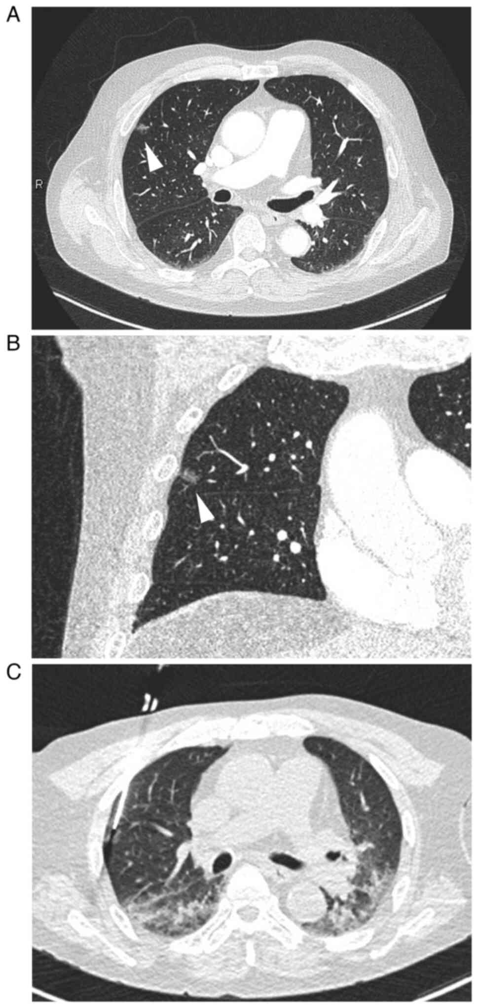
Chest CT revealed a focal GGO with a minimal solid area, measuring 11 mm in diameter, in the periphery of the right upper lobe (Fig. 1A and B). Pulmonary vessels and bronchioles were not involved. Since primary lung adenocarcinoma was suspected, CT-assisted percutaneous needle biopsy was performed (Fig. 1C).

Figure 1

CT images of the chest. (A) A focal ground glass opacity (GGO) was noted in the periphery of the upper lobe of the right lung (arrowhead) on contrast-enhanced CT. (B) The frontal section of contrast-enhanced CT showed irregularly shaped GGO (arrowhead). (C) CT image of the area for percutaneous lung biopsy. CT, computed tomography.

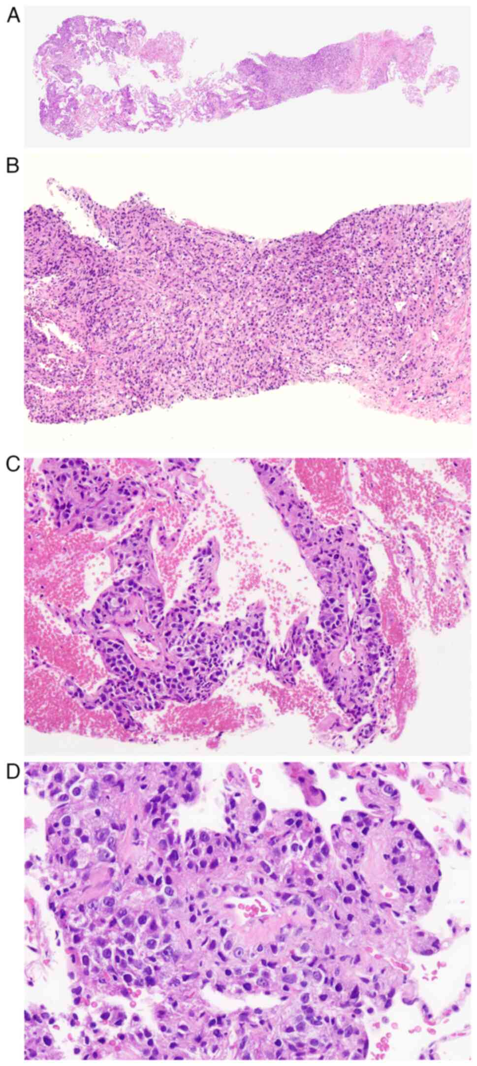
The biopsy specimen showed the proliferation of atypical cells in fibrous tissue and the alveolar septa (Fig. 2A). In fibrous tissue, atypical cells proliferated in sheet-like, trabecular, and vague glandular patterns, suggesting that the lesion was moderately to poorly differentiated adenocarcinoma (Fig. 2B). The surrounding alveolar wall was thickened by the invasion of the poorly differentiated component of adenocarcinoma into the interstitial space (Fig. 2B). Neither the lepidic growth of carcinoma cells within the alveolar space nor invasion into the alveolar space was observed. A tumor thrombus was not found in the blood or lymphatic vessels (Fig. 2C), and there was no stenosis or re-canalization of vessels. The perivascular infiltration of macrophages and fibrocellular intimal proliferation were not present.

Figure 2

Histology of the biopsy specimen. (A) Loupe image of the biopsy specimen (HE; magnification, x0.5). (B) The proliferation of atypical cells in fibrous tissue. Cells proliferated in sheet and trabecular patterns (HE; magnification x100). (C) Thickening of the alveolar septa and the extension of carcinoma cells into the interstitial space (HE; magnification x100). (D) Carcinoma cells proliferated around the vessel. No tumor thrombus was found in the blood or lymphatic vessels. Neither the perivascular infiltration of macrophages nor fibrocellular intimal proliferation was observed (HE; magnification x200). HE, hematoxylin and eosin.

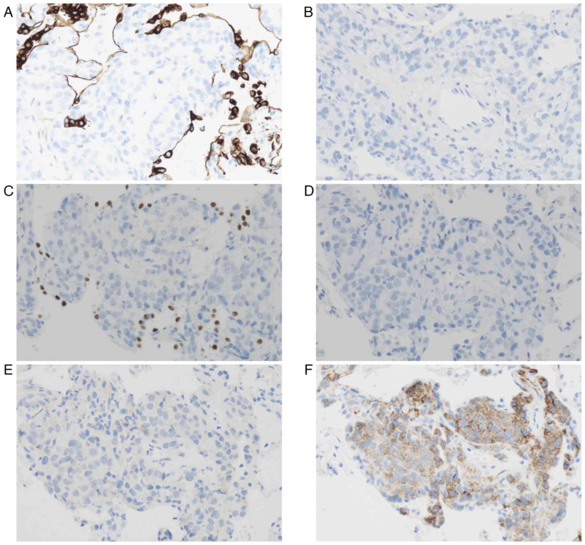
The phenotype of carcinoma cells was examined by immunohistochemistry (IHC). Carcinoma cells were negative for cytokeratin 7 (CK7) and CK20 (Fig. 3A and B) as well as thyroid transcription factor-1 (TTF1) and CDX2 (Fig. 3C and D). Therefore, primary lung adenocarcinoma and metastatic gastric cancer were considered to be unlikely. Carcinoma cells showed a faint positive reaction with PSA (Fig. 3E) and strong positive reaction with P504S (Fig. 3F). Based on these findings, the patient was diagnosed with metastatic carcinoma from prostate cancer. IHC with CK7 and TTF1 also clearly showed the subepithelial proliferation of carcinoma cells (Fig. 3A and C).

Figure 3

Immunohistochemistry of the biopsy specimen. (A) Carcinoma cells were negative for CK7. (B) Carcinoma cells were negative for CK20. (C) Cells were negative for thyroid transcription factor-1. (D) Cells were negative for CDX2. (E) Cells were faintly positive for prostate-specific antigen. (F) Cells showed clear positive reaction for P504S. Magnification x200. CK, cytokeratin.

After the diagnosis, ADT was continued, and the patient is being followed up with careful observations. The condition has remained stable, and new lesions were not detected in the lungs 4 months since the diagnosis. Furthermore, there have been no abnormalities in laboratory data.

Discussion

Metastatic carcinoma and malignant neoplasms generally present as nodular shadows in the lungs; however, GGO may be encountered in cases of lung metastasis (1). Diffuse and patchy GGO are caused by diffuse thickening of the alveolar wall due to lymphangitic carcinomatosis or PTTM, and the most frequent primary site of carcinoma is the gastrointestinal tract. Focal GGO is rarely found in metastatic carcinoma and malignant neoplasms, but has been documented in cases of malignant melanoma (2-4), malignant phyllodes tumor (5), tongue cancer (6), thyroid cancer (7), and prostate cancer (8-10), including the present case (Table I). Since it was not feasible to distinguish metastatic lesions from primary lung adenocarcinoma based solely on radiological findings, a diagnosis was made by a histological examination of partially resected lung or biopsy specimens in the reported cases. In the present case, which had a previous history of metachronous prostate and gastric cancers, CT-assisted percutaneous needle biopsy was performed for a definitive diagnosis of the lesion.

Table I

Reported cases of metastatic neoplasms presenting with focal ground glass opacity.

Table I

Reported cases of metastatic neoplasms presenting with focal ground glass opacity.

AuthorAge, yearsSexSize, mmSpecimenHistology(Refs.)
Malignant melanoma      
     Kang MJ56M11VATSSubepithelial proliferation of melanoma cells(2)
     Okita R56F28VATSSubepithelial proliferation of melanoma cells(3)
     Mizuuchi H57M10WRProliferation in a ‘lepidic-like’ fashion(4)
Malignant phyllodes tumor      
     Nakamura S71F21VATSSubepithelial proliferation of neoplastic cells(5)
Tongue cancer      
     Haro A22M24WRInvasion and destruction of alveoli and hemorrhage(6)
Thyroid cancer      
     Ryuko T68M8VATSLepidic growth of carcinoma cells(7)
Prostate cancer      
     Lubin DJ62M14ResectionLymphatic and perilymphatic growth of carcinoma cells(8)
     Osaki Y67MNDAutopsyPTTM(9)
     Katayama S81MMicronodularTBLB/AutopsyPTTM(10)
     Present case75M11PNBSubepithelial proliferation of carcinoma cells-

[i] F, female; M, male; ND, not described; PNB, percutaneous needle biopsy; PTTM, pulmonary tumor thrombotic microangiopathy; TBLB, transbronchial lung biopsy; VATS, video-assisted thoracic surgery; WR, wedge resection.

In biopsy specimens, it is important to confirm whether the lesion is primary lung adenocarcinoma. A benign lesion of interstitial pneumonia and other benign lesions are also carefully differentiated. If metastatic adenocarcinoma is suspected, the primary site needs to be identified by IHC with specific markers. In the present case, carcinoma cells were negative for CK7 and TTF1, suggesting that primary lung adenocarcinoma was unlikely. Furthermore, a negative reaction for CDX2 indicated that gastric cancer was unlikely. The confirmation of metastatic prostate cancer was reached by positive reactions for PSA and P504S. The majority of prostate cancers are negative for CK7 and CK20(12). The expression of PSA may be attenuated by long-term ADT (13).

GGO is caused by various pathological conditions, such as interstitial fibrosis, inflammatory cell infiltration, thrombosis, and the lepidic growth of carcinoma cells (1). In the present case, GGO was attributed to thickening of the alveolar walls due to the extension of carcinoma cells beneath alveolar epithelial cells. This extension was evident by IHC, and proliferating carcinoma cells were covered with alveolar epithelial cells, which were positive for CK7 and TTF1 (Fig. 3A and C). The subepithelial growth of carcinoma cells is similar to reported cases of malignant melanoma (2-4) and malignant phyllodes tumor (5). Lepidic growth similar to primary lung adenocarcinoma was previously documented in a case of metastatic thyroid cancer (7), and focal GGO was caused by the invasion and destruction of alveoli and hemorrhage in a case of metastatic tongue cancer (6).

Careful attention of reported cases of metastatic prostate cancer with focal GGO is needed (Table I). Two of the three cases were associated with PTTM (9,10), while one was associated with lymphangitic carcinomatosis (8). These conditions generally present with diffuse GGO. The clinical course of PTTM is rapid and fatal; therefore, an immediate diagnosis is essential. Respiratory failure, pulmonary hypertension, an elevated D-dimer level, and thrombocytopenia indicate PTTM in cases with a previous history of carcinoma (11). PTTM may be diagnosed using biopsy specimens. The pathological features of PTTM are a tumor thrombus, fibrocellular intimal proliferation, and the recanalization of vessels. The perivascular infiltration of macrophages may also be present because the inflammatory process plays an important role in the pathogenesis of PTTM (14,15). In the present case, these histological changes were not observed in the biopsy specimen. Respiratory failure was absent and there was neither a decrease in the platelet count nor an elevation in FDP. Although the patient's condition was stable, he needs to be followed up with careful attention to laboratory data and radiological findings.

Acknowledgements

Not applicable.

Funding

Funding: No funding was received.

Availability of data and materials

The datasets used during the present study are available from the corresponding author upon reasonable request.

Authors' contributions

RW, TY, KI, SU, NM, and HN participated in the diagnosis and treatment of the patient and wrote the first draft of this manuscript. RW and HN confirm the authenticity of all the raw data. All authors have read and approved the final manuscript.

Ethics approval and consent to participate

Ethics approval for the study was obtained from the Ethics Committee of Kashiwa Kousei General Hospital (approval no. 240025) dated February 26, 2024.

Patient consent for publication

Written informed consent was obtained from the patient to publish this case report and any accompanying images.

Competing interests

The authors declare that they have no competing interests.

References

1 

Seo JB, Im JG, Goo JM, Chung MJ and Kim MY: Atypical pulmonary metastases: Spectrum of radiologic findings. Radiographics. 21:403–417. 2001.PubMed/NCBI View Article : Google Scholar

2 

Kang MJ, Kim MA, Park CM, Lee CH, Goo JM and Lee HJ: Ground-glass nodules found in two patients with malignant melanomas: Different growth rate and different histology. Clin Imaging. 34:396–399. 2010.PubMed/NCBI View Article : Google Scholar

3 

Okita R, Yamashita M, Nakata M, Teramoto N, Bessho A and Mogami H: Multiple ground-glass opacity in metastasis of malignant melanoma diagnosed by lung biopsy. Ann Thorac Surg. 79:e1–e2. 2005.PubMed/NCBI View Article : Google Scholar

4 

Mizuuchi H, Suda K, Kitahara H, Shimamatsu S, Kohno M, Okamoto T and Maehara Y: Solitary pulmonary metastasis from malignant melanoma of the bulbar conjunctiva presenting as a pulmonary ground glass nodule: Report of a case. Thorac Cancer. 6:97–100. 2015.PubMed/NCBI View Article : Google Scholar

5 

Nakamura S, Goto T, Nara S, Kawahara Y, Yashiro S, Kano S, Hosokawa Y and Kamada H: Pure ground glass opacity (GGO) on chest CT: A rare presentation of lung metastasis of malignant phyllodes tumor. Breast Cancer. 27:1187–1190. 2020.PubMed/NCBI View Article : Google Scholar

6 

Haro A, Wakasu S, Takada K, Osoegawa A, Kamitani T, Tagawa T and Mori M: Pulmonary metastasis presenting as a ground glass opacity-like lesion with a thin-walled cavity: A case report. Int J Surg Case Rep. 60:287–290. 2019.PubMed/NCBI View Article : Google Scholar

7 

Ryuko T, Sano Y, Kitazawa R, Otani S, Sakao N and Mori Y: Lung metastasis from thyroid carcinoma showing a pure ground-glass nodule. Ann Thorac Surg. 114:e253–e256. 2022.PubMed/NCBI View Article : Google Scholar

8 

Lubin DJ, Holden SB, Rettig MB, Reiter RE, King CR, Lee JM, Wallace DW and Calais J: Prostate cancer pulmonary metastasis presenting as a ground-glass pulmonary nodule on 68Ga-PSMA-11 PET/CT. Clin Nucl Med. 44:e353–e356. 2019.PubMed/NCBI View Article : Google Scholar

9 

Osaki Y, Taoka R, Miyauchi Y and Sugimoto M: Docetaxel chemotherapy temporarily improved pulmonary tumor thrombotic microangiopathy induced by prostate cancer secreting carcinoembryonic antigen and carbohydrate antigen 19-9: A case report. Urol Case Rep. 29(101098)2020.PubMed/NCBI View Article : Google Scholar

10 

Katayama S, Takenaka T, Nakamura A, Sako S, Bessho A and Ohara N: Pulmonary tumor thrombotic microangiopathy induced by prostate cancer. Acta Med Okayama. 72:309–313. 2018.PubMed/NCBI View Article : Google Scholar

11 

Godbole RH, Saggar R and Kamangar N: Pulmonary tumor thrombotic microangiopathy: A systematic review. Pulm Circ. 9:1–13. 2019.PubMed/NCBI View Article : Google Scholar

12 

Dum D, Menz A, Völkel C, De Wispelaere N, Hinsch A, Gorbokon N, Lennartz M, Luebke AM, Hube-Magg C, Kluth M, et al: Cytokeratin 7 and cytokeratin 20 expression in cancer: A tissue microarray study on 15,424 cancers. Exp Mol Pathol. 126(104762)2022.PubMed/NCBI View Article : Google Scholar

13 

Ogreid P, Berner A, Dahl O, Rettedal E and Fossa SD: Tissue prostate-specific antigen and androgen receptor immunoreactivity in prostate cancer biopsies before, during and after neo-adjuvant androgen deprivation followed by radiotherapy. Eur Urol. 36:116–122. 1999.PubMed/NCBI View Article : Google Scholar

14 

Mantovani A, Allavena P, Sica A and Balkwill F: Cancer-related inflammation. Nature. 454:436–444. 2008.PubMed/NCBI View Article : Google Scholar

15 

Price LC, Wells AU and Wort SJ: Pulmonary tumour thrombotic microangiopathy. Curr Opin Pulm Med. 22:421–428. 2016.PubMed/NCBI View Article : Google Scholar

Related Articles

  • Abstract
  • View
  • Download
  • Twitter
Copy and paste a formatted citation
Spandidos Publications style
Wada R, Yamamoto T, Iwasaki K, Usui S, Matsubara N and Nomori H: Focal ground glass opacity of the lung in metachronous prostate and gastric cancer: A case report. Mol Clin Oncol 21: 54, 2024.
APA
Wada, R., Yamamoto, T., Iwasaki, K., Usui, S., Matsubara, N., & Nomori, H. (2024). Focal ground glass opacity of the lung in metachronous prostate and gastric cancer: A case report. Molecular and Clinical Oncology, 21, 54. https://doi.org/10.3892/mco.2024.2752
MLA
Wada, R., Yamamoto, T., Iwasaki, K., Usui, S., Matsubara, N., Nomori, H."Focal ground glass opacity of the lung in metachronous prostate and gastric cancer: A case report". Molecular and Clinical Oncology 21.2 (2024): 54.
Chicago
Wada, R., Yamamoto, T., Iwasaki, K., Usui, S., Matsubara, N., Nomori, H."Focal ground glass opacity of the lung in metachronous prostate and gastric cancer: A case report". Molecular and Clinical Oncology 21, no. 2 (2024): 54. https://doi.org/10.3892/mco.2024.2752
Copy and paste a formatted citation
x
Spandidos Publications style
Wada R, Yamamoto T, Iwasaki K, Usui S, Matsubara N and Nomori H: Focal ground glass opacity of the lung in metachronous prostate and gastric cancer: A case report. Mol Clin Oncol 21: 54, 2024.
APA
Wada, R., Yamamoto, T., Iwasaki, K., Usui, S., Matsubara, N., & Nomori, H. (2024). Focal ground glass opacity of the lung in metachronous prostate and gastric cancer: A case report. Molecular and Clinical Oncology, 21, 54. https://doi.org/10.3892/mco.2024.2752
MLA
Wada, R., Yamamoto, T., Iwasaki, K., Usui, S., Matsubara, N., Nomori, H."Focal ground glass opacity of the lung in metachronous prostate and gastric cancer: A case report". Molecular and Clinical Oncology 21.2 (2024): 54.
Chicago
Wada, R., Yamamoto, T., Iwasaki, K., Usui, S., Matsubara, N., Nomori, H."Focal ground glass opacity of the lung in metachronous prostate and gastric cancer: A case report". Molecular and Clinical Oncology 21, no. 2 (2024): 54. https://doi.org/10.3892/mco.2024.2752
Follow us
  • Twitter
  • LinkedIn
  • Facebook
About
  • Spandidos Publications
  • Careers
  • Cookie Policy
  • Privacy Policy
How can we help?
  • Help
  • Live Chat
  • Contact
  • Email to our Support Team