Spandidos Publications Logo
  • About
    • About Spandidos
    • Aims and Scopes
    • Abstracting and Indexing
    • Editorial Policies
    • Reprints and Permissions
    • Job Opportunities
    • Terms and Conditions
    • Contact
  • Journals
    • All Journals
    • Oncology Letters
      • Oncology Letters
      • Information for Authors
      • Editorial Policies
      • Editorial Board
      • Aims and Scope
      • Abstracting and Indexing
      • Bibliographic Information
      • Archive
    • International Journal of Oncology
      • International Journal of Oncology
      • Information for Authors
      • Editorial Policies
      • Editorial Board
      • Aims and Scope
      • Abstracting and Indexing
      • Bibliographic Information
      • Archive
    • Molecular and Clinical Oncology
      • Molecular and Clinical Oncology
      • Information for Authors
      • Editorial Policies
      • Editorial Board
      • Aims and Scope
      • Abstracting and Indexing
      • Bibliographic Information
      • Archive
    • Experimental and Therapeutic Medicine
      • Experimental and Therapeutic Medicine
      • Information for Authors
      • Editorial Policies
      • Editorial Board
      • Aims and Scope
      • Abstracting and Indexing
      • Bibliographic Information
      • Archive
    • International Journal of Molecular Medicine
      • International Journal of Molecular Medicine
      • Information for Authors
      • Editorial Policies
      • Editorial Board
      • Aims and Scope
      • Abstracting and Indexing
      • Bibliographic Information
      • Archive
    • Biomedical Reports
      • Biomedical Reports
      • Information for Authors
      • Editorial Policies
      • Editorial Board
      • Aims and Scope
      • Abstracting and Indexing
      • Bibliographic Information
      • Archive
    • Oncology Reports
      • Oncology Reports
      • Information for Authors
      • Editorial Policies
      • Editorial Board
      • Aims and Scope
      • Abstracting and Indexing
      • Bibliographic Information
      • Archive
    • Molecular Medicine Reports
      • Molecular Medicine Reports
      • Information for Authors
      • Editorial Policies
      • Editorial Board
      • Aims and Scope
      • Abstracting and Indexing
      • Bibliographic Information
      • Archive
    • World Academy of Sciences Journal
      • World Academy of Sciences Journal
      • Information for Authors
      • Editorial Policies
      • Editorial Board
      • Aims and Scope
      • Abstracting and Indexing
      • Bibliographic Information
      • Archive
    • International Journal of Functional Nutrition
      • International Journal of Functional Nutrition
      • Information for Authors
      • Editorial Policies
      • Editorial Board
      • Aims and Scope
      • Abstracting and Indexing
      • Bibliographic Information
      • Archive
    • International Journal of Epigenetics
      • International Journal of Epigenetics
      • Information for Authors
      • Editorial Policies
      • Editorial Board
      • Aims and Scope
      • Abstracting and Indexing
      • Bibliographic Information
      • Archive
    • Medicine International
      • Medicine International
      • Information for Authors
      • Editorial Policies
      • Editorial Board
      • Aims and Scope
      • Abstracting and Indexing
      • Bibliographic Information
      • Archive
  • Articles
  • Information
    • Information for Authors
    • Information for Reviewers
    • Information for Librarians
    • Information for Advertisers
    • Conferences
  • Language Editing
Spandidos Publications Logo
  • About
    • About Spandidos
    • Aims and Scopes
    • Abstracting and Indexing
    • Editorial Policies
    • Reprints and Permissions
    • Job Opportunities
    • Terms and Conditions
    • Contact
  • Journals
    • All Journals
    • Biomedical Reports
      • Information for Authors
      • Editorial Policies
      • Editorial Board
      • Aims and Scope
      • Abstracting and Indexing
      • Bibliographic Information
      • Archive
    • Experimental and Therapeutic Medicine
      • Information for Authors
      • Editorial Policies
      • Editorial Board
      • Aims and Scope
      • Abstracting and Indexing
      • Bibliographic Information
      • Archive
    • International Journal of Epigenetics
      • Information for Authors
      • Editorial Policies
      • Editorial Board
      • Aims and Scope
      • Abstracting and Indexing
      • Bibliographic Information
      • Archive
    • International Journal of Functional Nutrition
      • Information for Authors
      • Editorial Policies
      • Editorial Board
      • Aims and Scope
      • Abstracting and Indexing
      • Bibliographic Information
      • Archive
    • International Journal of Molecular Medicine
      • Information for Authors
      • Editorial Policies
      • Editorial Board
      • Aims and Scope
      • Abstracting and Indexing
      • Bibliographic Information
      • Archive
    • International Journal of Oncology
      • Information for Authors
      • Editorial Policies
      • Editorial Board
      • Aims and Scope
      • Abstracting and Indexing
      • Bibliographic Information
      • Archive
    • Medicine International
      • Information for Authors
      • Editorial Policies
      • Editorial Board
      • Aims and Scope
      • Abstracting and Indexing
      • Bibliographic Information
      • Archive
    • Molecular and Clinical Oncology
      • Information for Authors
      • Editorial Policies
      • Editorial Board
      • Aims and Scope
      • Abstracting and Indexing
      • Bibliographic Information
      • Archive
    • Molecular Medicine Reports
      • Information for Authors
      • Editorial Policies
      • Editorial Board
      • Aims and Scope
      • Abstracting and Indexing
      • Bibliographic Information
      • Archive
    • Oncology Letters
      • Information for Authors
      • Editorial Policies
      • Editorial Board
      • Aims and Scope
      • Abstracting and Indexing
      • Bibliographic Information
      • Archive
    • Oncology Reports
      • Information for Authors
      • Editorial Policies
      • Editorial Board
      • Aims and Scope
      • Abstracting and Indexing
      • Bibliographic Information
      • Archive
    • World Academy of Sciences Journal
      • Information for Authors
      • Editorial Policies
      • Editorial Board
      • Aims and Scope
      • Abstracting and Indexing
      • Bibliographic Information
      • Archive
  • Articles
  • Information
    • For Authors
    • For Reviewers
    • For Librarians
    • For Advertisers
    • Conferences
  • Language Editing
Login Register Submit
  • This site uses cookies
  • You can change your cookie settings at any time by following the instructions in our Cookie Policy. To find out more, you may read our Privacy Policy.

    I agree
Search articles by DOI, keyword, author or affiliation
Search
Advanced Search
presentation
Medicine International
Join Editorial Board Propose a Special Issue
Print ISSN: 2754-3242 Online ISSN: 2754-1304
Journal Cover
May-June 2022 Volume 2 Issue 3

Full Size Image

Sign up for eToc alerts
Recommend to Library

Journals

International Journal of Molecular Medicine

International Journal of Molecular Medicine

International Journal of Molecular Medicine is an international journal devoted to molecular mechanisms of human disease.

International Journal of Oncology

International Journal of Oncology

International Journal of Oncology is an international journal devoted to oncology research and cancer treatment.

Molecular Medicine Reports

Molecular Medicine Reports

Covers molecular medicine topics such as pharmacology, pathology, genetics, neuroscience, infectious diseases, molecular cardiology, and molecular surgery.

Oncology Reports

Oncology Reports

Oncology Reports is an international journal devoted to fundamental and applied research in Oncology.

Experimental and Therapeutic Medicine

Experimental and Therapeutic Medicine

Experimental and Therapeutic Medicine is an international journal devoted to laboratory and clinical medicine.

Oncology Letters

Oncology Letters

Oncology Letters is an international journal devoted to Experimental and Clinical Oncology.

Biomedical Reports

Biomedical Reports

Explores a wide range of biological and medical fields, including pharmacology, genetics, microbiology, neuroscience, and molecular cardiology.

Molecular and Clinical Oncology

Molecular and Clinical Oncology

International journal addressing all aspects of oncology research, from tumorigenesis and oncogenes to chemotherapy and metastasis.

World Academy of Sciences Journal

World Academy of Sciences Journal

Multidisciplinary open-access journal spanning biochemistry, genetics, neuroscience, environmental health, and synthetic biology.

International Journal of Functional Nutrition

International Journal of Functional Nutrition

Open-access journal combining biochemistry, pharmacology, immunology, and genetics to advance health through functional nutrition.

International Journal of Epigenetics

International Journal of Epigenetics

Publishes open-access research on using epigenetics to advance understanding and treatment of human disease.

Medicine International

Medicine International

An International Open Access Journal Devoted to General Medicine.

Journal Cover
May-June 2022 Volume 2 Issue 3

Full Size Image

Sign up for eToc alerts
Recommend to Library

  • Article
  • Citations
    • Cite This Article
    • Download Citation
    • Create Citation Alert
    • Remove Citation Alert
    • Cited By
  • Similar Articles
    • Related Articles (in Spandidos Publications)
    • Similar Articles (Google Scholar)
    • Similar Articles (PubMed)
  • Download PDF
  • Download XML
  • View XML
Case Report Open Access

Contrast‑induced encephalopathy following cerebral angiography: A case report

  • Authors:
    • Yicheng Liang
    • Xintong Liu
    • Haike Lu
    • Feng Wei
    • Zhu Zhang
    • Songbin Lin
  • View Affiliations / Copyright

    Affiliations: Department of Neurology, Guangdong Second Provincial General Hospital, Guangzhou, Guangdong 510220, P.R. China
    Copyright: © Liang et al. This is an open access article distributed under the terms of Creative Commons Attribution License.
  • Article Number: 16
    |
    Published online on: May 5, 2022
       https://doi.org/10.3892/mi.2022.41
  • Expand metrics +
Metrics: Total Views: 0 (Spandidos Publications: | PMC Statistics: )
Metrics: Total PDF Downloads: 0 (Spandidos Publications: | PMC Statistics: )
Cited By (CrossRef): 0 citations Loading Articles...

This article is mentioned in:


Abstract

The present study describes the case of patient with contrast‑induced encephalopathy following cerebrovascular angiography, and presents the clinical and imaging features, as well as the treatment and prognosis of this patient. Following digital subtraction angiography, cortical blindness and cognitive dysfunction were the main complaints of the patient. The emergency craniocerebral CT scan revealed hyperdense areas in the bilateral cerebellum, thalamus, sulcus and cistern, and a review of the CT scan 24 h following the procedure revealed that the hyperintense lesions were reduced or resolved in these areas. The patient obtained a good prognosis following treatment anti‑inflammatory and intracranial pressure reduction treatment. On the whole, the present study demonstrates that cognitive dysfunction may be a clinical manifestation of contrast‑induced encephalopathy. Thus, the earlier diagnosis and earlier treatment are crucial for the prognosis of patients.

Introduction

Contrast-induced encephalopathy (CIE) is an acute and reversible transient neurological disorder induced by a contrast agent. This occurs as a rare neurological complication following the intravascular injection of a contrast agent. The incidence of CIE is ~0.3-1%; however, this increases to 4% when hypertonic contrast agents are used (1). The clinical manifestations of CIE vary, such as intracranial hemorrhage, cortical blindness, epileptic seizure, etc.; however, cognitive dysfunction has not been reported to date, at least to the best of our knowledge. The present study describes the case of a patient with cognitive decline as the main clinical manifestation of CIE.

Case report

The patient described herein was a 57-year-old female, who presented with recurrent headaches for >1 month and was admitted to the Guangdong Second Provincial General Hospital. Although she had a 4-year history of hypertension, she was not taking anti-hypertensive drugs regularly. In 2016, she underwent right internal carotid artery stent implantation and left internal carotid aneurysm embolization in another hospital. Moreover, a left internal carotid artery dissection aneurysm was found without any positive signs upon a hospital physical examination. Subsequently, an auxiliary examination after admission revealed that the creatinine level was 79 µmol/l and the alanine aminotransferase level was 41 U/l. An abdominal ultrasonography revealed fatty liver 
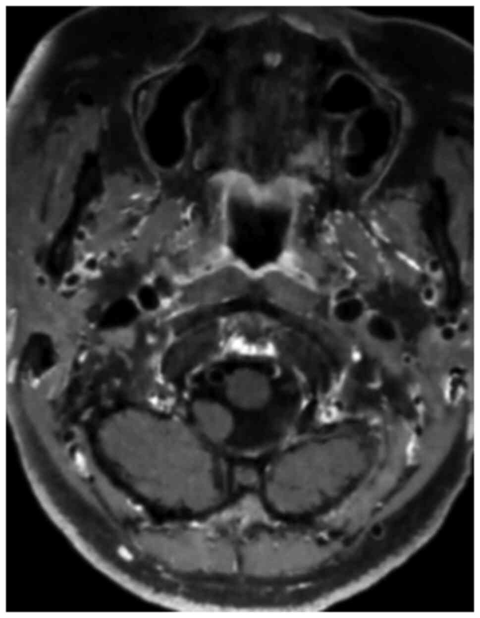
and slight effusion in the right kidney. Cerebral vascular wall imaging demonstrated the bilateral internal carotid artery with a double lumen in the C1 segment (Fig. 1) and a left internal carotid artery C6 segment aneurysm surgery was performed. A summary of the general information of the patient is presented in Table I.

Figure 1

Cerebral vascular wall imaging illustrating a bilateral internal carotid artery with a double lumen in the C1 segment.

Table I

General information of the patient.

Table I

General information of the patient.

Characteristic Features/information
SexFemale
Age57 years old
Physical examinationNo positive signs
Laboratory testsCreatinine level, 79 µmol/l
 Alanine aminotransferase level, 41 U/l
ImagingCerebral vascular wall imaging revealed a bilateral internal carotid artery with a double lumen in the C1 segment
Medical history 
     Chief complaintRecurrent headaches for >1 month
     Previous historyShe underwent right internal carotid artery stenting and left internal carotid artery aneurysm embolization 4 years prior

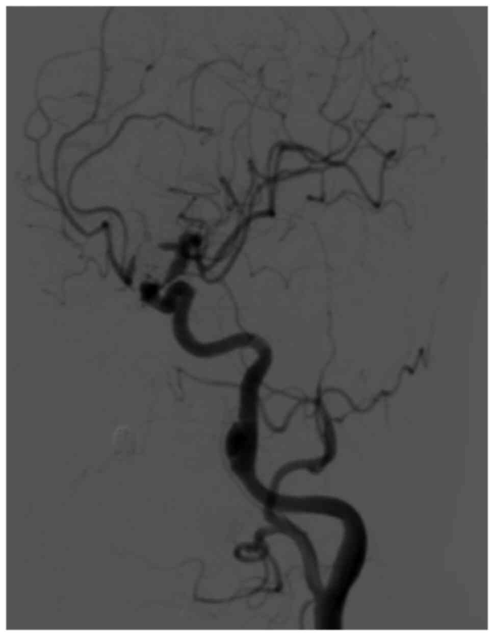
Nonetheless, bilateral internal carotid artery dissection was not excluded. A cerebral angiography was performed to determine whether there was dissection and whether further interventional treatment was required; the patient then underwent a cerebral angiography with 130 ml iohexol (the iodine concentration of the iohexol was 300 mg/ml) under local anesthesia, which revealed a dissecting aneurysm in the C1 segment of the left internal carotid artery (Fig. 2). 
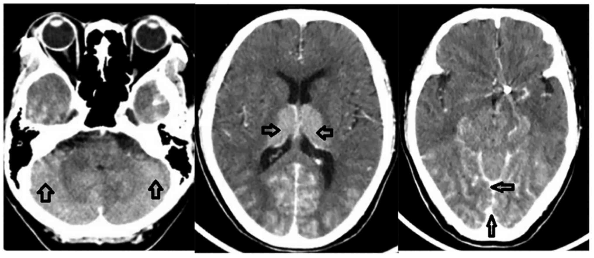
The surgery was successfully completed; however, the patient then suffered from headaches, vomiting and visual impairment, with a slight decrease in cognitive function 5 min later. A physical examination revealed that the visual acuity of both eyes had notably decreased, with only light sensation. The emergency craniocerebral CT scan then revealed an increased density in the bilateral cerebellum, thalamus, sulcus and cistern (Fig. 3). The probability of CIE was thus 
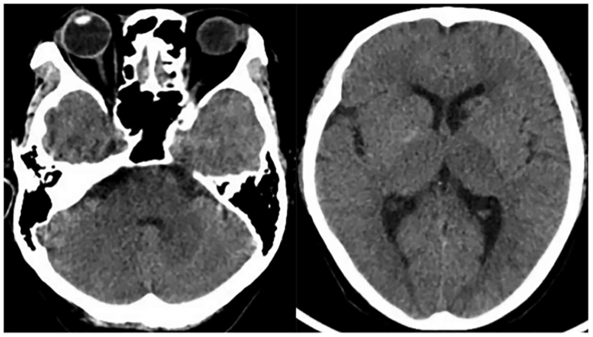
considered, due to the symptoms, signs and CT examination. Thus, the patient was administered anti-emetic (metoclopramide, 10 mg), anti-inflammatory (dexamethasone, 5 mg), vasospasm relief (nimodipine, 10 mg) and intracranial pressure reduction (mannitol, 25 g) treatment. The head CT scan then revealed the resolution of high-density in the aforementioned areas (Fig. 4) at 24 h after the surgery. On the third day after surgery, the visual acuity of the patient had gradually recovered; however, the development of cognitive function was not evident, which was manifested by occasional incomplete answers. The Mini-Mental State Examination score (2) was 16 points, and combined with the condition of illiteracy, the patient could be diagnosed with mild 
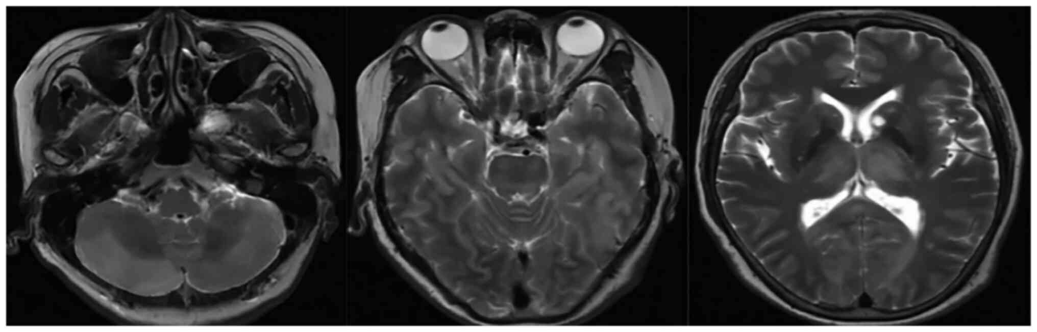
dementia. The MRI examination of the brain revealed multiple abnormal signal shadows in the bilateral thalamus, occipito-temporal lobe, corpus callosum, brainstem and bilateral cerebellar hemispheres (Fig. 5). Therefore, the administration of an intra-arterial injection of metoclopramide (10 mg) for anti-emetic treatment, dexamethasone (5 mg) for reducing the inflammatory response, nimodipine (10 mg) pump for vasospasm relief, mannitol (25 g) for intracranial pressure reduction and other treatments was continued. At 17 days after the surgery, the cognitive function of the patient gradually recovered and she was discharged from the hospital.

Figure 2

A dissecting aneurysm in the C1 segment of the left internal carotid artery.

Figure 3

Each of the three images from left to right indicate that the density of the bilateral cerebellum, thalamus and sulcus, cistern increased. Arrows indicate the dense areas.

Figure 4

Each of the two images from left to right indicate that the high-density shadow in the bilateral cerebellum, thalamus was attenuated 24 h after the surgery.

Figure 5

Each of the three images from left to right indicate multiple abnormal signal shadows in the bilateral cerebellar hemispheres, bilateral occipito-temporal lobe and bilateral thalamus.

Discussion

Contrast encephalopathy is an unusual neurological complication and its pathogenesis remains unclear. According to previous studies (3-5) the widely accepted theoretical mechanism is set around the destruction of the blood-brain barrier and the chemical properties of the contrast agents which may be as follows: i) the contrast medium disrupts the blood-brain barrier temporarily and enters the cerebrospinal fluid, increasing the osmotic pressure of the cerebrospinal fluid, and leading to impaired brain function. The majority of contrast agents have an osmolality range of 1.2-1.8 mOsm/l, compared with 0.3 mOsm/l for normal blood, which may open the normal endothelial cell tight junctions of the blood-brain barrier (6,7). ii) Contrast agents exert a direct toxic effect on nerve cells and affect their function. iii) Iodine contrast agents affect the secretion of functional substances regulating vasomotor and causes vasospasm. In this case, the cognitive decline of the patient is considered to be related to the neurological deficits caused by the edema of nerve cells. Moreover, the risk factors of CIE are also not clear. Some studies have suggested that the most common factors related to the occurrence of CIE include the male sex, an advanced age, hypertension, diabetes, renal function impairment, the contrast agent dose, etc. (8,9). CIE can occur at any age; however, patients who are older are more likely to suffer from this condition, although the incidence for each age group has not yet been determined. At the same time, the male sex and hypertension may be the main risk factors for CIE.

CIE may occur within minutes or hours of the application of the contrast agent. Generally, normal conditions will resume within 1-3 days; however, a small portion of patients do not return to a normal state until several weeks after the application of the contrast agent (8). There is as yet no clear explanation available for this phenomenon. In addition, very few fatal CIE cases have been reported (10). The clinical manifestations of CIE are diverse, such as intracranial hemorrhage, meningitis, cortical blindness, epileptic seizure, etc. (11). The typical imaging manifestations of CIE are brain parenchyma, subarachnoid contrast enhancement and brain edema (11); the majority of patients gradually return to a normal state. In the case presented herein, bilateral cortical blindness and cognitive decline occurred within a few minutes following digital subtraction angiography, and cortical blindness gradually recovered within 3 days after the surgery; however, the recovery of cognitive function was not evident. The occurrence of cortical blindness has been demonstrated in previous reports of CIE cases (8), although the occurrence of cognitive decline is very rare. In the present study, combined with the MRI results of the patient, it was considered that this cognitive decline was caused by neurological dysfunction. The head CT examination revealed that the density of the brain parenchyma, sulcus and cistern had increased; however, the head CT re-examination 24 h after the surgery indicated that the high-density shadow in the aforementioned areas was attenuated or had disappeared, which was in line with the typical imaging manifestations of CIE.

Although the majority of patients with CIE have a favorable outcome, often with a complete resolution of symptoms, once a diagnosis of CIE is made, aggressive treatment should be administered immediately to prevent irreversible events. Current treatment options include the promotion of contrast agent excretion, the reduction of cerebral edema, and anti-inflammatory and symptomatic supportive therapy (8). In the case described herein, the clinical and imaging manifestations of the patient gradually improved following the aforementioned combination of supportive treatments.

In conclusion, CIE is a rare neurological complication caused by contrast agents and generally presents as a transient, reversible neurologic disorder. The earlier diagnosis and treatment are crucial for the better prognosis of patients.

Acknowledgements

Not applicable.

Funding

Funding: No funding was received.

Availability of data and materials

The datasets used and/or analyzed during the current study are available from the corresponding author on reasonable request.

Authors' contributions

YL designed the study, and wrote, edited and reviewed the manuscript, and processed the figures. XL designed the study. HL identified the disease and provided guidance. FW provided the imaging data. ZZ collected the surgical data. SL managed the patient and provided the clinical data. All authors have read and approved the final manuscript. XL and FW confirm the authenticity of all the raw data.

Ethics approval and consent to participate

The institutional Review Board of Guangdong Second Provincial General Hospital approved the study.

Patient consent for publication

Not applicable.

Competing interests

The authors declare that they have no competing interests.

References

1 

Potsi S, Chourmouzi D, Moumtzouoglou A, Nikiforaki A, Gkouvas K and Drevelegas A: Transient contrast encephalopathy after carotid angiography mimicking diffuse subarachnoid haemorrhage. Neurol Sci. 33:445–448. 2012.PubMed/NCBI View Article : Google Scholar

2 

Folstein MF, Folstein SE and McHugh PR: ‘Mini-mental state’. A practical method for grading the cognitive state of patients for the clinician. J Psychiatr Res. 12:189–198. 1975.PubMed/NCBI View Article : Google Scholar

3 

Yu J and Dangas G: Commentary: New insights into the risk factors of contrast-induced encephalopathy. J Endovasc Ther. 18:545–546. 2011.PubMed/NCBI View Article : Google Scholar

4 

Junck L and Marshall WH: Neurotoxicity of radiological contrast agents. Ann Neurol. 13:469–484. 1983.PubMed/NCBI View Article : Google Scholar

5 

Rapoport S, Bookstein JJ, Higgins CB, Carey PH, Sovak M and Lasser EC: Experience with metrizamide in patients with previous severe anaphylactoid reactions to ionic contrast agents. Radiology. 143:321–325. 1982.PubMed/NCBI View Article : Google Scholar

6 

Rapoport SI and Levitan H: Neurotoxicity of X-ray contrast media. Relation to lipid solubility and blood-brain barrier permeability. Am J Roentgenol Radium Ther Nucl Med. 122:186–193. 1974.PubMed/NCBI View Article : Google Scholar

7 

Grainger R: Osmolality of intravascular radiological contrast media. Br J Radiol. 53:739–746. 1980.PubMed/NCBI View Article : Google Scholar

8 

Liu MR, Jiang H, Li XL and Yang P: Case report and literature review on low-osmolar, non-ionic iodine-based contrast-induced encephalopathy. Clin Interv Aging. 15:2277–2289. 2020.PubMed/NCBI View Article : Google Scholar

9 

Chu YT, Lee KP, Chen CH, Sung PS, Lin YH, Lee CW, Tsai LK, Tang SC and Jeng JS: Contrast-induced encephalopathy after endovascular thrombectomy for acute ischemic stroke. Stroke. 51:3756–3759. 2020.PubMed/NCBI View Article : Google Scholar

10 

Zhao W, Zhang J, Song Y, Sun L, Zheng M, Yin H, Zhang J, Wang W and Han J: Irreversible fatal contrast-induced encephalopathy: A case report. BMC Neurol. 19(46)2019.PubMed/NCBI View Article : Google Scholar

11 

Leong S and Fanning NF: Persistent neurological deficit from iodinated contrast encephalopathy following intracranial aneurysm coiling. A case report and review of the literature. Interv Neuroradiol. 18:33–41. 2012.PubMed/NCBI View Article : Google Scholar

Related Articles

  • Abstract
  • View
  • Download
Copy and paste a formatted citation
Spandidos Publications style
Liang Y, Liu X, Lu H, Wei F, Zhang Z and Lin S: Contrast‑induced encephalopathy following cerebral angiography: A case report. Med Int 2: 16, 2022.
APA
Liang, Y., Liu, X., Lu, H., Wei, F., Zhang, Z., & Lin, S. (2022). Contrast‑induced encephalopathy following cerebral angiography: A case report. Medicine International, 2, 16. https://doi.org/10.3892/mi.2022.41
MLA
Liang, Y., Liu, X., Lu, H., Wei, F., Zhang, Z., Lin, S."Contrast‑induced encephalopathy following cerebral angiography: A case report". Medicine International 2.3 (2022): 16.
Chicago
Liang, Y., Liu, X., Lu, H., Wei, F., Zhang, Z., Lin, S."Contrast‑induced encephalopathy following cerebral angiography: A case report". Medicine International 2, no. 3 (2022): 16. https://doi.org/10.3892/mi.2022.41
Copy and paste a formatted citation
x
Spandidos Publications style
Liang Y, Liu X, Lu H, Wei F, Zhang Z and Lin S: Contrast‑induced encephalopathy following cerebral angiography: A case report. Med Int 2: 16, 2022.
APA
Liang, Y., Liu, X., Lu, H., Wei, F., Zhang, Z., & Lin, S. (2022). Contrast‑induced encephalopathy following cerebral angiography: A case report. Medicine International, 2, 16. https://doi.org/10.3892/mi.2022.41
MLA
Liang, Y., Liu, X., Lu, H., Wei, F., Zhang, Z., Lin, S."Contrast‑induced encephalopathy following cerebral angiography: A case report". Medicine International 2.3 (2022): 16.
Chicago
Liang, Y., Liu, X., Lu, H., Wei, F., Zhang, Z., Lin, S."Contrast‑induced encephalopathy following cerebral angiography: A case report". Medicine International 2, no. 3 (2022): 16. https://doi.org/10.3892/mi.2022.41
Follow us
  • Twitter
  • LinkedIn
  • Facebook
About
  • Spandidos Publications
  • Careers
  • Cookie Policy
  • Privacy Policy
How can we help?
  • Help
  • Live Chat
  • Contact
  • Email to our Support Team