Introduction
Ovarian cancer is the fourth leading cause of cancer
mortalities worldwide (1).
Cisplatin and its analogs are first-line chemotherapeutic agents
for the treatment of human ovarian cancer (2,3).
Although the mechanisms involved in cisplatin resistance have yet
to be comprehensively explored (4), decreased cell detoxication and
defects in intracellular or extracellular survival/apoptotic
pathways have been suggested to play a role in the development of
resistance to cisplatin (5).
Survivin (SVV) is a unique member of the inhibitor
of apoptosis (IAP) protein family. Abnormal IAP during homeostasis
is considered a critical step in the initiation of cancer. SVV is
an anticancer target due to its high expression in numerous types
of cancer. SVV is expressed in ovarian cancer and correlates with
its clinicopathological, surgical and apoptosis-related parameters
(6). SVV mRNA expression levels
correlate with the clinical stage, differentiation grade and lymph
node metastasis, but not with histological type in ovarian cancer
(7). Serum SVV reflects the
peritoneal metastasis of serous ovarian cancer and may be useful as
a prognostic biomarker (8).
Nuclear SVV expression is a strong independent prognostic marker
for poor clinical outcomes in epithelial ovarian carcinoma
(9), however, a previous study
demonstrated that nuclear SVV expression predicts improved outcome
in pre-chemotherapy patients with metastatic ovarian cancer
(10). In addition, among ovarian
cancer patients receiving a taxol/platinum-based regimen, SVV
expression correlates with a lower clinical or pathological
complete remission rate, indicative of a direct link between SVV
expression and tumor cell susceptibility to taxol (11). Nuclear SVV expression is a positive
prognostic factor in taxane-platinum-treated ovarian cancer
patients (12).
Notably, downregulation of SVV appears to be an
effective gene therapy approach in the treatment of ovarian cancer
(13). SVV and Granzyme B-induced
apoptosis may function as potent agents for the treatment of
primary and recurrent human ovarian carcinoma (14). SVV is important in resistance to
chemotherapy and radiotherapy in ovarian cancer and in the
progression of ovarian tumors and provides a pivotal prognostic
implication for epithelial ovarian carcinomas (15). To date, evidence has confirmed the
role and possible mechanisms of SVV in cisplatin-resistant ovarian
cancer cells (15). In the present
study, using a loss-of-function approach, we investigated the
effects of adenovirus-mediated knockdown of SVV on the expression
of pro-caspase-3, cleaved caspase-3, PCNA and MMP-2 in A2780/CP
cells by real-time PCR and western blot analysis. Proliferation was
measured by MTT assay, invasive potential by Transwell, and cell
apoptosis by FITC-Annexin V and propidium iodide (PI) were
performed for the functional analysis of A2780/CP cells following
infection with ad5-SVV.
Materials and methods
Materials
The A2780/CP cell line was obtained from the
Institute of Biochemistry and Cell Biology (Shanghai, China).
Adenovirus-mediated SVV shRNA vector, negative control vector and
virion-packaging elements were purchased from Genechem (Shanghai,
China). Primers for SVV, PCNA, cleaved caspase-3 and MMP-2 were
synthesized by Advanced Biotechnologies, Inc. (Columbia, MD, USA).
Antibodies were from Santa Cruz Biotechnology, Inc. (Santa Cruz,
CA, USA).
Drugs and reagents
Cisplatin was purchased from Sigma-Aldrich (St.
Louis, MO, USA). Dulbecco’s Modified Eagle’s medium (DMEM) and
fetal bovine serum (FBS) were purchased from Thermo Fisher
Scientific (Waltham, MA, USA); TRIzol reagent and Lipofectamine
2000 were from Invitrogen (Carlsbad, CA, USA); M-MLV Reverse
Transcriptase was from Promega Corp. (Madison, WI, USA); SYBR-Green
Master Mixture was from Takara Bio, Inc. (Shiga, Japan). A cell
apoptosis kit, PI, RNase A and Annexin V-FITC were obtained from
KeyGEN Biology (Nanjing, China).
Cell culture and transfection
A2780/CP cells were cultured in DMEM supplemented
with 10% heat-inactivated FBS, 100 U/ml penicillin and 100 μg/ml
streptomycin. Cells were placed in a humidified atmosphere
containing 5% CO2 at 37°C. Ad5-SVV and negative control
adenovirus were used to transfect A2780/CP cells. Cells were
subcultured at a 1:5 dilution in 300 μg/ml G418-containing medium.
Positive stable transfectants were selected and expanded for
further analysis. The clone in which Ad5-SVV vectors were
transfected was designated as Ad5-SVV, the negative control vectors
transfected were designated as NC, and A2780/CP cells were
designated as CON.
Quantitative real-time PCR
To quantitatively determine the mRNA expression
level of SVV in A2780/CP cell lines, real-time PCR was performed.
Total RNA from each clone was extracted with TRIzol according to
the manufacturer’s instructions. Reverse transcription was
performed using M-MLV and cDNA amplification was carried out using
a SYBR Green Master Mix kit according to the manufacturer’s
instructions. The SVV gene was amplified using specific
oligonucleotide primers and the human GAPDH gene was used as an
endogenous control. PCR primer sequences were as follows: SVV,
5′-ACCAGGTGAGAAGTGAGGGA-3′ and 5′-AACAGTAGAGGAGCCAGGGA-3′; PCNA,
5′-CCATCC TCAAGAAGGTGTTGG-3′ and 5′-GTGTCCCATATCCGC AATTTTAT-3′;
cleaved caspase-3, 5′-AGAGGGGATCGTTG TAGAAG-3′ and
5′-GTTGCCACCTTTCGGTTAAC-3′; MMP-2, 5′-GGCCCTGTCACTCCTGAGAT-3′ and
5′-GGC ATCCAGGTTATCGGGGA-3′; GAPDH, 5′-CAACGAATT TGGCTACAGCA-3′ and
5′-AGGGGTCTACATGGCAA CTG-3′. Data were analyzed using the
comparative Ct method (2−ΔΔCt). Three separate
experiments were performed for each clone.
Western blot analysis
A2780/CP cells were harvested and extracted using
lysis buffer (Tris-HCl, SDS, mercaptoethanol, glycerol). Cell
extracts were boiled for 5 min in loading buffer and then equal
amounts of cell extracts were separated by 15% SDS-PAGE. Separated
protein bands were transferred onto polyvinylidene fluoride
membranes and the membranes were blocked in 5% skimmed milk powder.
Primary antibodies against SVV, PCNA, pro-caspase-3, cleaved
caspase-3 and MMP-2 were diluted according to the manufacturer’s
instructions and applied to membranes overnight at 4°C. Following
this, horseradish peroxidase-linked secondary antibodies were added
at a dilution ratio of 1:1,000 and incubated at room temperature
for 2 h. Membranes were washed with PBS three times and the
immunoreactive bands were visualized using ECL-PLUS kit according
to the manufacturer’s instructions. Relative protein levels in
various cell lines were normalized to GAPDH concentration. Three
separate experiments were performed for each clone.
Cell proliferation assay
Cell proliferation was analyzed with the MTT assay.
Briefly, cells infected with ad5-SVV virus were incubated in
96-well plates at a density of 1×10⁵ cells/well with
DMEM supplemented with 10% FBS. Cells were treated with 20 μl MTT
dye at 0, 24, 48, 72, 96 and 120 h and then incubated with 150 μl
DMSO for 5 min. The color reaction was measured at 570 nm with an
enzyme immunoassay analyzer (Bio-Rad, Hercules, CA, USA).
Proliferation activity was calculated for each clone.
Transwell invasion assay
Transwell filters were coated with matrigel (3.9
μg/μl, 60–80 μl) on the upper surface of a polycarbonic membrane
(diameter, 6.5 mm; pore size, 8 μm). Following incubation at 37°C
for 30 min, the matrigel solidified and served as the extracellular
matrix for analysis of tumor cell invasion. Harvested cells
(1×10⁵) in 100 μl DMEM were added into the upper
compartment of the chamber. A total of 200 μl conditioned medium
derived from NIH3T3 cells was used as a source of chemoattractant
and was placed in the bottom compartment of the chamber. Following
24 h incubation at 37°C with 5% CO2, the medium was
removed from the upper chamber. The non-invaded cells on the upper
side of the chamber were scraped off with a cotton swab. Cells that
had migrated from the matrigel into the pores of the inserted
filter were fixed with 100% methanol, stained with hematoxylin and
mounted and dried at 80°C for 30 min. The number of cells invading
through the matrigel was counted in three randomly selected visual
fields from the central and peripheral portion of the filter using
an inverted microscope (x200 magnification). Each assay was
repeated three times.
Cell apoptosis analysis
To detect cell apoptosis, A2780/CP cells treated
with ad5-SVV virus were trypsinized, washed with cold PBS and
resuspended in binding buffer according to the manufacturer’s
instructions. FITC-Annexin V and PI were added to the fixed cells
for 20 min in the dark at room temperature. Following this, Annexin
V binding buffer was added to the mixture prior to fluorescence
measurement by FACsort flow cytometer. Cell apoptosis was analyzed
using CellQuest software (BD Biosciences, CA, USA). Three separate
experiments were performed for each clone.
Statistical analysis
Results of each experiment were presented as the
mean ± SD, where applicable. Statistically significant difference
in each assay was determined by SPSS version 11.5. Difference in
each group was tested for significance using the ANOVA analysis of
variance. P<0.05 was considered to indicate a statistically
significant difference.
Results
Effect of SVV knockdown on PCNA, cleaved
caspase-3 and MMP-2 expression
To examine whether SVV was knocked down by ad5-SVV
and its effect on PCNA, cleaved caspase-3 and MMP-2 mRNA expression
in A2780/CP cells, the expression levels of SVV, PCNA, cleaved
caspase-3 and MMP-2 mRNA were measured by real-time PCR. As is
evident in Fig. 1A, expression
levels of SVV, PCNA and MMP-2 mRNA were decreased, while the
expression level of cleaved caspase-3 was increased in ad5-SVV
compared with the NC and CON groups (each P<0.01). Western blot
analysis of SVV protein expression, revealed a marked
downregulation in the ad5-SVV group compared with the NC and CON
groups (P<0.01; Fig. 1B and
C).
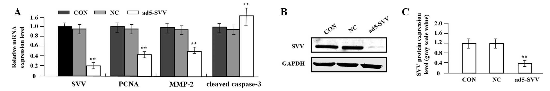 | Figure 1Effect of SVV knockdown on PCNA,
cleaved caspase-3 and MMP-2 expression. (A) Expression levels of
SVV, PCNA, cleaved caspase-3 and MMP-2 mRNA were measured in
A2780/CP cells by real-time PCR, demonstrating that expression of
SVV, PCNA and MMP-2 mRNA was decreased, while that of cleaved
caspase-3 was increased in ad5-SVV compared with NC and CON groups
(each **P<0.01). (B and C) Western blot analysis
demonstrates that the expression level of SVV protein was markedly
downregulated in ad5-SVV compared with that in the NC and CON
groups in A2780/CP cells (**P<0.01). CON, A2780/CP
cells; NC, transfected negative control vectors; Ad5-SVV,
transfected Ad5-SVV vectors. SVV, survivin; PCNA, proliferating
cell nuclear antigen; MMP-2 matrix metalloproteinase-2. |
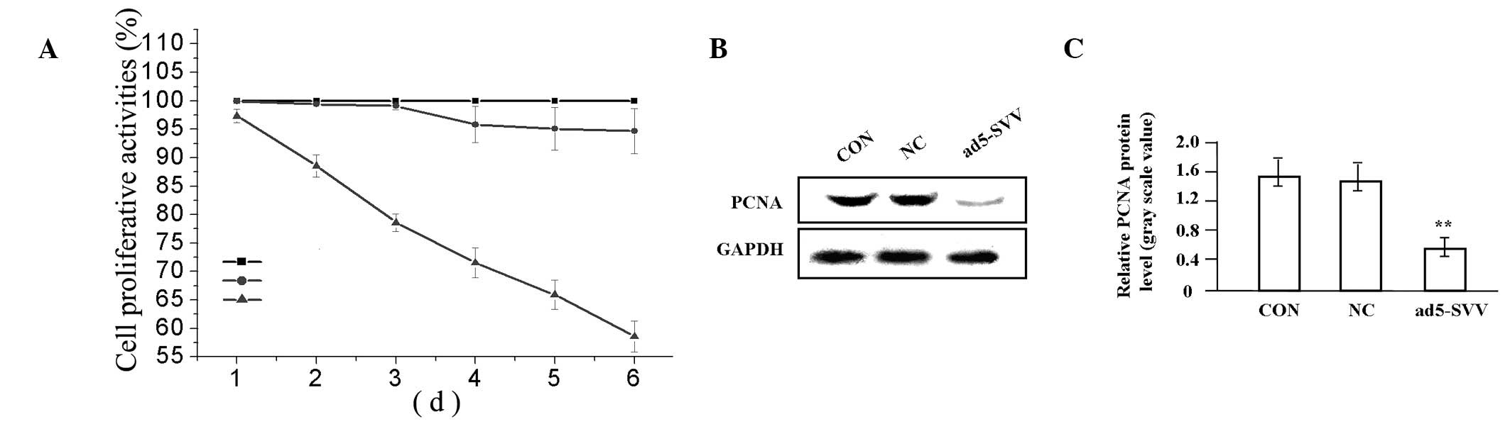
Effect of SVV knockdown on A2780/CP cell
proliferation and cisplatin sensitivity
The effect of SVV knockdown on proliferation and
cisplatin sensitivity to A2780/CP cells by MTT was investigated.
The results demonstrate that SVV knockdown was identified to
significantly inhibit the proliferation of A2780/CP cells in a
time-dependent manner, compared with NC and CON groups (Fig. 2A). PCNA is important for a number
of essential cell processes, tumor progression and the outcome of
anticancer treatment. To determine whether SVV knockdown suppressed
endogenous PCNA expression through translational repression, the
expression of PCNA protein was examined by western blot analysis,
which revealed a decreased amount of PCNA protein in the ad5-SVV
compared with the NC and CON groups (P<0.01; Fig. 2B and C).
Following this, we pretreated A2780/CP cells in
ad5-SVV, NC and CON groups with various concentrations of cisplatin
(0.01, 0.1, 1, 10 and 100 μg/ml) and investigated the effect of SVV
knockdown on cisplatin sensitivity to A2780/CP cells. Knockdown of
SVV markedly enhanced the inhibitory effect and chemotherapeutic
sensitivity of cisplatin on A2780/CP cells (each P<0.01)
(Table I).
 | Table IEffect of SVV knockdown on
chemotherapeutic sensitivity of cisplatin to A2780/CP cells
indicated by MTT assay (%) |
Table I
Effect of SVV knockdown on
chemotherapeutic sensitivity of cisplatin to A2780/CP cells
indicated by MTT assay (%)
| Concentration of
cisplatin (μg/ml) |
|---|
|
|
|---|
| Group | 0.01 | 0.1 | 1 | 10 | 100 |
|---|
| CON | 100±0.00 | 99.56±0.88 | 88.63±0.75 | 46.72±0.43 | 39.87±0.56 |
| NC | 98.22±0.15a | 97.83±0.43a | 86.76±0.42a | 45.55±0.56a | 38.34±0.25a |
| ad5-SVV | 78.56±0.47b | 71.28±0.52b | 40.05±0.36b | 32.63±0.37b | 24.57±0.27b |
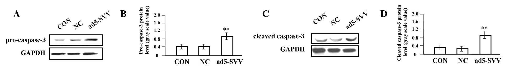
Effect of SVV knockdown on A2780/CP cell
apoptosis
To determine the effect of SVV knockdown on A2780/CP
cell apoptosis, flow cytometric analysis with FITC/PI-Annexin V
staining was performed. As demonstrated in Fig. 3A, compared with NC and CON, the
number of apoptotic cells significantly increased and morphological
changes, including nuclear condensation, nuclear debris, foam and
apoptotic body formation, were identified in the ad5-SVV group. In
addition, the apoptotic index of A2780/CP cells in the ad5-SVV
group was markedly higher than the NC and CON groups (P<0.01;
Fig. 3B and C). Western blot
analysis was performed to investigate the effect of SVV knockdown
on the endogenous expression of pro-caspase-3 and cleaved caspase-3
protein. As shown in Fig. 4,
pro-caspase-3 and cleaved caspase-3 expression was identified as
significantly elevated in the ad5-SVV group, compared with the NC
and CON groups (each P<0.01).
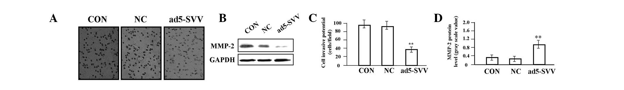
Effect of SVV knockdown on A2780/CP cell
invasion
To determine the effect of SVV knockdown on A2780/CP
cell invasion, a Transwell assay was performed. Invasive and
metastatic potential in the Transwell assay was determined on the
basis of the ability of cells to invade a matrix barrier containing
laminin and type IV collagen, the major components of the basement
membrane. Representative micrographs of Transwell filters are shown
in Fig. 5A. The invasive potential
of A2780/CP cells was markedly decreased in the ad5-SVV group,
compared with the NC and CON groups (P<0.01; Fig. 5C). Western blot analysis was
performed to investigate the effect of SVV knockdown on the
endogenous expression of MMP-2 protein. As demonstrated in Fig. 5B and D, MMP-2 expression was
identified as significantly reduced in the ad5-SVV group, compared
with the NC and CON groups (P<0.01).
Discussion
The present study demonstrates that shRNA-based
knockdown of SVV inhibits proliferation and invasion and induces
apoptosis in cisplatin-resistant ovarian cancer cells (A2780/CP).
Several studies have demonstrated that the SVV pathway, regulated
by other factors, is involved in cell apoptosis in ovarian cancer.
Luteinizing hormone (16), FSH
(17) and ovarian-specific
promoter-2 (18) affect the
sensitivity of ovarian cancer cells to chemotherapy and inhibit
apoptosis via upregulation of the SVV pathway. Activation of the
SVV pathway by interleukin-4 leads to cancer proliferation
(19). shRNA targeting against SVV
has potential for the treatment of ovarian cancer (20). YM155, a novel small-molecule SVV
inhibitor, has demonstrated antitumor activities in human cancer
cells (21).
Previous studies have indicated that SVV antisense
oligonucleotides induce apoptosis in the drug-resistant ovarian
cancer cell line (COC1/DDP) (22).
AKT/mTOR/SVV signaling is involved in epithelial ovarian cancer
development and cisplatin/paclitaxel-resistant chemotherapy and the
downregulation of AKT/SVV may be an effective antitumor therapy
(23–25). The present findings have
demonstrated that knockdown of SVV enhanced the inhibitory effect
and the chemotherapeutic sensitivity of cisplatin to A2780/CP cells
and are in agreement with previous reports (22,23),
suggesting that SVV is a potential target for therapeutic
anticancer drugs. Moreover, accumulating evidence indicates that
combinations of targeting SVV with additional compounds, including
herbal dietary antioxidants (26),
methylseleninic acid (27) and
paclitaxel (28), is more
effective in the chemoprevention and/or chemotherapy of ovarian and
other types of cancer.
PCNA is a nuclear protein expressed in proliferating
cells and may be required for maintaining cell proliferation.
Protein levels are used as a marker for the cell proliferation of
overall survival (29). MMP-2 is a
key enzyme involved in the degradation of type IV collagen and high
levels of MMP-2 in tissues is correlated with tumor growth and
invasion (30). Caspase-3 is
important in the proteolytic cleavage of cell proteins responsible
for progression to apoptosis and has the potential to predict the
clinical response to chemotherapy (31). The present study has demonstrated a
marked decrease in PCNA and MMP-2 expression, but an increase in
pro-caspase-3 and cleaved caspase-3 in group ad5-SVV compared with
the NC and CON groups in A2780/CP cells. These observations
indicate that the knockdown of SVV may inhibit proliferation and
invasion and induce apoptosis in cisplatin-resistant ovarian cancer
cells via the downregulation of PCNA and MMP-2 expression and the
upregulation of caspase-3 expression.
To the best of our knowledge, the present study has
demonstrated that knockdown of SVV enhances cisplatin-induced
proliferative activities, induces cell apoptosis and inhibits the
invasive potential in A2780/CP cells. Knockdown of SVV contributes
to antitumor activity in cisplatin-resistant ovarian cancer cells
via the downregulation of PCNA and MMP-2 and upregulation of
caspase-3 expression, rendering SVV a potential target for
therapeutic anticancer drugs.
References
|
1
|
Eltabbakh G and Awtrey C: Current
treatment for ovarian cancer. Expert Opin Pharmacother. 2:109–124.
2001. View Article : Google Scholar
|
|
2
|
McKeage M: New-generation platinum drugs
in the treatment of cisplatin-resistant cancers. Expert Opin
Investig Drugs. 14:1033–1046. 2005. View Article : Google Scholar : PubMed/NCBI
|
|
3
|
Tewari K, Mehta R, Burger R, et al:
Emerging drugs for ovarian cancer. Expert Opin Emerg Drugs.
10:413–442. 2005. View Article : Google Scholar
|
|
4
|
Chen H, Hardy TM and Tollefsbol TO:
Epigenomics of ovarian cancer and its chemoprevention. Front Genet.
2:672011. View Article : Google Scholar : PubMed/NCBI
|
|
5
|
Lee S, Choi E, Jin C, et al: Activation of
PI3K/Akt pathway by PTEN reduction and PIK3CA mRNA amplification
contributes to cisplatin resistance in an ovarian cancer cell line.
Gynecol Oncol. 97:26–34. 2005. View Article : Google Scholar : PubMed/NCBI
|
|
6
|
Ferrandina G, Legge F, Martinelli E, et
al: Survivin expression in ovarian cancer and its correlation with
clinico-pathological, surgical and apoptosis-related parameters. Br
J Cancer. 92:271–277. 2005.
|
|
7
|
Liguang Z, Peishu L, Hongluan M, et al:
Survivin expression in ovarian cancer. Exp Oncol. 29:121–125.
2007.PubMed/NCBI
|
|
8
|
No JH, Jeon YT, Kim YB, et al:
Quantitative detection of serum survivin and its relationship with
prognostic factors in ovarian cancer. Gynecol Obstet Invest.
71:136–140. 2011. View Article : Google Scholar : PubMed/NCBI
|
|
9
|
Qian X, Xi X and Li L: Nuclear survivin is
associated with malignant potential in epithelial ovarian
carcinoma. Appl Immunohistochem Mol Morphol. 19:126–132. 2011.
View Article : Google Scholar : PubMed/NCBI
|
|
10
|
Kleinberg L, Flørenes VA, Silins I, et al:
Nuclear expression of survivin is associated with improved survival
in metastatic ovarian carcinoma. Cancer. 109:228–238. 2007.
View Article : Google Scholar : PubMed/NCBI
|
|
11
|
Zaffaroni N, Pennati M, Colella G, et al:
Expression of the anti-apoptotic gene survivin correlates with
taxol resistance in human ovarian cancer. Cell Mol Life Sci.
59:1406–1412. 2002. View Article : Google Scholar : PubMed/NCBI
|
|
12
|
Felisiak-Golabek A, Rembiszewska A,
Rzepecka IK, et al: Nuclear survivin expression is a positive
prognostic factor in taxane-platinum-treated ovarian cancer
patients. J Ovarian Res. 4:202011. View Article : Google Scholar
|
|
13
|
Ma X, Wang S, Zhou J, et al: Induction of
apoptosis in human ovarian epithelial cancer cells by antisurvivin
oligonucleotides. Oncol Rep. 14:275–279. 2005.PubMed/NCBI
|
|
14
|
Caldas H, Jaynes FO, Boyer MW, et al:
Survivin and Granzyme B-induced apoptosis, a novel anticancer
therapy. Mol Cancer Ther. 5:693–703. 2006. View Article : Google Scholar : PubMed/NCBI
|
|
15
|
Zhang B, Pan JS, Liu JY, et al: Effects of
chemotherapy and/or radiotherapy on survivin expression in ovarian
cancer. Methods Find Exp Clin Pharmacol. 28:619–625. 2006.
View Article : Google Scholar : PubMed/NCBI
|
|
16
|
Zhang Z, Liao H, Chen X, et al:
Luteinizing hormone upregulates survivin and inhibits apoptosis in
ovarian epithelial tumors. Eur J Obstet Gynecol Reprod Biol.
155:69–74. 2011. View Article : Google Scholar : PubMed/NCBI
|
|
17
|
Huang Y, Jin H, Liu Y, et al: FSH inhibits
ovarian cancer cell apoptosis by up-regulating survivin and
down-regulating PDCD6 and DR5. Endocr Relat Cancer. 18:13–26. 2010.
View Article : Google Scholar : PubMed/NCBI
|
|
18
|
Tu CH, Liu WP, Dong M, et al: Protection
of CHO cells by transfer of survivin driven by ovarian-specific
promoter OSP-2. Mol Biol Rep. 38:2323–2328. 2011. View Article : Google Scholar : PubMed/NCBI
|
|
19
|
Roca H, Craig MJ, Ying C, et al: IL-4
induces proliferation in prostate cancer PC3 cells under
nutrient-depletion stress through the activation of the JNK-pathway
and survivin up-regulation. J Cell Biochem. 113:1569–1580.
2012.PubMed/NCBI
|
|
20
|
Xing J, Jia CR, Wang Y, et al: Effect of
shRNA targeting survivin on ovarian cancer. J Cancer Res Clin
Oncol. 138:1221–1229. 2012. View Article : Google Scholar : PubMed/NCBI
|
|
21
|
Nakahara T, Kita A, Yamanaka K, et al:
Broad spectrum and potent antitumor activities of YM155, a novel
small-molecule survivin suppressant, in a wide variety of human
cancer cell lines and xenograft models. Cancer Sci. 102:614–621.
2011. View Article : Google Scholar
|
|
22
|
Zheng F, Ruan F, Xie XK, et al: Apoptosis
of drug-resistant human ovarian carcinoma cell line COC1/DDP
induced by survivin antisense oligonucleotides. Chin Med J (Engl).
119:1572–1575. 2006.PubMed/NCBI
|
|
23
|
Zhang HY, Zhang PN and Sun H: Aberration
of the PI3K/AKT/mTOR signaling in epithelial ovarian cancer and its
implication in cisplatin-based chemotherapy. Eur J Obstet Gynecol
Reprod Biol. 146:81–86. 2009. View Article : Google Scholar : PubMed/NCBI
|
|
24
|
Weng D, Song X, Xing H, et al: Implication
of the Akt2/survivin pathway as a critical target in paclitaxel
treatment in human ovarian cancer cells. Cancer Lett. 273:257–265.
2009. View Article : Google Scholar : PubMed/NCBI
|
|
25
|
Xing H, Weng D, Chen G, et al: Activation
of fibronectin/PI-3K/Akt2 leads to chemoresistance to docetaxel by
regulating survivin protein expression in ovarian and breast cancer
cells. Cancer Lett. 261:108–119. 2008. View Article : Google Scholar : PubMed/NCBI
|
|
26
|
Raj MH, Abd Elmageed ZY, Zhou J, et al:
Synergistic action of dietary phyto-antioxidants on survival and
proliferation of ovarian cancer cells. Gynecol Oncol. 110:432–438.
2008. View Article : Google Scholar : PubMed/NCBI
|
|
27
|
Azrak RG, Frank CL, Ghadersohi A, et al:
Silencing survivin results in synergy between methylseleninic acid
and paclitaxel against skov3 ovarian cancer cells. Cancer Biol
Ther. 7:1901–1908. 2008. View Article : Google Scholar : PubMed/NCBI
|
|
28
|
Vivas-Mejia PE, Rodriguez-Aguayo C, Han
HD, et al: Silencing survivin splice variant 2B leads to antitumor
activity in taxane - resistant ovarian cancer. Clin Cancer Res.
17:3716–3726. 2011. View Article : Google Scholar : PubMed/NCBI
|
|
29
|
Reitmaier M, Rudlowski C, Biesterfeld S,
et al: Comparative studies on the biological significance of the
marker for proliferation Ki-67-antigen and PCNA in primary ovarian
carcinoma. Zentralbl Gynakol. 122:361–367. 2000.(In German).
|
|
30
|
Brinckerhoff CE, Rutter JL and Benbow U:
Interstitial collagenases as markers of tumor progression. Clin
Cancer Res. 6:4823–4830. 2000.PubMed/NCBI
|
|
31
|
Flick MB, O’Malley D, Rutherford T, et al:
Apoptosis-based evaluation of chemosensitivity in ovarian cancer
patients. J Soc Gynecol Investig. 11:252–259. 2004. View Article : Google Scholar : PubMed/NCBI
|