Introduction
Acute pancreatitis (AP) is an inflammatory disease
involving normal pancreatic gland that exhibits a variety of
clinical manifestations. Although mild AP has a low morbidity and
mortality rate, severe acute pancreatitis (SAP) is a necrotizing
disease involving remote organ systems. SAP occupies 20% of the
patients with AP, and 15–25% of SAP patients will eventually die
(1). SAP is initiated by the
premature activation of digestive enzymes (trypsin, elastase and
lipase) within the pancreatic acinar cells, leading to
self-digestion of pancreatic tissue (2). During the early phase of SAP,
inflammatory responses occur in the ductal cells and give rise to
systemic inflammatory response syndrome (SIRS), leading to
long-distance organ damage and multiple organ dysfunction syndrome
(MODS) (3). The most common and
serious SIRS is pancreatitis-associated lung injury, which is
characterized by increased endothelial and epithelial barrier
permeability, with exudate leaking into the alveolar space and
interstitial lung, thus leading to pulmonary edema and gas exchange
dysfunction (4). As shown in a
previous study, inflammatory mediators play an important role in
the pathogenesis of pancreatitis-associated acute lung injury
(ALI). These mediators include tumor necrosis factor-α (TNF-α),
interleukin (IL)-1β, -6 and -10, granulocyte macrophage-colony
stimulating factor (GM-CSF) and neuropeptide substance P (SP)
(5). All of these mediators
represent potential targets for therapeutic intervention.
Mesenchymal stem cells (MSCs) are multipotent adult
stem cells, mainly found in the bone marrow, that differentiate
into a variety of cell types, including chondrocytes, myoblasts,
endothelial, epithelial, nerve and alveolar epithelial cells. Bone
marrow-derived MSCs possess immunomodulatory properties and are
differentiated into alveolar endothelial cells to replace damaged
alveolar endothelial cells. Furthermore, an allograft of MSCs has
been shown to inhibit the inflammatory response and to decrease
collagen content in the injured lung tissues, thus diminishing the
incidence of pulmonary fibrosis (6). All these advantages are beneficial to
the cell replacement therapy for the treatment of diseases of the
respiratory system.
In this study, we explored the protective role of
MSCs in rats with SAP and the possible mechanisms involved. We also
investigated the pathology and function of lung tissue in rats with
SAP, as well as the regulation of inflammatory mediators and
possible mechanisms involved.
Materials and methods
Experimental animals
Sprague-Dawley (SD) rats, weighing 250–300 g [Batch
No. SCXK (Shanghai) 2007-0005], were provided by the Shanghai SLK
Experimental Animal Co. Two rats of 4 weeks of age, weighing 60–80
g were used for the isolation of bone marrow-derived MSCs. The
guidelines for animal ethics were followed in all the care and
handling of the animals.
Cultivation and identification of
MSCs
MSCs were isolated from bone marrow. The male SD
rats of 4 weeks of age were sacrificed by cervical dislocation, and
their bilateral lower limb was amputated under sterile and
anaesthetic conditions. The tibia and femur were exposed with the
muscles knocked out. The bone marrow cavity was washed with
DMEM-F12 nutrient solution until the whitening of the backbone. The
bone marrow cell suspension was acquired under sterile conditions
in mixed nutrient medium with 10% fetal calf serum and DMEM-F12,
and was inoculated with DMEM-F12 nutrient solution at
6×10⁵/cm2 density with the supernatant
discarded. The bone marrow cells were cultured by differential
adherence methods, and the situations of cell adherence were
observed with no exchange of medium within 48 h. When the cells
grew to 90% confluence at the bottom of the culture flask, they
were passaged at 1:2 or 1:3 according to the cell growth. At the
third generation, cells were harvested, digested with trypsin and
counted for reserve. The cellular identification of the expression
of MSC surface makers, including CD90, CD29, CD34 and CD45, was
performed by flow cytometry. The third generation MSCs were induced
to differentiate into adipogenic and osteoblast cells for
identification. These third generation MSCs were used for the
following transplant experiments.
Experimental model
SD rats, weighing 250–300 g, were selected for the
experiment. AP was induced by sodium taurocholate (7). Rats were anesthetized by an
intraperitoneal injection of 3% pentobarbital sodium at a dose of
30 mg/kg. The biliopancreatic duct was cannulated through the
duodenum, and the hepatic duct was closed with a small bulldog
clamp. The SAP model was established by a retrograde infusion of 5%
sodium taurocholate (Sigma, St. Louis, MO, USA) into the
biliopancreatic duct. In the sham-operated group (SO), SD rats
received a retrograde infusion of sterile saline.
Experimental design
Seventy-five male SD rats were randomly divided into
the SO group (n=25), SAP group (n=25) and MSC group (n=25). Rats
with AP were randomly divided into SAP group and MSC treatment
group, which, 2 h after the surgery, were injected with l ml medium
(SAP group) or l ml medium with cell suspension (1×10⁶
MSCs) (MSC group). Rats in the SO group were injected with medium
with an equivalent volume at the same time-point. Rats were
sacrificed 1, 3, 6, 12 and 24 h after the establishment of the
model, and then the serum amylase (AMS) levels, IL-1, IL-10, TNF-α,
macrophage inflammatory protein (MIP) and other inflammation
factors were detected. Pancreatic and lung tissues were
harvested.
Cell marking
Third generation MSCs were added with sterile DAPI
(final concentration 50 g/l) and incubated at 37°C for 12 h. After
repeated washing with PBS to remove DAPI, the cells were collected
after one more centrifugation. DMEM-F12 medium (1 ml) supplemented
with 10% fetal calf serum containing 1×10⁶ MSCs was
injected into the rats with SAP through the caudal vein at 2 h.
Quantitative reverse
transcriptase-polymerase chain reaction (RT-PCR)
The total RNA was extracted with the use of a TRIzol
kit (Invitrogen, Carlsbad, CA, USA) and converted to cDNA with a
first strand cDNA synthesis kit (Fermentas, Burlington, Canada).
Quantitative RT-PCR was performed using SYBR-Green SuperMix-UDG
(Invitrogen). The primer sequences used for PCR were: TNF-α
forward, 5′-ACTCCCAGAAAAGCAAGCAA-3′ and reverse,
5′-CGAGCAGGAATGAGAAGAGG-3′; SP forward, 5′-AGAGGAAATCGGTGCCAACG-3′
and reverse, 5′-TAATCCAAAGAACTGCTGAGG-3′; β-actin forward,
5′-CACCCGCGAGTACAACCTTC-3′ and reverse, 5′-CCCATACCCACCATCACACC-3′.
PCR amplification was performed at an annealing temperature of 50°C
for 2 min (UDG incubation), at 95°C for 2 min, followed by 40
cycles of denaturing at 95°C for 20 sec and annealing at 60°C for
30 sec. All reactions were performed in triplicate. Melting curve
analysis was constructed to ensure the specificity of quantitative
PCR. Data were expressed as TNF-α or SP mRNA levels relative to the
β-actin gene.
Detection for wet-to-dry ratio of lung
tissue
Water content in lung tissue samples was measured to
estimate the severity of pulmonary edema. As soon as the rats were
sacrificed, their right middle lobe lung tissues were removed.
Fresh lung tissue samples were weighed. After saline rinse, the
samples were placed in an oven at 160°C for 48 h and dried to
constant weight and were then reweighed. The moisture content of
the lung was expressed as wet weight/dry weight, presenting the
degree of pulmonary edema.
Observation of lung morphology
Lung tissue rat samples were fixed with 4%
paraformaldehyde, and embedded in paraffin. The paraffin-embedded
tissues were then sectioned into slices (5 μm thick). After H&E
staining, the morphological changes in lung tissue were observed
under a microscope.
Determination of lung myeloperoxidase
(MPO) activity
Lung tissue samples were homogenized by freeze
thawing and sonication. The supernatant after centrifugation was
used for the assay. In 20 μl of supernatant, 10 μl of
tetramethylbenzidine (final concentration, 1.6 mmol/l) and 70 μl of
H2O2 (final concentration, 3.0 mmol/l) were
added to make reaction solution. MPO activity was assessed at the
wavelength of 630 nm in a spectrophotometer for 2 min. The
absorbance difference was recorded at the first 30 and 90 sec, and
this value represents the changes in MPO enzyme activity in the
granules of polymorphonuclear neutrophils (PMNs).
Measurement of serum AMS levels
Serum AMS levels were measured with an Automatic
Biochemistry Analyzer CL-7300 (Shimadzu Corp., Kyoto, Japan) to
confirm the successful induction of SAP.
Statistical analysis
Data are expressed as the means ± SD. ANOVA was used
to analyze the differences between the groups. All statistical
analyses were performed using the SPSS software (version 13.0 for
Windows). P<0.05 denoted statistically significant
differences.
Results
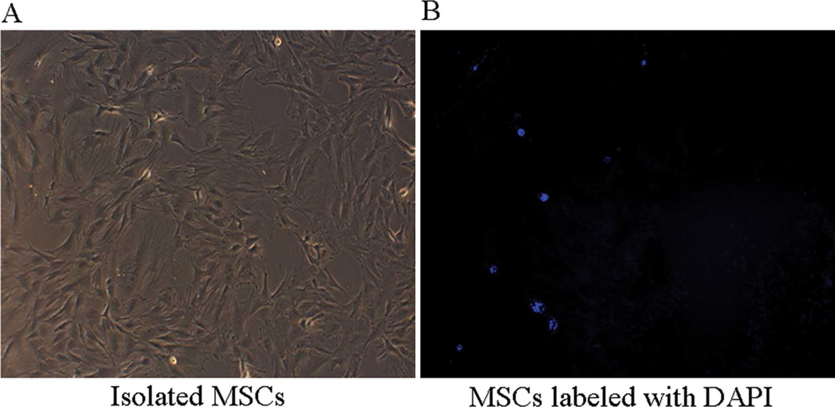
Observation of MSCs
The third generation MSCs were observed under an
inverted microscope. A small amount of cell adhesion was found at
24 h after incubation in a flat-bottom plate, which was
significantly increased when the incubation time was prolonged to
48 h. Most cells were spindle-shaped when observed under a
microscope. The primary generation grew in lumps and reached 90%
confluence after incubation for 2 weeks (Fig. 1A).
Tracing of MSCs by DAPI
The third generation MSCs were incubated with DAPI
in the culture medium the day before the allograft, followed by the
sacrificing of rats and the isolation of lung tissue. The tissue
samples were observed under a microscope, and the blue stained
cells were immigrated MSCs (Fig.
1B).
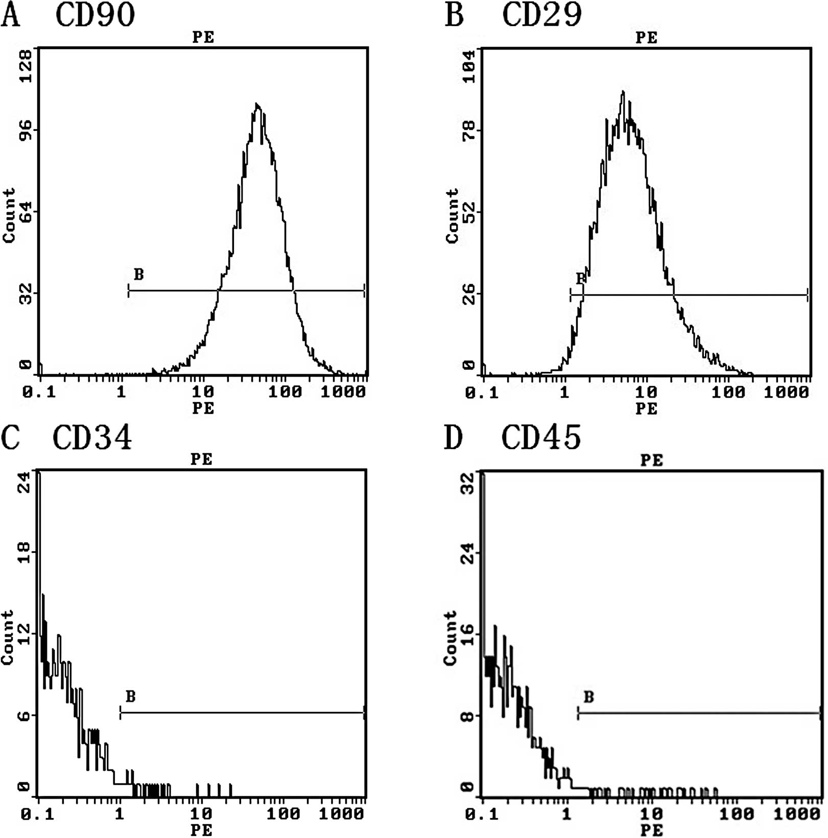
Detection of surface markers on MSCs by
flow cytometry
The cell surface markers on third generation MSCs
were detected by flow cytometry. The positive rates of CD90 and
CD29 were 98.6% (Fig. 2A) and
97.5% (Fig. 2B), respectively.
However, the expression of CD34 and CD45 in MSCs was low, with
positive rates of 0.56% (Fig. 2C)
and 0.79% (Fig. 2D), respectively.
This demonstrated that the purity of the MSCs was >95%.
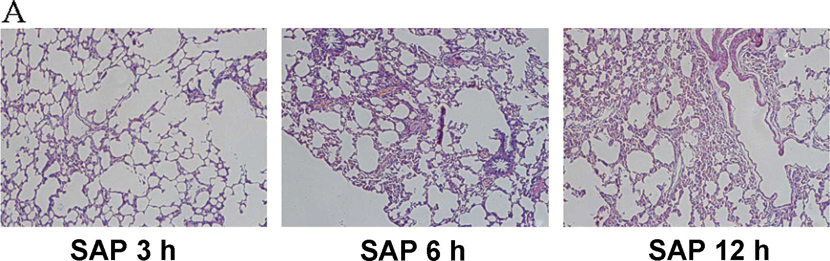
MSC transplantation attenuates pulmonary
edema and inflammation in rats with SAP
In the SAP group, the pancreatitis-associated ALI
was demonstrated by histology, characterized by alveolar edema,
hemorrhaging, pulmonary interstitial edema, inflammatory cell
infiltration, thicker alveolar septum and damage to the normal
structure of the lung tissues. After MSC transplantation, the
alveolar structure had improved, with thin walls, less infiltration
of inflammatory cells and decreased tissue edema (Fig. 3).
Pulmonary edema was examined by measuring the water
content in the lung tissue samples and expressed as lung wet-to-dry
ratio. The lung wet-to-dry ratio showed no significant difference
at 1 h after MSC transplantation, but was significantly decreased
at 3, 6, 12 and 24 h after MSC transplantation (P<0.05)
(Table I).
 | Table IPulmonary edema scores (lung
wet-to-dry ratio) in the different groups (means ± SD). |
Table I
Pulmonary edema scores (lung
wet-to-dry ratio) in the different groups (means ± SD).
| Group | 1 h after
modeling | 3 h after
modeling | 6 h after
modeling | 12 h after
modeling | 24 h after
modeling |
|---|
| SO group | 4.23±0.17 | 5.78±0.10 | 5.88±0.180 | 6.69±0.13 | 6.02±0.15 |
| SAP group | 5.21±0.13 | 6.23±0.18a | 6.62±0.331a | 6.95±0.42a | 6.32±0.23a |
| MSC group | 4.93±0.15 | 5.74±0.10b | 5.85±0.160b | 6.03±0.30a,b | 6.21±0.25a,b |
There was no significant difference in MPO activity
in the lung tissue at 1 h between the groups. On the contrary, in
the MSC group, the MPO activity was decreased significantly
compared to the SAP group at 3, 6, 12 and 24 h (P<0.05)
(Table II).
| Table IIChanges in myeloperoxidase content in
lung tissue in the different groups (means ± SD). |
Table II
Changes in myeloperoxidase content in
lung tissue in the different groups (means ± SD).
| Group | 1 h after
modeling | 3 h after
modeling | 6 h after
modeling | 12 h after
modeling | 24 h after
modeling |
|---|
| SO group | 1.01±0.05 | 1.03±0.06 | 1.06±0.07 | 1.08±0.11 | 1.05±0.08 |
| SAP group | 1.26±0.10 | 3.60±0.30a | 8.62±0.58a | 10.82±0.59a | 9.76±0.42a |
| MSC group | 1.18±0.06 | 2.25±0.11a,b | 4.26±0.30a,b | 3.12±0.49a,b | 3.89±0.38a,b |
Serum AMS in the SAP group was significantly higher
than that in the SO group at each time-point (P<0.05). After MSC
transplantation, serum AMS decreased significantly compared to the
SAP group at each time-point (P<0.01) (Table III).
| Table IIISerum amylase levels at each
time-point in the different groups (U/l). |
Table III
Serum amylase levels at each
time-point in the different groups (U/l).
| Group | 1 h after
modeling | 3 h after
modeling | 6 h after
modeling | 12 h after
modeling | 24 h after
modeling |
|---|
| SO group | 1,452±93 | 3,463±310 | 3,874±230 | 3,782±382 | 3,521±140 |
| SAP group | 3,123±246 | 5,509±890a | 8,800±1120a | 10,249±1,392a | 6,724±1,031a |
| MSC group | 2,956±109 | 4,766±556a,b | 5,482±887a,b | 6,724±1,024a,b | 3,108±228a,b |
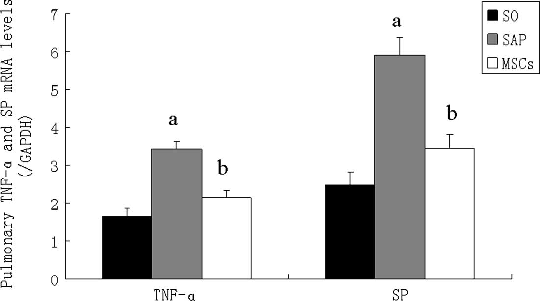
Transplanted MSCs decrease the mRNA
levels of TNF-α and SP in lung tissues of SAP rats
The mRNA expression levels of TNF-α and SP were
markedly higher in the SAP group than in the SO group. The
transplanted MSCs increased the expression levels of TNF-α and SP
mRNA in rats with SAP (Fig.
4).
Discussion
In the present study, we established an experimental
model of sodium taurocholate-induced SAP that is characterized by
systemic inflammation and MODS, particularly
pancreatitis-associated lung injury. Compared to the controls, rats
with SAP had increased pulmonary edema, higher MPO content in the
lung tissue and higher serum AMS levels. The intravenous
transplantation of MSCs attenuated pulmonary edema and inflammatory
infiltration in the rats with SAP, and reduced the mRNA levels of
TNF-α and SP in the lung tissues of the rats with SAP.
Pancreatitis-associated lung injury is the most
frequent and serious systemic complication of SAP (8), which is characterized by pulmonary
edema, inflammatory infiltration in pulmonary tissues and increased
inflammatory mediators (9).
Pancreatitis-associated lung injury exhibits a phenotype with
diffuse alveolar damage, microvascular injury, type I pneumocyte
necrosis and an influx of inflammatory cells, leading to injured
endothelial and epithelial cells in lung tissues (10).
Recently, a certain study demonstrated that
transplanted MSCs have strong immunoregulatory effects and reduce
inflammation and damage to pancreatic tissue in a rat model of AP,
as evidenced by the reduced expression of inflammatory mediators
and cytokines (11). MSCs have
been shown to have anti-inflammatory effects in a variety of
diseases, such as inflammatory bowel disease (12), hepatitis (13) and peritonitis (14).
In our study, we found that transplanted MSCs
reduced pancreatitis-associated lung injury in a rat model,
attenuated pulmonary edema, and reduced inflammatory mediators in
lung tissue, such as TNF-α and SP. TNF-α and SP have been
introduced as two important factors in adjusting and controlling
pancreatitis-associated lung injury. TNF-α expression plays an
important role in the pathogenesis of ALI. Pancreatic
inflammation-associated elastase activation induces TNF-α-mediated
lung injury, and lung injury is markedly reduced in TNF-α-deficient
animals (15). TNF-α also potently
stimulates matrix metalloproteinase-9 (MMP-9) release from PMNs,
and induces PMN transmigration and resultant alveolar-capillary
leakage in SAP (16). SP is the
gene product of preprotachykinin-A, a type of neural peptide with a
regulatory function in different phases of inflammation. In
pancreatitis-associated lung injury, the integrity of epidermal and
alveolar cells in the lungs is destroyed, leading to damaged
vacuolar membranes in the alveoli and leaking capillaries, and
subsequently pulmonary interstitial edema. Thus, SP plays an
important pro-inflammatory role in regulating the severity of AP
and pancreatitis-associated lung injury (17). Our study demonstrates that MSC
transplantation significantly decreases TNF-α and SP mRNA levels,
indicating that the severity of lung injury may be attenuated in
rats with SAP.
MSCs also have an antioxidant effect and promote
antioxidant components, such as superoxide dismutase and
glutathione peroxidase (18),
exhibiting a protective function against oxidative damage-induced
apoptosis (19). Considering the
oxidative stress effect of MPO activity, MSCs exhibit antioxidant
effects, partly through the downregulation MPO activity, in rats
with SAP. Furthermore, MPO activity is a marker of local leukocyte
sequestration (20). Our study
suggests that MSC transplantation attenuates
pancreatitis-associated lung injury by inhibiting the production of
inflammatory cytokines and leukocyte infiltration in lung
tissues.
MSCs have a protective effect on lung injury in
diseases other than SAP (21,22),
indicating that MSCs have similar therapeutic targets in lung
injury. However, multiple factors and inflammatory mediators
participate in pancreatitis-associated lung injury. Therefore,
further research is required to elucidate the exact mechanism
behind the protective effect of MSCs on lung injury. A recent study
showed that in chronic inflammation and lung injury, MSCs were
converted into AT II epithelial cells, performing a tissue repair
function (23). Whether this
mechanism plays a role in our model requires further study.
In conclusion, MSC transplantation attenuates
pulmonary edema and inflammation, and decreases the mRNA expression
of TNF-α and SP in lung tissues of rats with
pancreatitis-associated lung injury.
Acknowledgements
This study was funded by the Medical and Health
Research Project of the Nanjing Military Region (No. 08Z029).
References
|
1
|
Carroll JK, Herrick B, Gipson T and Lee
SP: Acute pancreatitis: diagnosis, prognosis, and treatment. Am Fam
Physician. 75:1513–1520. 2007.PubMed/NCBI
|
|
2
|
Shah AU, Sarwar A, Orabi AI, et al:
Protease activation during in vivo pancreatitis is dependent on
calcineurin activation. Am J Physiol Gastrointest Liver Physiol.
297:G967–G973. 2009. View Article : Google Scholar : PubMed/NCBI
|
|
3
|
Bhatia M: Acute pancreatitis as a model of
SIRS. Front Biosci. 14:2042–2050. 2009. View Article : Google Scholar : PubMed/NCBI
|
|
4
|
Zhou MT, Chen CS, Chen BC, Zhang QY and
Andersson R: Acute lung injury and ARDS in acute pancreatitis:
mechanisms and potential intervention. World J Gastroenterol.
16:2094–2099. 2010. View Article : Google Scholar : PubMed/NCBI
|
|
5
|
Bhatia M and Moochhala S: Role of
inflammatory mediators in the pathophysiology of acute respiratory
distress syndrome. J Pathol. 202:145–156. 2004. View Article : Google Scholar : PubMed/NCBI
|
|
6
|
Aggarwal S and Pittenger MF: Human
mesenchymal stem cells modulate allogeneic immune cell responses.
Blood. 105:1815–1822. 2005. View Article : Google Scholar : PubMed/NCBI
|
|
7
|
Pereda J, Sabater L, Cassinello N, et al:
Effect of simultaneous inhibition of TNF-alpha production and
xanthine oxidase in experimental acute pancreatitis: the role of
mitogen activated protein kinases. Ann Surg. 240:108–116. 2004.
View Article : Google Scholar
|
|
8
|
Browne GW and Pitchumoni CS:
Pathophysiology of pulmonary complications of acute pancreatitis.
World J Gastroenterol. 12:7087–7096. 2006.PubMed/NCBI
|
|
9
|
Pezzilli R, Bellacosa L and Felicani C:
Lung injury in acute pancreatitis. JOP. 10:481–484. 2009.PubMed/NCBI
|
|
10
|
Tomashefski JF Jr: Pulmonary pathology of
the adult respiratory distress syndrome. Clin Chest Med.
11:593–619. 1990.PubMed/NCBI
|
|
11
|
Jung KH, Song SU, Yi T, et al: Human bone
marrow-derived clonal mesenchymal stem cells inhibit inflammation
and reduce acute pancreatitis in rats. Gastroenterology.
140:998–1008. 2011. View Article : Google Scholar : PubMed/NCBI
|
|
12
|
Sánchez L, Gutierrez-Aranda I, Ligero G,
et al: Enrichment of human ESC-derived multipotent mesenchymal stem
cells with immunosuppressive and anti-inflammatory properties
capable to protect against experimental inflammatory Bowel disease.
Stem Cells. 29:251–262. 2011.
|
|
13
|
Kubo N, Narumi S, Kijima H, Mizukami H,
Yagihashi S, Hakamada K and Nakane A: Efficacy of adipose
tissue-derived mesenchymal stem cells for fulminant hepatitis in
mice induced by concanavalin A. J Gastroenterol Hepatol.
27:165–172. 2012. View Article : Google Scholar : PubMed/NCBI
|
|
14
|
Choi H, Lee RH, Bazhanov N, Oh JY and
Prockop DJ: Anti-inflammatory protein TSG-6 secreted by activated
MSCs attenuates zymosan-induced mouse peritonitis by decreasing
TLR2/NF-κB signaling in resident macrophages. Blood. 118:330–338.
2011.PubMed/NCBI
|
|
15
|
Jaffray C, Yang J, Carter G, Mendez C and
Norman J: Pancreatic elastase activates pulmonary nuclear factor
kappa B and inhibitory kappa B, mimicking pancreatitis-associated
adult respiratory distress syndrome. Surgery. 128:225–231. 2000.
View Article : Google Scholar
|
|
16
|
Keck T, Balcom JH IV, Fernández-del
Castillo C, Antoniu BA and Warshaw AL: Matrix metalloproteinase-9
promotes neutrophil migration and alveolar capillary leakage in
pancreatitis-associated lung injury in the rat. Gastroenterology.
122:188–201. 2002. View Article : Google Scholar : PubMed/NCBI
|
|
17
|
Bhatia M, Saluja AK, Hofbauer B, et al:
Role of substance P and the neurokinin 1 receptor in acute
pancreatitis andpancreatitis-associated lung injury. Proc Natl Acad
Sci USA. 95:4760–4765. 1998. View Article : Google Scholar : PubMed/NCBI
|
|
18
|
Zhuo W, Liao L, Xu T, Wu W, Yang S and Tan
J: Mesenchymal stem cells ameliorate ischemia-reperfusion-induced
renal dysfunction by improving the antioxidant/oxidant balance in
the ischemic kidney. Urol Int. 86:191–196. 2011. View Article : Google Scholar : PubMed/NCBI
|
|
19
|
Liu L, Cao JX, Sun B, et al: Mesenchymal
stem cells inhibition of chronic ethanol-induced oxidative damage
via upregulation of phosphatidylinositol-3-kinase/Akt and
modulation of extracellular signal-regulated kinase 1/2 activation
in PC12 cells and neurons. Neuroscience. 167:1115–1124. 2010.
View Article : Google Scholar
|
|
20
|
Li ZF, Xia XM, Huang C, Zhang S, Zhang J
and Zhang AJ: Emodin and baicalein inhibit pancreatic stromal
derived factor-1 expression in rats with acute pancreatitis.
Hepatobiliary Pancreat Dis Int. 8:201–208. 2009.PubMed/NCBI
|
|
21
|
Yang H, Wen Y, Bin J, Hou-You Y and
Yu-Tong W: Protection of bone marrow mesenchymal stem cells from
acute lung injury induced by paraquat poisoning. Clin Toxicol
(Phila). 49:298–302. 2011. View Article : Google Scholar : PubMed/NCBI
|
|
22
|
Saito S, Nakayama T, Hashimoto N, et al:
Mesenchymal stem cells stably transduced with a dominant-negative
inhibitor of CCL2 greatly attenuate bleomycin-induced lung damage.
Am J Pathol. 179:1088–1094. 2011. View Article : Google Scholar : PubMed/NCBI
|
|
23
|
Wu L, Wang G, Qu P, Yan C and Du H:
Overexpression of dominant negative peroxisome
proliferator-activated receptor-γ (PPARγ) in alveolar type II
epithelial cells causes inflammation and T-cell suppression in the
lung. Am J Pathol. 178:2191–2204. 2011.
|