Introduction
Thyroid cancer (TC) is the most common type of
endocrine malignancy (1); ~90% of
thyroid cancer cases are of poorly differentiated poorly
differentiated thyroid carcinoma, the prevalence of which is
increasing (2–4). Following treatment with specific
methods, including surgery, radioactive iodine and
thyroid-stimulating hormone inhibition, the majority of patients
exhibit an improvement. However, there remains a risk of tumor
recurrence and metastasis (5–7).
Therefore, it is necessary to understand the occurrence and
development of TC, in order to identify specific effective targets
for diagnosis and treatment.
MicroRNAs (miRNAs/miRs) are non-coding single-
stranded small molecule RNAs that exert their biological function
by inhibiting the transcription and translation of target genes,
via binding to the 3′ non-coding region of the target mRNA
(8,9). Previous studies have demonstrated
that miRNAs are closely associated with the occurrence and
development of numerous tumors, including ovarian cancer, liver
cancer, stomach cancer and breast cancer (10–13).
Therefore, miRNAs have attracted increasing attention in tumor
research.
Previous studies have reported that differentially
expressed miRNAs in the tissues of patients with TC may be screened
by gene chip technology (14,15).
These findings indicated that, in the tissues of patients with TC,
the expression levels of miR-26b-5p are significantly lower
compared with in normal tissues; in addition, it was revealed that
miR-26b-5p may be associated with the occurrence and development of
TC. Therefore, it was be hypothesized that miR-26b-5p may be a
clinically relevant target for the treatment of TC.
However, the molecular mechanism underlying the
anti-TC effect of miR-26b-5p remains unclear. A previous study
using TargetScan predicted that the target gene for miR-26b-5p is
glycogen synthase kinase-3β (Gsk-3β) (16). It is well documented that the
Gsk-3β/β-catenin pathway serves an important role in the
development of TC (17,18).
In the present study, the expression levels of
miR-26b-5p were detected in the tissues of patients with TC, and
the association between miR-26b-5p and clinicopathological features
was examined. Additionally, the inhibitory effect of miR-26b-5p on
B-CPAP TC cells and its mechanism based on the Gsk-3β/β-catenin
pathway were investigated. The results suggested that miR-26b-5p
was downregulated in TC tissues and that miR-26b-5p expression may
be associated with lymph node metastasis. In addition, miR-26b-5p
was able to inhibit the proliferation, migration and invasion of
TC, which may be associated with the Gsk-3β/β-catenin pathway.
Materials and methods
Chemicals and materials
A total of 67 patients with TC undergoing surgical
treatment at the Provincial Hospital of Shandong University were
enrolled between February 2017 and September 2017. TC and
corresponding paracarcinoma tissues were collected. TC was
confirmed by pathological diagnosis and written informed consent
was obtained from all patients. The 67 samples were collected and
stored in liquid nitrogen until further use. The samples were
collected from 17 men and 50 women, with an average age of 45 years
(age range, 18–75 years).
RNAiso reagent, Prime Script 1st Strand cDNA
Synthesis kit for reverse transcription (RT) and SYBR Premix Ex Taq
for quantitative PCR (qPCR) were purchased from Takara Bio, Inc.
Fetal bovine serum (FBS) and RPMI-1640 medium were purchased from
Thermo Fisher Scientific, Inc. The Cell Counting Kit-8 (CCK-8) was
purchased from Dojindo. Anti-GAPDH (cat. no. sc-32233), anti-Gsk-3β
(cat. no. sc-71186), anti-phosphorylated (p) GSK-3β (cat. no.
sc-81495) and anti-β-catenin (cat. no. sc-65480) were purchased
from Santa Cruz Biotechnology, Inc. The lentiviral vectors were
purchased from GeneChem, Inc. The Dual-Glo™ Luciferase Assay System
and Lipofectamine 2000 were purchased from Promega Corporation.
Detection of miR-26b-5p
expression
The expression levels of miR-26b-5p were detected in
tissues from patients with TC by RT-qPCR, using the aforementioned
materials. Total RNA was extracted from fresh frozen tissue using
the RNAiso RNA extraction kit, according to the manufacturer's
protocol, and total RNA concentration and purity were determined
using a UV spectrophotometer. The primers were synthesized by
Takara Bio, Inc. The miR-26b-5p primers were designed based on the
sequence 5′-UCAAGUAAUUCAGGAUAGGU-3. U6 was used as an internal
reference gene and its primer sequences were as follows: Forward
5′-GGAACGATACAGAGAAGATTAGC-3′; and reverse
5′-TGGAACGCTTCACGAATTTGCG-3′. cDNA synthesis was conducted using an
RT kit in a 10 µl volume, according to the manufacturer's protocol.
The RT temperature protocol was as follows: 37°C for 1 h and 85°C
for 5 min. qPCR was performed on a Roche LightCycler 480 II
instrument (Roche Molecular Diagnostics) as follows: denaturation
(95°C, 30 sec), PCR reaction for 45 cycles (95°C for 5 sec, 60°C
for 30 sec), melting (95°C for 5 sec, 60°C for 1 min), cooling
(50°C, 30 sec). The final experimental data were standardized to
the internal standard U6 and were analyzed using the
2−∆∆Cq method (19):
ΔCq=CqmiRNA-Cqinternal reference,
ΔΔCq=ΔCqSample-ΔCqMax. The maximum value of
all data (ΔCqMax) was used to normalize and correct the
samples.
Cell culture
The B-CPAP cell line was donated by Professor
Kennichi Kakudo (The Human Pathology Laboratory of Wakayama Medical
University (Wakayama, Japan) (20)
and the 293 cell line was purchased from GeneChem, Inc. The B-CPAP
cell line was verified using the short-tandem repeat (STR)
profiling. Briefly, DNA was extracted using Axygen genomic
extraction kit and amplified using 20-STR amplification scheme
(Shanghai Zhong Qiao Xin Zhou Biotechnology Co., Ltd.). The test of
STR loci and sex gene Amelogenin was performed using an ABI 3730XL
analyzer (Applied Biosystems; Thermo Fisher Scientific, Inc.). The
cells were cultured in RPMI-1640 (B-CPAP) or Dulbecco's modified
Eagle's medium (Sigma-Aldrich; Merck KGaA) growth medium
supplemented with 10% FBS, 100 U/ml penicillin and 100 µg/ml
streptomycin at 37°C in a thermostatic incubator containing 5%
CO2. All experiments were performed on cells in the
logarithmic growth phase.
Cell infection
miR-26b-5p mimics on a lentiviral vector were
designed and synthesized by Shanghai GeneChem Co., Ltd., based on
the following sequence: 5′-UUCAAGUAAUUCAGGAUAGGU-3′. Once the cells
reached 80% confluence in 96-well plates, B-CPAP cells were
infected with a miR-26b-5p-overexpressing lentivirus or an empty
vector at a multiplicity of infection value of 50. After 12 h, the
virus solution was aspirated and replaced with fresh medium. The
fluorescence efficiency was observed after 48–72 h, and the
infection efficiency was detected by RT-qPCR, and stably
overexpressing cells and negative control (NC) were selected for
subsequent experiments.
CCK-8 cell viability assay
Following digestion of miR-26b-5p-overexpressing
cells and negative control cells with trypsin, the cells were
adjusted to 5,000 cells/well and seeded in 96-well plates at 37°C
in an incubator containing 5% CO2 saturated humidity.
Cells were cultured for 24, 48, 72 and 96 h, and viability was
detected using the CCK-8 assay. Briefly, 10 µl CCK-8 and 90 µl
fresh medium were added to each well. After 90 min at 37°C, optical
density values were determined at 450 nm using a microplate reader.
The experiment was repeated at least three times.
Cell migration assay
Cell migration was detected using a Transwell assay.
miR-26b-5p-overexpressing B-CPAP cells and negative control B-CPAP
cells were re-suspended (1×10⁵ cells/ml) in RPMI-1640
medium, and a 200 µl cell suspension was added to the upper chamber
of Transwell plates. In addition, 600 µl complete medium containing
5% FBS was added to the lower chamber. After culturing for 18 h,
the cells in the lower chamber were washed twice with PBS, fixed
with 95% ethanol at 37°C for 15 min, and stained with hematoxylin
at 37°C for 10 min. The cells were then washed three times with
water, air-dried and observed using light microscopy
(magnification, ×200).
Cell invasion assay
Cell invasion was detected using a Matrigel assay.
Matrigel was thawed at 4°C overnight and thoroughly mixed with
serum-free medium to obtain a 1:7 ratio on ice. Briefly, 70 µl
diluted Matrigel was added to the Transwell plates and incubated at
37°C for 30 min, in order to polymerize the Matrigel.
miR-26b-5p-overexpressing B-CPAP cells and NC B-CPAP cells were
re-suspended (1×10⁵ cells/ml) in RPMI-1640 medium.
Briefly, a 200 µl cell suspension was added to the upper chamber
and 600 µl complete medium containing 10% FBS was added to the
lower chamber. After culturing for 18 h, the chamber was washed
twice with PBS and the cells in the lower chamber were fixed with
95% ethanol at 37°C for 15 min. The cells were then stained with
hematoxylin at 37°C for 10 min and the chamber was washed with
water three times. After natural air-drying, the chamber membranes
were sealed with slides, and observed under an inverted
light-transmitting microscope (magnification, ×200). In total, five
fields were randomly selected to count the number of cells.
Dual-luciferase enzyme assay
TargetScan (release no. 3.1; Whitehead Institute for
Biomedical Research) was used to predict target genes of
miR-26b-5p. The results revealed Gsk-3β to be a potential target,
and this was verified via a dual-luciferase enzyme assay. Briefly,
293 cells (at a density of 4×10⁵ cells/ml) were seeded
in 24-well plates and cultured for 24 h. The luciferase reporter
plasmids containing wild-type or mutant 3′-untranslated regions
(UTRs) of Gsk-3β (Promega Corporation) were transfected into cells
alongside miR-26b-5p mimics using Lipofectamine 2000 (transfection
of 1 µg plasmid) at 37°C. After ~48 h incubation at 37°C, firefly
and Renilla luciferase activities were assessed. Normalized
relative light units represent firefly luciferase
activity/Renilla luciferase activity.
Western blot analysis
The protein expression levels of Gsk-3β, p-Gsk-3β
and β-catenin in both miR-26b-5p-overexpressing and NC B-CPAP cells
were detected via western blotting. Cellular proteins were
extracted by using RIPA (Beijing Solarbio Science and Technology
Co., Ltd.) containing phosphorylase inhibitors, and protein
quantification was performed using a bicinchoninic acid kit.
Subsequently, equal amounts of proteins (30 µg) were separated
using 10% SDS-PAGE, and were transferred to polyvinylidene
difluoride membranes (EMD Millipore), which were blocked with 5%
milk at 37°C for 1 h. The membranes were then incubated overnight
at 4°C with the following primary polyclonal antibodies: anti-GAPDH
(1:1,000), anti-Gsk-3β (1:1,000), anti-p-GSK-3β (1:1,000) and
anti-β-catenin (1:1,000). Membranes were then incubated with a
horseradish peroxidase-conjugated goat anti-rabbit secondary
antibody (cat. no. abs20002; Absin Bioscience, Inc.; 1:5,000) at
37°C for 1 h. The membranes were imaged using enhanced
chemiluminescence (ProteinSimple) and analyzed using Photoshop 6.0
(Adobe Systems, Inc.) and protein expression was normalized to
GAPDH (dilution, 1:1,000).
Statistical analysis
All experiment was repeated at least 3 times, and
data were analyzed using SPSS 22.0 statistical software (IBM
Corp.), and a receiver operating characteristic (ROC) curve was
analyzed and drawn using Prism 7 (GraphPad Software, Inc.).
Measurement results are expressed as the mean ± standard deviation.
Student's t-test was used to compare two groups, whereas Spearman's
correlation analysis was used to analyze the correlation between
tumor stage and miR-26b-5p expression. P<0.05 was considered to
indicate a statistically significant difference.
Results
Association between miR-26b-5p
expression and clinicopathological features in TC
As shown in Table
I, no significant differences in miR-26b-5p expression levels
were identified between patients of different ages and sexes, nor
was it associated with tumor size. In addition, Spearman's
correlation analysis identified no significant correlation between
miR-26b-5p expression and Tumor-Node-Metastasis (TNM) stage
(21). The relative expression
levels of miR-26b-5p in TC tissues (5.18±2.92) were lower compared
with in paracarcinoma tissues (7.15±2.99) in the 67 patients
(P<0.05; Table I).
Additionally, the expression levels of miR-26b-5p were
significantly lower in patients with lymph node metastasis compared
with in patients without lymph node metastasis (P<0.05).
 | Table I.Expression levels of miR-26b-5p in
patients with thyroid cancer. |
Table I.
Expression levels of miR-26b-5p in
patients with thyroid cancer.
| Parameters | Cases | Expression of
miR-26b-5p (mean ± SD) | P-value |
|---|
| Sex |
|
| 0.439 |
|
Male | 17 | 4.70±2.48 |
|
|
Female | 50 | 5.34±3.06 |
|
| Age (years) |
|
| 0.473 |
|
≥45 | 37 | 5.41±3.56 |
|
|
<45 | 30 | 4.89±3.20 |
|
| Tumor size
(cm) |
|
| 0.916 |
| ≥1 | 48 | 5.20±3.03 |
|
|
<1 | 19 | 5.12±2.72 |
|
| Lymph node
metastasis |
|
| 0.015 |
|
Yes | 39 | 4.45±1.92 |
|
| No | 28 | 6.19±3.72 |
|
| TNM
stagea |
|
| 0.473 |
| I | 35 | 4.37±0.80 |
|
| II | 13 | 3.87±0.80 |
|
|
III | 19 | 4.60±0.49 |
|
| Tissue |
|
| <0.001 |
|
Cancer | 67 | 5.18±2.92 |
|
|
Normal | 67 | 7.15±2.99 |
|
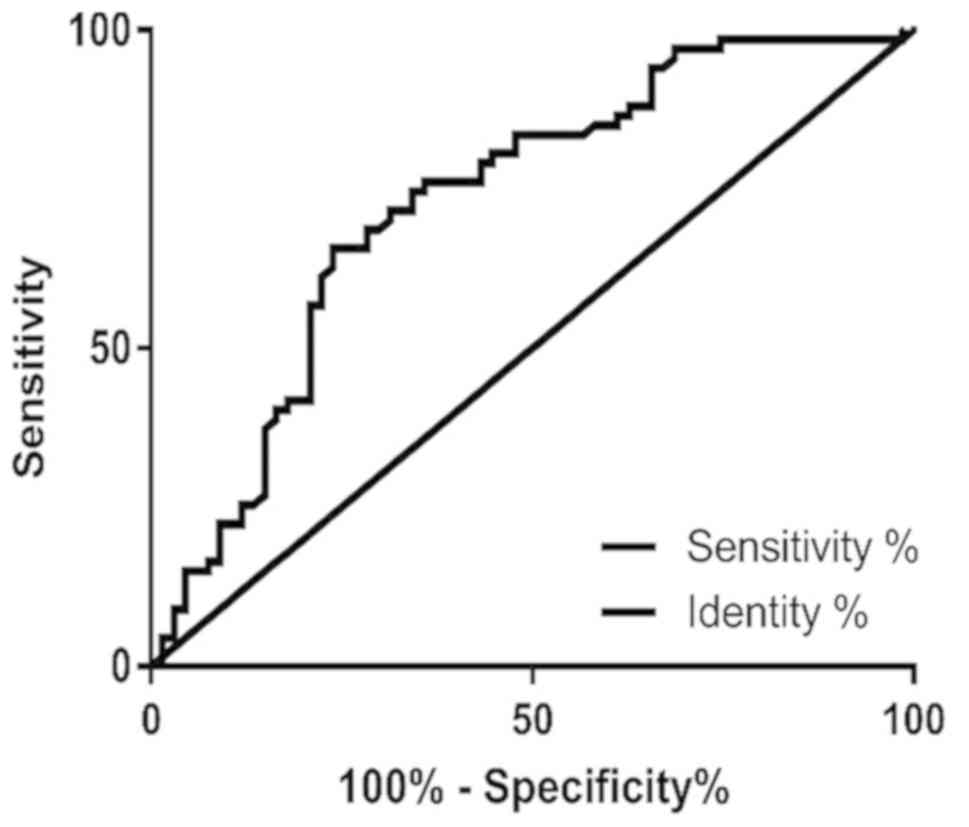
As presented in Fig.
1, the area under the curve value was 0.7287 (95% confidence
interval: 0.6422–0.8152; specificity value: 65.67%; sensitivity
value: 76.12%), which suggested that miR-26b-5p has high
specificity and sensitivity in the clinical diagnosis of TC.
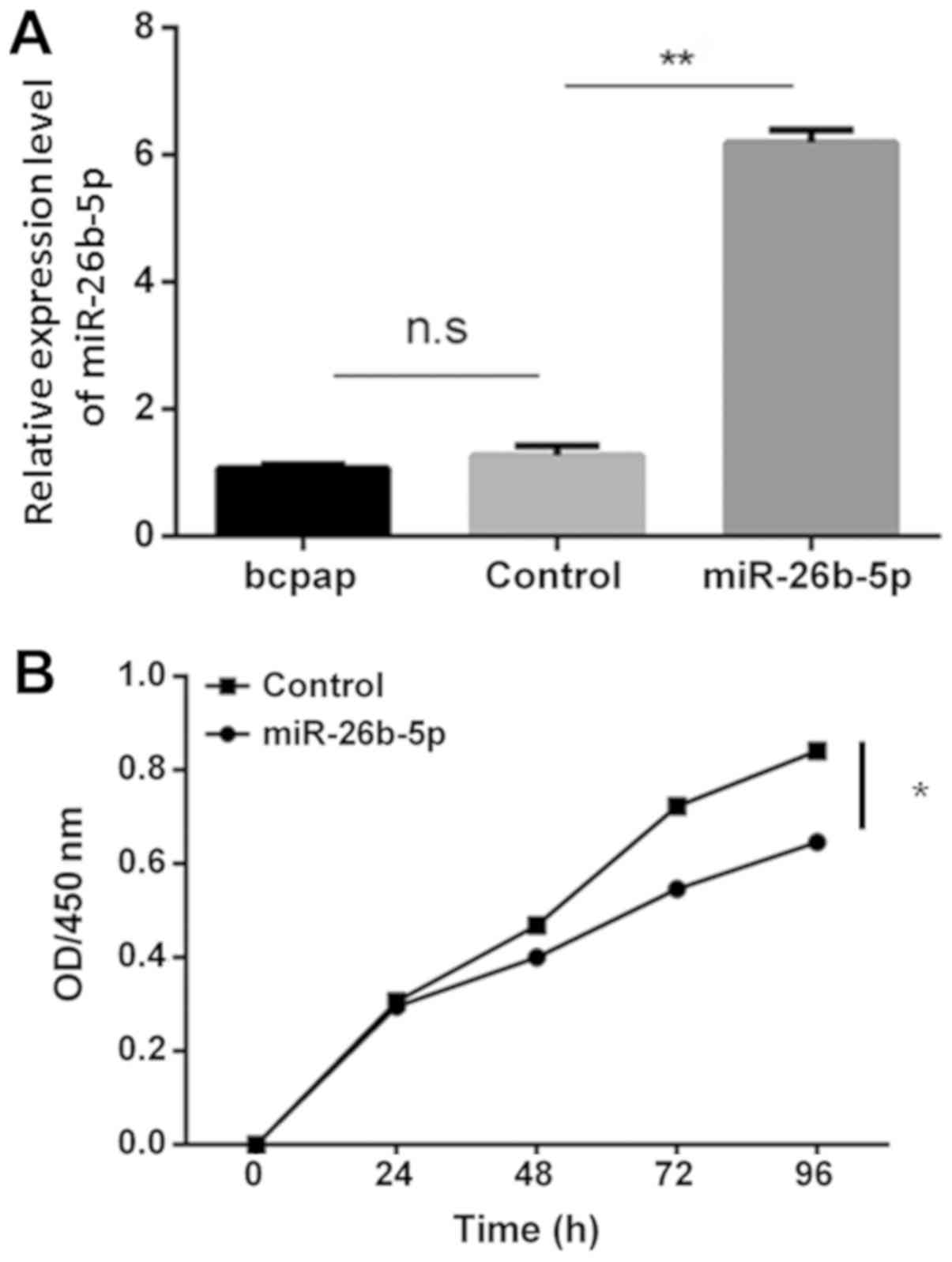
miR-26b-5p overexpression inhibits
B-CPAP cell proliferation
Successful cell transfection was confirmed using
RT-qPCR (Fig. 2A); no significant
difference was detected in the expression levels of miR-26b-5p
between the blank control group and the empty vector control group
(P>0.05). Conversely, the expression levels of miR-26b-5p were
significantly increased in the miR-26b-5p-overexpressing cells
compared with in the blank control and empty vector control groups
(P<0.05). Furthermore, after 24 h, cell proliferation was
reduced in the miR-26b-5p overexpression group compared with in the
control group, thus suggesting that miR-26b-5p inhibited B-CPAP
cell proliferation (P<0.05; Fig.
2B).
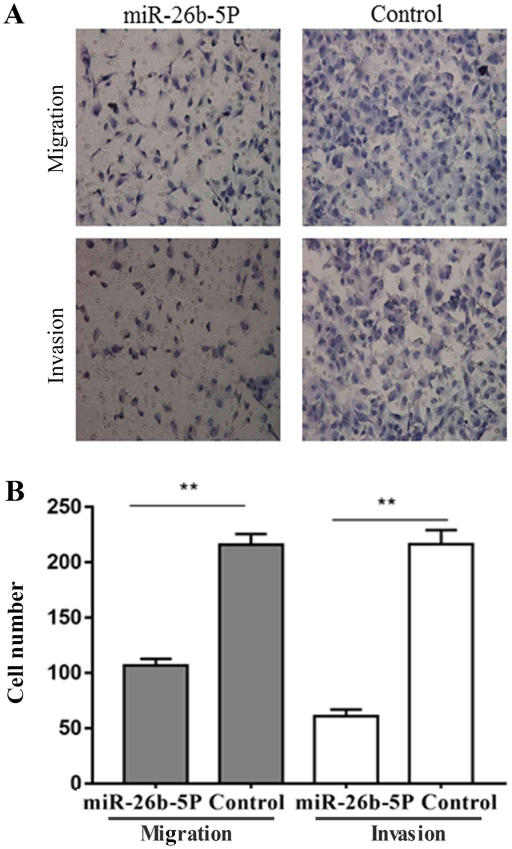
miR-26b-5p overexpression inhibits
B-CPAP cell migration and invasion
The results of the migration and invasion assays are
presented in Fig. 3. Briefly, the
number of migratory and invasive B-CPAP cells in the control group
was 216±5.6 and 216.3±7.5, respectively. However, the number of
migratory and invasive B-CPAP cells was decreased in the miR-26b-5p
overexpression group compared with in the control group, to
106.7±3.5 and 61±3.5, respectively (P<0.001). These results
suggested that miR-26b-5p overexpression inhibited the migration
and invasion of B-CPAP cells.
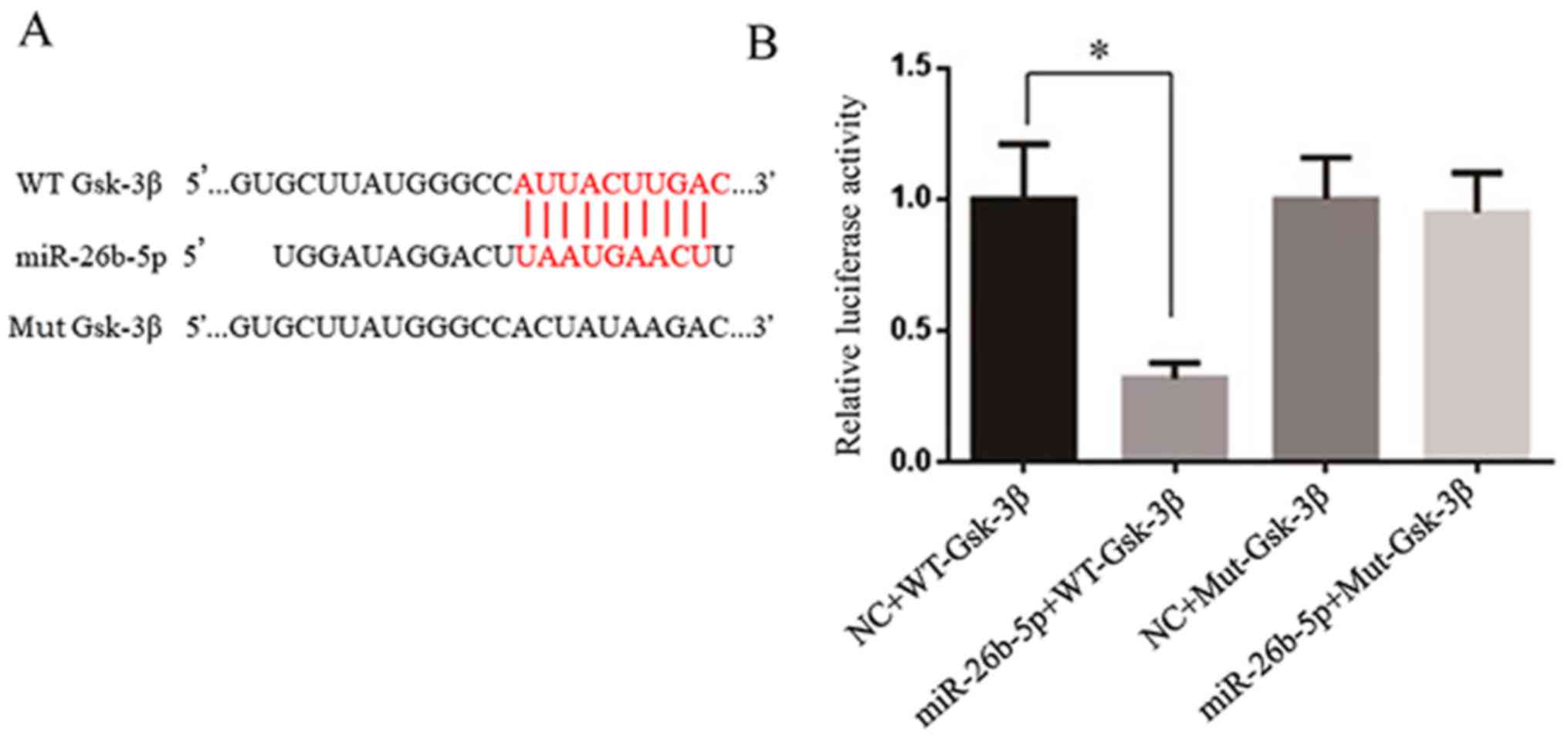
miR-26b-5p directly targets
Gsk-3β
TargetScan was used to predict target genes of
miR-26b-5p; Gsk-3β was predicted as a target gene, since the 3′-UTR
of Gsk-3β contains numerous binding sites for miR-26b-5p (Fig. 4A). As presented in Fig. 4B, the luciferase reporter assay
demonstrated that miR-26b-5p significantly inhibited the luciferase
activity in the wild-type Gsk-3β group in vitro. Conversely,
mutations in the binding site restored luciferase activity.
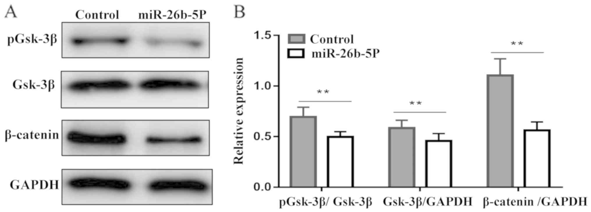
miR-26b-5p downregulates the protein
expression levels of pGsk-3β and β-catenin in B-CPAP cells
The results of western blotting are presented in
Fig. 5. In the control group,
β-catenin was highly expressed, whereas, in the miR-26b-5p
overexpression group, the protein expression levels of β-catenin
were significantly decreased (P<0.01). In addition, pGsk-3β in
the miR-26b-5p overexpression group was decreased compared with in
the control group (P<0.01). These results indicated that
miR-26b-5p may exert antitumor activity by downregulating the
expression of β-catenin and pGsk-3β proteins in B-CPAP cells.
Discussion
miRNAs may bind to specific target genes, and are
involved in the regulation of tumor growth, differentiation,
apoptosis and other processes (22,23).
Numerous miRNAs have been reported to be involved in the
development of thyroid cancer, including miR-21, miR-338-3p,
miR-221 and miR-146b-5p (24–27).
Liu et al (28) revealed
that miR-363-3p inhibits the proliferation, migration and invasion
of TC by downregulating the expression of the target gene
phosphatidylinositol-4,5- biphosphate 3-kinase catalytic subunit α.
Zhang et al (29) suggested
that miR-146a may inhibit the expression of target gene protein
kinase Cε, and upregulated miR-146a expression promotes the
apoptosis of TC cells. In addition, Yin et al (30) demonstrated that miR-195 may inhibit
the proliferation and migration of B-CPAP and K1 TC cells by
downregulating the expression of target genes cyclin D1 and
fibroblast growth factor 2. Cheng et al (31) observed that overexpression of
miR-150 inhibits the proliferation and invasion of tumor cells, and
proposed that its target gene is Rho-associated coiled-coil
containing protein kinase 1. These results suggested that certain
miRNAs are expressed at low levels in the majority of tumors and
under physiological conditions, and may serve a role in inhibiting
tumor proliferation and migration. Therefore, the overexpression of
miRNAs may provide a novel strategy for the clinical treatment and
diagnosis of thyroid cancer.
In the present study, the expression levels of
miR-26b-5p were significantly downregulated in TC compared with in
adjacent tissues. This result suggested that detecting the
expression levels of miR-26b-5p may be used to distinguish cancer
from normal thyroid tissue. In addition, the present results
demonstrated that pre-operative detection of miR-26b-5p expression
levels combined with biopsy results may be helpful in the diagnosis
of TC. There was no significant association between miR-26b-5p
expression and the sex, age, tumor size and TNM stage of patients;
however, the expression levels of miR-26b-5p were significantly
higher in TC tissues without lymph node metastasis compared with in
tissues with lymph node metastasis.
To further examine the biological role of miR-26b-5p
in TC, miR-26b-5p was overexpressed in the TC cell line B-CPAP.
Cytological experiments demonstrated that miR-26b-5p overexpression
may inhibit the proliferation, migration and invasion of B-CPAP
cells, thus suggesting that miR-26b-5p may serve a negative
regulatory role in the development of TC, which is consistent with
previous studies. For example, Wu et al (16) reported that miR-26b-5p is
downregulated in lung cancer, and overexpression of miR-26b-5p
promotes tumor proliferation and inhibits apoptosis. Kato et
al (32) demonstrated that
miR-26a/b inhibits prostate cancer migration and invasion by
regulating La-related protein 1. Kozubek et al (33) revealed that miR-26b-5p expression
is downregulated in melanoma, which may affect the progression of
melanoma by regulating the mitogen-activated protein kinase
pathway.
However, the molecular mechanism underlying the
inhibitory effects of miR-26b-5p on TC remains unclear. TargetScan
was used to predict the target gene of miR-26b-5p; Gsk-3β was
identified as a potential target gene. The dual-luciferase enzyme
assay confirmed that Gsk-3β may be a target of miR-26b-5p. Gsk-3β
is an evolutionarily conserved serine/threonine kinase, which acts
on numerous signaling proteins, structural proteins and
transcription factors to regulate cell differentiation,
proliferation, survival and apoptosis (34). Phosphorylation at different sites
can regulate the activity of Gsk-3β: Phosphorylation at the 9th
serine can inhibit the activity of Gsk-3β, and tyrosine
phosphorylation at position 216 can enhance its activity (35). Gsk-3β substrates are widely
involved in various physiological cell processes, and Gsk-3β serves
a tumor-suppressing role in numerous types of cancer. Farago et
al (36) demonstrated that
following inhibition of Gsk-3β activity, the Wnt pathway is
activated to promote breast cancer cell proliferation. Jiang et
al (37) demonstrated that the
expression levels of pGsk-3βSer9 are increased in lung
cancer tissues, and that the expression levels of
pGsk-3βSer9 are negatively correlated with lung cancer
prognosis. Gsk-3β is a key kinase that determines the
phosphorylation of β-catenin. An increase in Gsk-3β activity may
promote the phosphorylation of β-catenin, which is degraded by the
proteasome, resulting in antitumor effects. The anticancer effect
of Gsk-3β is associated with obstruction of the Wnt signaling
pathway, which is important for cell proliferation, differentiation
and apoptosis (38). Further
studies have demonstrated that the Wnt signaling pathway is
involved in cell migration and invasion, and its regulation is
associated with matrix metalloproteinase-7 (39), C-X-C motif chemokine receptor 4b
and 7b (40). In the present
study, the results demonstrated that the protein expression levels
of pGsk-3βSer9 and β-catenin were significantly
decreased in the miR-26b-5p overexpression group compared with in
the empty vector group, so decreased phosphorylation could increase
its activation. These findings suggested that miR-26b-5p may
inhibit the proliferation, migration and invasion of B-CPAP cells
via the Gsk-3β/β-catenin pathway. Lv et al (41) demonstrated that miR-26a is
consistently downregulated in TC specimens, whereas the
upregulation of miR-26a in TC cells lines induces G2
phase cell cycle arrest by targeting and downregulating CDC28
protein kinase regulatory subunit 2 (CKS2), further research
indicated that the negative regulatory effects are related to the
CKS2 downstream genes cyclin A, Bcl2 like 1 and AKT. Lin et
al (42) demonstrated that
CKS2 regulates AKT and Gsk-3β phosphorylation. The present study
suggested that miR-26b-5p may directly regulate Gsk-3β; this may be
associated with the mechanism underlying the effects of miR-26.
In conclusion, the present results suggested that
miR-26b-5p was downregulated in TC tissues and was associated with
lymph node metastasis. In addition, miR-26b-5p overexpression was
able to inhibit the proliferation, migration and invasion of TC
cells; these effects may be associated with the Gsk-3β/β-catenin
pathway. The present results further demonstrated that miR-26b-5p
may provide a novel basis for the auxiliary diagnosis of TC,
providing better individualized treatment. However, the mechanism
by which miR-26b-5p is involved in tumorigenesis and development
remains unclear and requires further study.
Acknowledgements
Not applicable.
Funding
This work was supported financially by the Shandong
Key Research and Development Plan (grant no. 2017GSF18120) and the
Jinan Science and Technology Development Plan (grant no.
201704105).
Availability of data and materials
The datasets used and/or analyzed during the current
study are available from the corresponding author on reasonable
request.
Authors' contributions
AZ, TW, and LL conceived and designed the project
and prepared the manuscript. AZ, GC, XC, CZ and HX conducted the
cell experiments. MQ and XC analyzed the data. All authors read and
approved the manuscript.
Ethics approval and consent to
participate
The study was approved by the Ethics Committee of
The Shandong Provincial Hospital Affiliated to Shandong University,
and all participants provided written informed consent.
Patient consent for publication
Not applicable.
Competing interests
The authors declare that they have no competing
interests.
References
|
1
|
Randle RW, Bushman NM, Orne J, Balentine
CJ, Wendt E, Saucke M, Pitt SC, Macdonald CL, Connor NP and Sippel
RS: Papillary thyroid cancer: The good and bad of the ‘Good
Cancer’. Thyroid. 27:902–907. 2017. View Article : Google Scholar : PubMed/NCBI
|
|
2
|
Garau LM, Rubello D, Ferretti A, Boni G,
Volterrani D and Manca G: Sentinel lymph node biopsy in small
papillary thyroid cancer. A review on novel surgical techniques.
Endocrine. 62:340–350. 2018. View Article : Google Scholar : PubMed/NCBI
|
|
3
|
Yan S, Zhao W, Wang B and Zhang L:
Preoperative injection of carbon nanoparticles is beneficial to the
patients with thyroid papillary carcinoma: From a prospective study
of 102 cases. Medicine (Baltimore). 97:e113642018. View Article : Google Scholar : PubMed/NCBI
|
|
4
|
Paulsson JO, Zedenius J and Juhlin CC:
Papillary thyroid carcinoma with pleomorphic tumor giant cells in a
pregnant woman-a case report. BMC Endocr Disord. 18:462018.
View Article : Google Scholar : PubMed/NCBI
|
|
5
|
Yongfu Z, Ziyu L, Chen L and Jingchao X:
Cervical mass as the initial manifestation of occult papillary
thyroid carcinoma: Report of three cases. J Cancer Res Ther. 14
(Suppl):S544–S548. 2018. View Article : Google Scholar : PubMed/NCBI
|
|
6
|
Hardin H, Helein H, Meyer K, Robertson S,
Zhang R, Zhong W and Lloyd R: Thyroid cancer stem-like cell
exosomes: Regulation of EMT via transfer of lncRNAs. Lab Invest.
98:1133–1142. 2018. View Article : Google Scholar : PubMed/NCBI
|
|
7
|
Ma B, Wei W, Xu W, Wang Y, Guan H, Fan J,
Zhao Z, Wen D, Yang S, Wang Y, et al: Surgical confirmation of
incomplete treatment for primary papillary thyroid carcinoma by
percutaneous thermal ablation: A retrospective case review and
literature review. Thyroid. 28:1134–1142. 2018. View Article : Google Scholar : PubMed/NCBI
|
|
8
|
Henn D, Abu-Halima M, Falkner F, Wermke D,
Meme LG, Kühner C, Keller A, Kneser U, Meese E, Schmidt V, et al:
Micro-RNA regulated pro-angiogenic signaling in arteriovenous loops
in patients with combined vascular and soft tissue
reconstructions-reisiting the nutrient flap concept. Plast.
Reconstr. Surg. 142:12018.
|
|
9
|
Gomes A, da Silva IV, Rodrigues CMP,
Castro RE and Soveral G: The emerging role of microRNAs in
aquaporin regulation. Front Chem. 6:2382018. View Article : Google Scholar : PubMed/NCBI
|
|
10
|
Nigita G, Distefano R, Veneziano D, Romano
G, Rahman M, Wang K, Pass H, Croce CM, Acunzo M and Nana-Sinkam P:
Tissue and exosomal miRNA editing in non-small cell lung cancer.
Sci Rep. 8:102222018. View Article : Google Scholar : PubMed/NCBI
|
|
11
|
Uen Y, Wang J, Wang C, Jhang Y, Chung J,
Tseng T, Sheu M and Lee S: Mining of potential microRNAs with
clinical correlation-regulation of syndecan-1 expression by
miR-122-5p altered mobility of breast cancer cells and possible
correlation with liver injury. Oncotarget. 9:28165–28175. 2018.
View Article : Google Scholar : PubMed/NCBI
|
|
12
|
Liu DT, Yao HR, Li YY, Song YY and Su MY:
MicroRNA-19b promotes the migration and invasion of ovarian cancer
cells by inhibiting the PTEN/AKT signaling pathway. Oncol Lett.
16:559–565. 2018.PubMed/NCBI
|
|
13
|
Hersi HM, Raulf N, Gaken J, Folarin N and
Tavassoli M: MicroRNA-9 inhibits growth and invasion of head and
neck cancer cells and is a predictive biomarker of response to
plerixafor, an inhibitor of its target CXCR4. Mol Oncol.
12:2023–2041. 2018. View Article : Google Scholar : PubMed/NCBI
|
|
14
|
Liu X, He M, Hou Y, Liang B, Zhao L, Ma S,
Yu Y and Liu X: Expression profiles of microRNAs and their target
genes in papillary thyroid carcinoma. Oncol Rep. 29:1415–1420.
2013. View Article : Google Scholar : PubMed/NCBI
|
|
15
|
Yang Z, Yuan Z, Fan Y, Deng X and Zheng Q:
Integrated analyses of microRNA and mRNA expression profiles in
aggressive papillary thyroid carcinoma. Mol Med Rep. 8:1353–1358.
2013. View Article : Google Scholar : PubMed/NCBI
|
|
16
|
Wu T, Chen W, Liu S, Lu H, Wang H, Kong D,
Huang X, Kong Q, Ning Y and Lu Z: Huaier suppresses proliferation
and induces apoptosis in human pulmonary cancer cells via
upregulation of miR-26b-5p. FEBS Lett. 588:2107–2114. 2014.
View Article : Google Scholar : PubMed/NCBI
|
|
17
|
Mei JY, Zhang MJ, Wang YY and Liu YH: The
positive clinical therapeutically effects of Escin on advanced
thyroid cancer. Cancer Med. 6:937–943. 2017. View Article : Google Scholar : PubMed/NCBI
|
|
18
|
Abbosh PH and Nephew KP: Multiple
signaling pathways converge on beta-catenin in thyroid cancer.
Thyroid. 15:551–561. 2005. View Article : Google Scholar : PubMed/NCBI
|
|
19
|
Livak KJ and Schmittgen TD: Analysis of
relative gene expression data using real-time quantitative PCR and
the 2 (-Delta Delta C(T)) method. Methods. 25:402–408. 2001.
View Article : Google Scholar : PubMed/NCBI
|
|
20
|
Wakasa T, Li Y, Bai Y, Liu Z, Ozaki T,
Mori I, Miyauchi A, Kakudo K and Nakamura M: Up-regulation of
urinary-type plasminogen activator correlates with high-risk
papillary thyroid carcinoma with BRAF(V600E) mutation and its
possible molecular mechanism. Pathol Res Pract. 210:733–738. 2014.
View Article : Google Scholar : PubMed/NCBI
|
|
21
|
Edge SB and Compton CC: The American Joint
Committee on Cancer: The 7th edition of the AJCC cancer staging
manualand the future of TNM. Ann Surg Oncol. 17:1471–1474. 2010.
View Article : Google Scholar : PubMed/NCBI
|
|
22
|
Ambros V: MicroRNA pathways in flies and
worms: Growth, death, fat, stress, and timing. Cell. 113:673–676.
2003. View Article : Google Scholar : PubMed/NCBI
|
|
23
|
Farazi TA, Hoell JI, Morozov P and Tuschl
T: MicroRNAs in human cancer. Adv Exp Med Biol. 774:1–20. 2013.
View Article : Google Scholar : PubMed/NCBI
|
|
24
|
Menon MP and Khan A: Micro-RNAs in thyroid
neoplasms: molecular, diagnostic and therapeutic implications. J
Clin Pathol. 62:978–985. 2009. View Article : Google Scholar : PubMed/NCBI
|
|
25
|
Visone R, Russo L, Pallante P, De Martino
I, Ferraro A, Leone V, Borbone E, Petrocca F, Alder H, Croce CM and
Fusco A: MicroRNAs (miR)-221 and miR-222, both overexpressed in
human thyroid papillary carcinomas, regulate p27Kip1 protein levels
and cell cycle. Endocr Relat Cancer. 14:791–798. 2007. View Article : Google Scholar : PubMed/NCBI
|
|
26
|
Sui GQ, Fei D, Guo F, Zhen X, Luo Q, Yin S
and Wang H: MicroRNA-338-3p inhibits thyroid cancer progression
through targeting AKT3. Am J Cancer Res. 7:1177–1178.
2017.PubMed/NCBI
|
|
27
|
Lima CR, Geraldo MV, Fuziwara CS, Kimura
ET and Santos MF: miRNA-146b-5p upregulates migration and invasion
of different Papillary Thyroid Carcinoma cells. BMC Cancer.
16:1082016. View Article : Google Scholar : PubMed/NCBI
|
|
28
|
Liu J, Li Q, Li R, Ren P and Dong S:
MicroRNA-363-3p inhibits papillary thyroid carcinoma progression by
targeting PIK3CA. Am J Cancer Res. 7:148–158. 2017.PubMed/NCBI
|
|
29
|
Zhang X, Li D, Li M, Ye M, Ding L, Cai H,
Fu D and Lv Z: MicroRNA-146a targets PRKCE to modulate papillary
thyroid tumor development. Int J Cancer. 134:257–267. 2014.
View Article : Google Scholar : PubMed/NCBI
|
|
30
|
Yin Y, Hong S, Yu S, Huang Y, Chen S, Liu
Y, Zhang Q, Li Y and Xiao H: miR-195 inhibits tumor growth and
metastasis in papillary thyroid carcinoma cell lines by targeting
CCND1 and FGF2. Int J Endocrinol. 2017:61804252017. View Article : Google Scholar : PubMed/NCBI
|
|
31
|
Cheng L, Zhou R, Chen M, Feng L and Li H:
MicroRNA-150 targets Rho-associated protein kinase 1 to inhibit
cell proliferation, migration and invasion in papillary thyroid
carcinoma. Mol Med Rep. 16:2217–2224. 2017. View Article : Google Scholar : PubMed/NCBI
|
|
32
|
Kato M, Goto Y, Matsushita R, Kurozumi A,
Fukumoto I, Nishikawa R, Sakamoto S, Enokida H, Nakagawa M,
Ichikawa T and Seki N: MicroRNA-26a/b directly regulate La-related
protein 1 and inhibit cancer cell invasion in prostate cancer. Int
J Oncol. 47:710–718. 2015. View Article : Google Scholar : PubMed/NCBI
|
|
33
|
Kozubek J, Ma Z, Fleming E, Duggan T, Wu
R, Shin DG and Dadras SS: In-depth characterization of microRNA
transcriptome in melanoma. PLoS One. 8:e726992013. View Article : Google Scholar : PubMed/NCBI
|
|
34
|
Ikeda S, Kishida S, Yamamoto H, Murai H,
Koyama S and Kikuchi A: Axin, a negative regulator of the Wnt
signaling pathway, forms a complex with GSK-3beta and beta-catenin
and promotes GSK-3beta-dependent phosphorylation of beta-catenin.
EMBO J. 17:1371–1384. 1998. View Article : Google Scholar : PubMed/NCBI
|
|
35
|
Forde JE and Dale TC: Glycogen synthase
kinase 3: A key regulator of cellular fate. Cell Mol Life Sci.
64:1930–1944. 2007. View Article : Google Scholar : PubMed/NCBI
|
|
36
|
Farago M, Dominguez I, Landesman-Bollag E,
Xu X, Rosner A, Cardiff RD and Seldin DC: Kinase-inactive glycogen
synthase kinase 3beta promotes Wnt signaling and mammary
tumorigenesis. Cancer Res. 65:5792–5801. 2005. View Article : Google Scholar : PubMed/NCBI
|
|
37
|
Jiang Y, Miao J, Wang D, Zhou J, Liu B,
Jiao F, Liang J, Wang Y, Fan C and Zhang Q: MAP30 promotes
apoptosis of U251 and U87 cells by suppressing the LGR5 and
Wnt/β-catenin signaling pathway, and enhancing Smac expression.
Oncol Lett. 15:5833–5840. 2018.PubMed/NCBI
|
|
38
|
Clevers H: Wnt/beta-catenin signaling in
development and disease. Cell. 127:469–480. 2006. View Article : Google Scholar : PubMed/NCBI
|
|
39
|
Chen XJ, Meng J, Yue W, Yu J, Yang J, Yao
Z and Zhang L: Fibulin-3 suppresses Wnt/β-catenin signaling and
lung cancer invasion. Carcinogenesis. 35:1707–1716. 2014.
View Article : Google Scholar : PubMed/NCBI
|
|
40
|
Aman A and Piotrowski T: Wnt/beta-catenin
and Fgf signaling control collective cell migration by restricting
chemokine receptor expression. Dev Cell. 15:749–761. 2008.
View Article : Google Scholar : PubMed/NCBI
|
|
41
|
Lv M, Zhang X, Li M, Chen Q, Ye M, Liang
W, Ding L, Cai H, Fu D and Lv Z: miR-26a and its target CKS2
modulate cell growth and tumorigenesis of papillary thyroid
carcinoma. PLoS One. 8:e675912013. View Article : Google Scholar : PubMed/NCBI
|
|
42
|
Lin L, Fang Z, Lin H, Lin H, You H, Wang
J, SU H, Wang F and Zhang ZY: Depletion of Cks1 and Cks2 expression
compromises cell proliferation and enhance chemotherapy-induced
apoptosis in HepG2 cells. Oncol Rep. 35:26–32. 2016. View Article : Google Scholar : PubMed/NCBI
|