Introduction
Fatty acids are structural components of the
cellular membrane, and a source of energy. Numerous studies have
been conducted on fatty acids to analyze their effect on diseases
and health (1,2). Consumption of a high amount of
unsaturated fatty acids is thought to be one of the main risk
factors for certain cancers. Fatty acids, such as docosahexaenoic
acid (DHA), eicosapentaenoic acid (EPA), linoleic acid (LA), and
palmitic acid (PA), have various cellular roles. Especially,
polyunsaturated fatty acids play a key role in inflammatory
processes (3). In addition, some
fatty acids suppress cancer growth in vitro and in
vivo (4–10). A mixture of fatty acids
(EPA+arachidonic acid (AA) or DHA+AA) decreases the viability and
proliferation of breast cancer cell lines (MDA-MB-231 and MCF7)
(11). Saturated fatty acids (PA
and stearic acid) also induce death in human cancer cells (12,13).
Not only fatty acids, but also fatty acid-analogues have been shown
to be potent in anti-cancer therapies (14). However, the mechanism of the
multifunctional effects of fatty acids is not clear.
Polyunsaturated fatty acids are oxidized by
non-enzymatic or enzymatic reactions. In non-enzymatic reaction,
lipid peroxidation is an autoxidation process initiated by the
attack of free radicals, such as reactive oxygen and nitrogen
species (·OH and ONOO−). After a radical chain reaction,
various bioactive oxidized products are produced from fatty acids
(15). Paradoxically, these
products exhibit both pro- and anti-inflammatory effects. The
oxidized
1-palmitoyl-2-arachidonoyl-sn-glycero-3-phosphocholine
(OxPAPC) physiologically exerts the multiple functions (15,16).
15-Deoxy-Δ12,14-prostaglandin
J2, an oxidized fatty acid, induces death in breast
cancer cells (17). Synthetic
ether phospholipid hexadecyl azelaoyl phosphatidylcholine causes
mitochondrial dysfunction and apoptosis in HL-60 cells (18). 13-Hydroxy ocatadecadienoic acid is
a highly potent inducer of reactive oxygen species (ROS) production
in human aortic endothelial cells (19). DHA hydroperoxide is a potential
inducer of apoptosis via mitochondrial dysfunction in human
neuroblastoma SH-SY5Y cells (20).
In contrast, 15-deoxy-Δ12,14-prostaglandin J2
protects SIN-1-induced death in PC12 cells (21). EPA, but not AA, attenuates
dexamethasone-induced cell apoptosis in murine bone marrow-derived
mesenchymal stem cells (22). The
mixture of OxEPA and OxDHA ameliorates the loss of viability
induced by 2,2′, 4,4′-tetrabromodiphenyl ether in HepG2 cells
(23). Oleic acid (0.1–1.5 mM) has
a protective effect against the cytotoxicity induced by
tert-butyl hydroperoxide, a oxidative stress inducer, in
mouse 3T3-L1 fibroblasts, while DHA (>0.1 mM) enhanced this
cytotoxicity (24). However, the
relationship between oxidized fatty acids and cell death is
controversial.
In this study, we aimed to investigate how oxidized
fatty acids affect the viability and death of cultured cells. We
showed that of all the fatty acids used in this study, OxDHA had
the most anti-proliferative effect in cultured cells. Moreover,
OxDHA, but not DHA induced apoptosis through caspase activation in
THP-1 cells (human monocytic leukemia cell line). Among fatty
acids, DHA has the most number of double bonds, and is most
susceptible to oxidation. These results suggested that oxidized
products from highly unsaturated fatty acids have a potent
anti-proliferative and pro-apoptotic activity. The switch between
cell survival and cell death may be regulated by the activity
and/or number of oxidized products from polyunsaturated fatty
acids.
Materials and methods
Cell culture
THP-1 (ATCC) and DLD-1 (Cell Resource Center) cells
were cultured in RPMI medium (Invitrogen/Life technologies, Tokyo,
Japan) supplemented with 10% fetal bovine serum (GE Healthcare Life
Sciences, Tokyo, Japan), penicillin (Nacalai Tesque, Kyoto, Japan),
streptomycin (Nacalai Tesque, Kyoto, Japan), and amphotericin B (GE
Healthcare Life Sciences, Tokyo, Japan) at 37°C with 5%
CO2.
Preparation of human lymphocytes
Cell culture analysis, using peripheral blood
mononuclear cells (PBMC) from healthy volunteers, was approved by
the Institutional Review of Committee of Seikei University.
Informed consent was obtained from volunteers in accordance with
the Declaration of Helsinki. Peripheral blood samples from four
healthy volunteers were transferred into polystyrene centrifuge
tubes containing EDTA and then gently mixed. The same volume of PBS
was added into the tubes. The diluted blood samples (3 ml each)
were layered on 3 ml of Lymphosepar I (Immuno-Biological
Laboratories, Gunma, Japan). Lymphocytes were isolated by
centrifugation at room temperature for 30 min at 1,800 rpm (400 ×
g). The lymphocyte layer was collected and washed with PBS, and
then resuspended in RPMI-1640 medium supplemented with 10% fetal
bovine serum, penicillin, streptomycin, and amphotericin B.
Lymphocytic blastogenesis was induced by treatment with 20 µg/ml
phytohemagglutinin-P (PHA) (Sigma-Aldrich, Tokyo, Japan). PHA is
able to stimulate lymphocyte proliferation by binding on the cell
surface and activating RNA and protein synthesis.
Oxidized fatty acid preparation in an
in vitro cell free system
Oxidized fatty acids were produced by autoxidation
as described previously (25).
DHA, EPA, LA, and PA were purchased from Funakoshi (Tokyo, Japan).
2.5 mg of DHA, EPA, LA, or PA in 500 µl of ethanol was transferred
to a glass tube, and dried under a gentle stream of nitrogen. The
fatty acid residue was allowed to autoxidize for 2 days in the
presence of the gaseous mixture of 80% N2 and 20%
O2 at 25°C in a closed aluminum bag (Fig. 1). Then, the oxidized fatty acids
were suspended in PBS containing 10% ethanol.
Treatment of cells with fatty acids or
oxidized fatty acids
Cells were seeded at a concentration of
0.5-1×10⁵ cells/ml, incubated for the indicated times,
and then treated with the fatty acids or oxidized fatty acids at
the indicated concentrations. We used the concentrations of fatty
acids used in our experiments (0.6 to 5 µg/ml), based on the
previous reference (26) described
the human plasma concentrations of DHA.
Cellular viability of cells treated
with oxidized fatty acids
Cell viability was measured using a Cell Counting
Kit-8 (CCK-8, Dojindo Laboratories, Kumamoto, Japan), according to
the manufacture's protocol. The cells were treated with oxidized
fatty acids. Then, the CCK-8 reagent was added and the cells were
incubated for 4 h at 37°C. Absorbance was measured at 450 nm using
a microplate reader (Food Mark, Bio-Lad, CA, USA).
Caspase-3/7 activation assay
To determine the activity of caspase-3/7, CellEvent™
Caspase-3/7 Green ReadyProbes reagent (Invitrogen/Life
Technologies, Tokyo, Japan) was used. This reagent consists of a
four-amino acid peptide (DEVD) conjugated to a nucleic acid-binding
dye. After 1 h of incubation, the fluorescence of the nuclei was
observed using a fluorescence microscope (Leica DM IL LED; Leica
Microsystems, Wetzlar, Germany).
Propidium iodide exclusion assay
Cell death was determined by the ability of the
cells to exclude propidium iodide (PI) (Sigma-Aldrich). THP-1 cells
were treated with vehicle, DHA, or OxDHA in the absence or presence
of 20 µM zVAD-fmk (American Peptide Company, CA, USA), and stained
with 50 µg/ml PI. The cells were then analyzed using the Cell Lab
Quanta™ SC flow cytometer (Beckman Coulter, Brea, CA, USA).
Determination of apoptotic cells
treated with OxDHA
Apoptotic cell death assay was performed by Hoechst
33342 and propidium iodide staining, as described previously
(27). The cells were treated with
oxidized fatty acids for the indicated times. The cells were
stained with Hoechst 33342 (1 µg/ml) (Dojindo Laboratories,
Kumamoto, Japan) and propidium iodide (1 µg/ml), and observed under
a fluorescence microscope (Leica DM IL LED; Leica Microsystems).
The apoptotic cell death was determined by morphological changes of
chromatin (condensed and/or fragmented chromatin).
Assessment of apoptotic cell death by
Annexin V staining
After treatment with OxDHA, THP-1 cells were washed
with PBS and resuspended in a staining solution containing the
Annexin V-Cy3 reagent (BioVision, CA, USA) in 1×binding buffer.
After a 15-min incubation in the dark at room temperature, the
cells were washed with HEPES buffer, and observed using a
fluorescence microscope (Leica DM IL LED; Leica Microsystems).
Statistical analysis
The data shown in the figures represent the mean
values ± standard deviation of three or four independent
determinations. Data were evaluated statistically by one-way
analysis of variance followed by the Tukey's honest significant
difference test. The statistical significance at different P-values
is indicated in each figure.
Results
Oxidized polyunsaturated fatty acids
inhibit proliferation of cultured cells
Previous studies showed that polyunsaturated fatty
acids inhibit cancer cell growth in vitro (28). We first investigated the effect of
fatty acids and oxidized fatty acids on the proliferation of
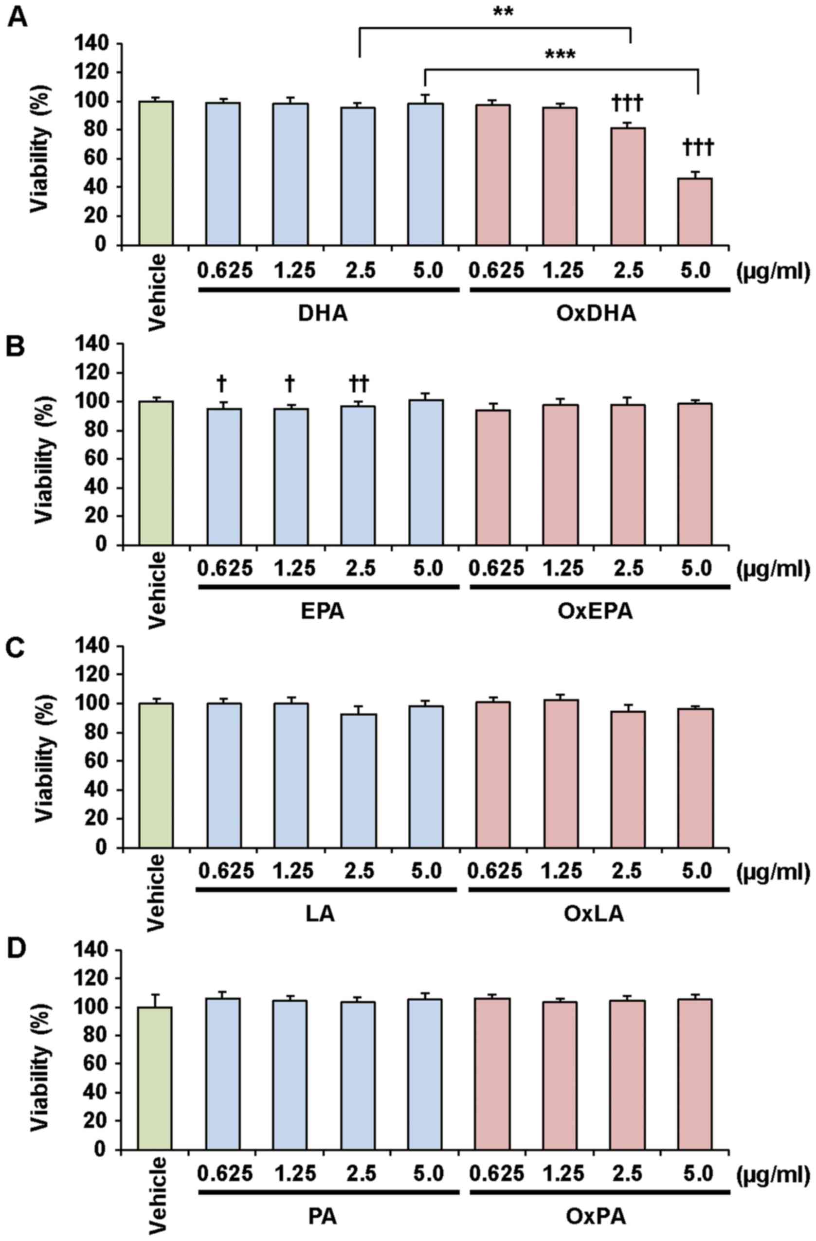
various cultured cells, as determined by the CCK-8 assay (Figs. 2 and 3). Treatment with OxDHA significantly
decreased the proliferation of THP-1 cells in a dose-dependent
manner (Fig. 2A). Native DHA
slightly decreased cell proliferation at high concentrations
(>2.5 µg/ml DHA). OxEPA also decreased the proliferation of
THP-1 cells dose-dependently, but EPA (except for 5.0 µg/ml EPA)
did not (Fig. 2B). OxLA, as well
as OxEPA, slightly decreased the proliferation of THP-1 cells
dose-dependently, but LA (except for 5.0 µg/ml LA) did not
(Fig. 2C). Neither PA nor OxPA
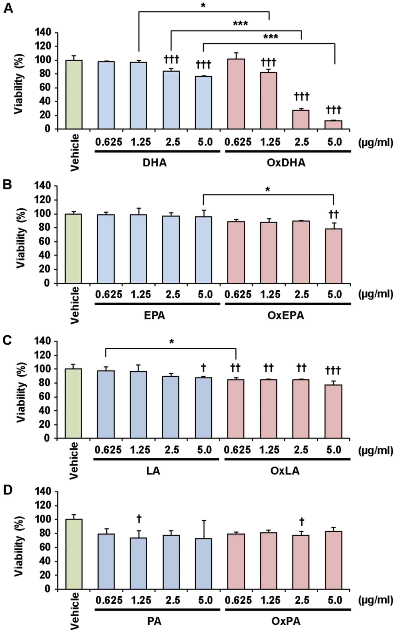
inhibited the proliferation of THP-1 cells (Fig. 2D). As shown in Fig. 3, OxDHA but not DHA inhibited the
proliferation of the DLD-1 cells. Proliferation in DLD-1 cells was
hardly inhibited by EPA, LA, OxEPA, and OxLA, even at high
concentrations (5.0 µg/ml) (Figs. 3B
and 3C). PA and OxPA hardly decreased the proliferation of
DLD-1 cells at all concentrations (Fig. 3C). As shown in Figs. 2 and 3, OxDHA had the most anti-proliferative
effect among these fatty acids. These results indicated that the
anti-proliferative effect of oxidized fatty acids is responsible
for the activity and/or number of oxidized products.
 | Figure 2.Effect of FA and OxFA on THP-1 cell
proliferation. (A) Effect of DHA or OxDHA on cell proliferation.
THP-1 cells were treated with DHA or OxDHA at the indicated
concentrations for 24 h. Cell growth was determined by a Cell
Counting Kit-8 assay, according to the manufacturer's protocol. (B)
Effect of EPA or OxEPA on cell proliferation. (C) Effect of LA or
OxLA on cell proliferation. (D) Effect of PA or OxPA on cell
proliferation. n=3-4. †P<0.05,
††P<0.01, †††P<0.001 vs. vehicle;
*P<0.05, ***P<0.001. FA, fatty acid; Ox, oxidized; DHA,
docosahexaenoic acid; EPA, eicosapentaenoic; LA, linoleic acid; PA,
palmitic acid. |
Oxidized DHA, but not DHA induces
death of THP-1 cells
As shown above, treatment of cells with oxidized
unsaturated fatty acids resulted in a decrease in their
proliferation. To investigate whether the oxidized fatty acids
induced death in the cultured cells, the THP-1 cells were analyzed
using the propidium iodide (PI) exclusion assay. PI can only enter
the dead cells, as their membranes are disrupted. As described in
Fig. 4A, samples treated with
OxDHA, but not with DHA contained PI-positive cells (dead cells).
OxDHA-induced cell death was significantly suppressed by a
pan-caspase inhibitor (zVAD-fmk) (Fig.
4A). As presented in Fig. 4B,
the anti-proliferative effect of OxDHA was partially suppressed by
zVAD-fmk (Fig. 4B). Palmitic acid
did not inhibit cell proliferation in the presence of zVAD-fmk
(Fig. 4C). In both the cell
proliferation and the PI exclusion assay, the OxDHA-induced
decrease of viability was suppressed by pre-treatment with
zVAD-fmk. These results suggest that OxDHA decreased cell
proliferation through the activation of caspases, followed by cell
death.
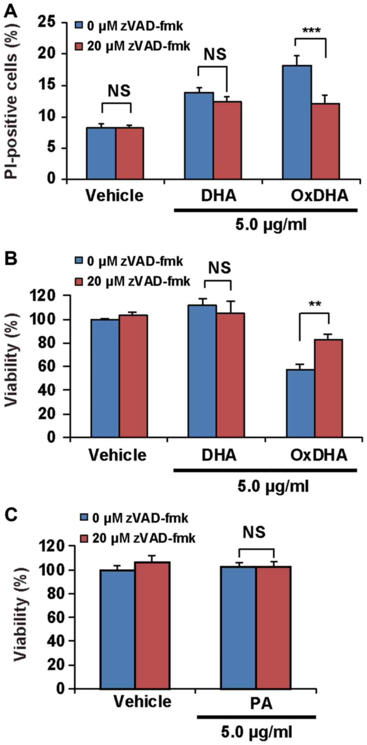 | Figure 4.OxDHA induces cell death through
caspase activation in THP-1 cells. (A) THP-1 cells were treated
with vehicle, DHA, or OxDHA in the presence or absence of zVAD-fmk
for 24 h. Subsequently, cell death was assessed by PI staining. The
THP-1 cells treated with vehicle, DHA, or OxDHA were analyzed for
cell death using flow cytometry. (B) THP-1 cells were treated with
vehicle, DHA, or OxDHA in the presence or absence of zVAD-fmk for
24 h. Cell growth was determined by a CCK-8 assay. n=3-4. (C) THP-1
cells were treated with vehicle or PA in the presence or absence of
zVAD-fmk for 24 h. Cell growth was determined by CCK-8 assay. n=4.
**P<0.01, ***P<0.001. Ox, oxidized; DHA, docosahexaenoic
acid; PI, propidium iodide; CCK-8, Cell Counting Kit-8; PA,
palmitic acid. |
Oxidized DHA, but not DHA induces
apoptosis in THP-1 cells
During early apoptosis, phosphatidylserine on the
outer membrane of cells was exposed. As shown in Fig. 5A, the analysis using the Annexin
V-Cy3 reagent, which stains phosphatidylserine residues, showed
that the number of Annexin V-positive cells was high in the
OxDHA-treated samples (Fig. 5A).
Apoptosis is generally accompanied by chromatin shrinkage,
chromatin condensation, and nuclear fragmentation. We analyzed the
changes in chromatin morphology in THP-1 cells treated with OxDHA.
As shown in Fig. 5B, THP-1 cells
treated with OxDHA but not DHA or PA exhibited typical apoptotic
features, such as chromatin condensation and fragmentation
(Fig. 5B). These results suggested
that OxDHA induced apoptotic cell death.
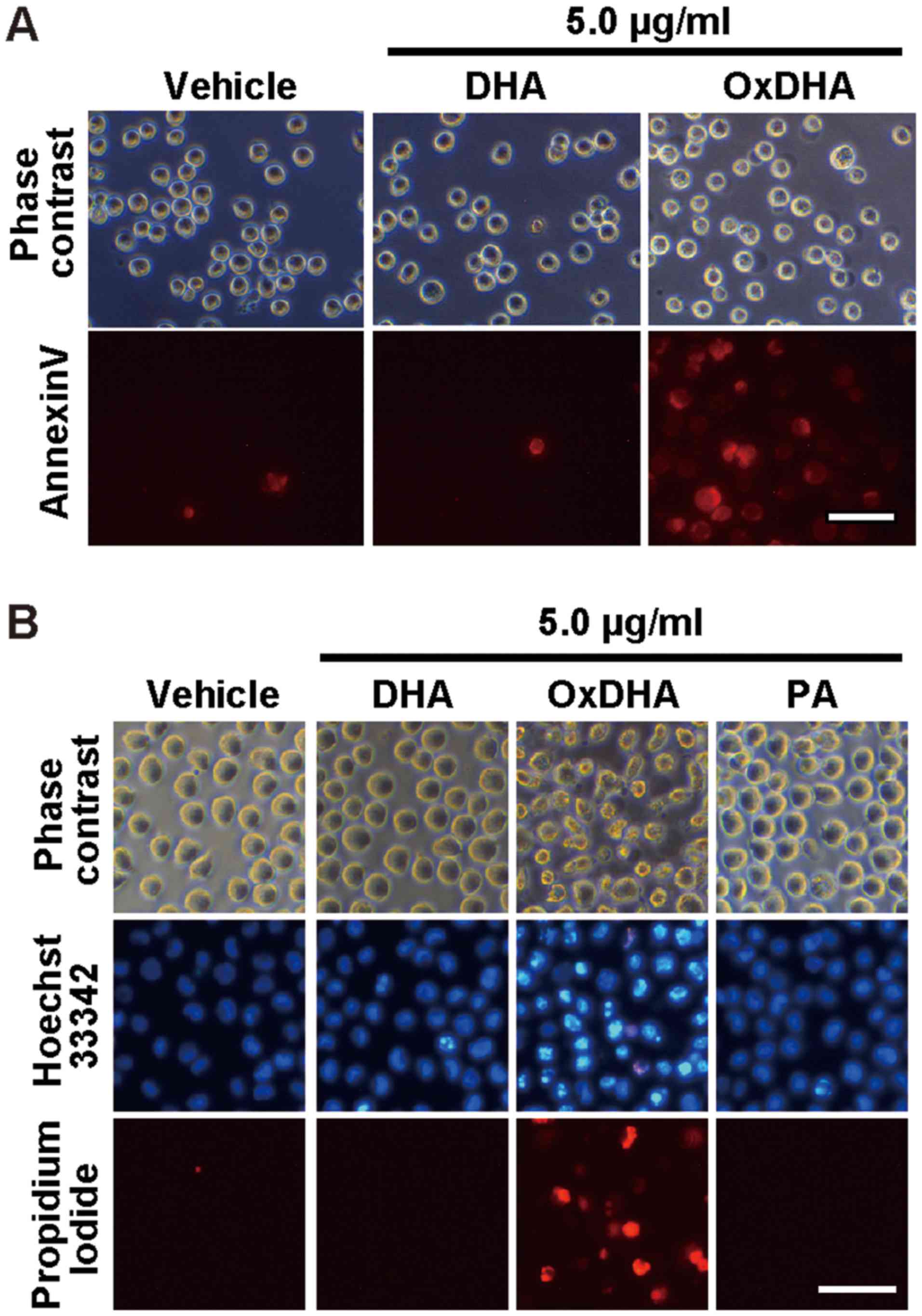 | Figure 5.OxDHA induces apoptosis of THP-1
cells. (A) THP-1 cells were treated with vehicle, DHA, or OxDHA for
24 h at the indicated concentrations. Subsequently, cell death was
assessed using Annexin V assay. (B) Apoptotic nuclear morphological
changes of THP-1 cells treated with OxDHA. THP-1 cells were treated
with vehicle, DHA, OxDHA, or PA for 48 h at the indicated
concentrations. Then, the cells were stained with Hoechst 33342
Propidium Iodide and observed under a fluorescence microscope.
Apoptotic cells had condensed or fragmented chromatin. Scale bar,
50 µm. Ox, oxidized; DHA, docosahexaenoic acid; PA, palmitic
acid. |
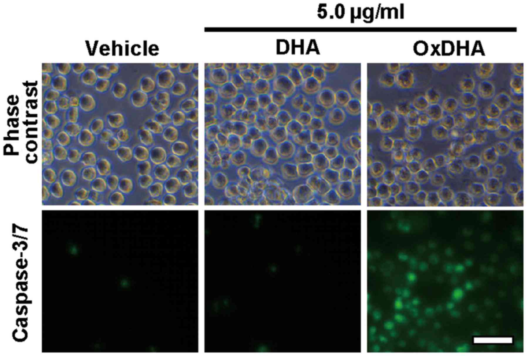
Oxidized DHA but not DHA induces
caspase-3/7 activation in THP-1 cells
Fig. 4 showed that
OxDHA-induced cell death was inhibited by a pan-caspase inhibitor.
Next, we investigated whether the members of the caspase family are
required for the induction of apoptosis by OxDHA. Caspase-3/7 were
activated in THP-1 cells treated with OxDHA; this was not observed
in THP-1 cells treated with DHA and vehicle (Fig. 6). These results indicate that
caspase-3/7 activation is involved in apoptotic cell death induced
by OxDHA.
Discussion
In this study, the oxidized products from
unsaturated fatty acids (DHA, EPA, and LA) exhibited an
anti-proliferative activity in cultured human cells. Among the
oxidized fatty acids, OxDHA had the most significant
anti-proliferative activity. The numbers of double bonds of DHA,
EPA, LA, or PA are 6, 5, 2, or 0, respectively (Fig. 1A). DHA has the most number of
double bonds, and is most susceptible to oxidation. DHA has six
double bonds and bis-allylic methylene groups, which have
several possible positions for hydrogen abstraction. Therefore, DHA
can be easily oxidized into highly active products in the air as
well as under intracellular conditions. These chemical properties
are the reason why OxDHA produces more toxic products (e.g.,
various hydroperoxides, aldehydes, and other products) than OxEPA
and OxLA. Among these fatty acids, OxDHA has the most number of
highly active oxidized products.
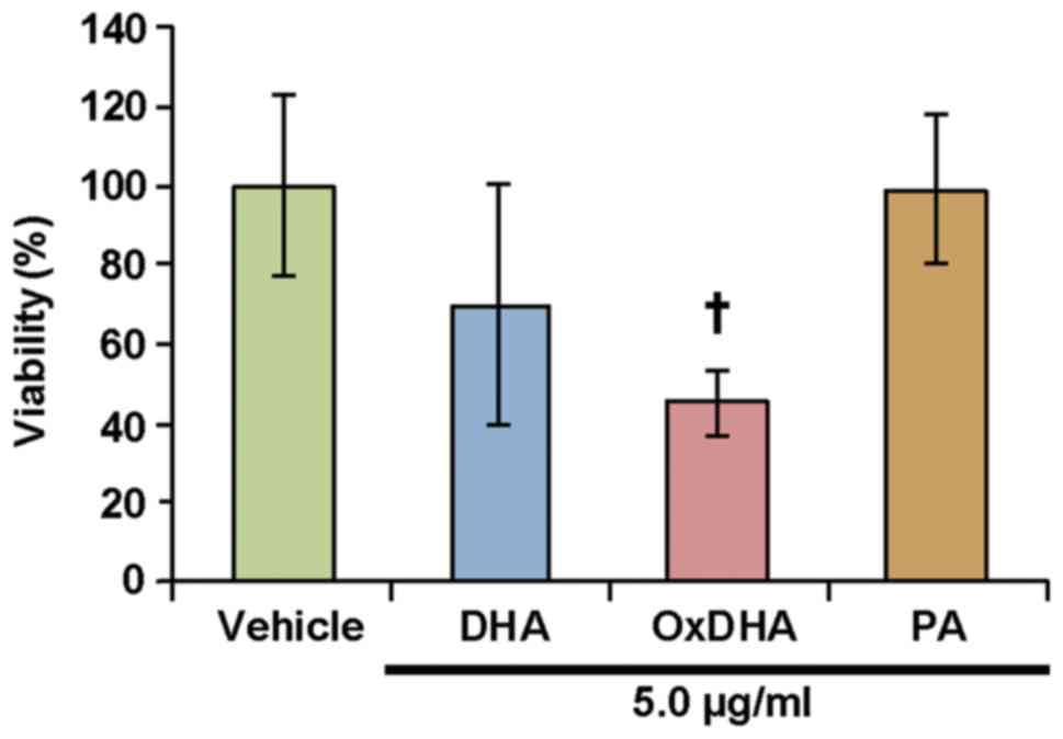
Suspension cells (THP-1 cells and lymphocytes) were
sensitive to native and oxidized fatty acids (Figs. 2 and 7). However, only OxDHA induced a loss of
viability of DLD-1 cells, which are adherent cells. Neither DHA nor
OxDHA inhibited proliferation in Human Hepatoma HepG2 cells (data
not shown). These results suggest that the sensitivity of blood
cells to oxidized fatty acids is higher than those of solid cancer
cells. In general, human leukemia cell lines are highly sensitive
to ROS-generating agents.
As mentioned previously, OxDHA has the most potent
anti-proliferative activity. DHA has the most number of double
bonds; therefore, it has the most unsaturated fatty acids and
sensitivity to ROS. In contrast, saturated fatty acids, such as PA,
are resistant to oxidation when exposed to ROS. As shown in
Figs. 2 and 3, neither PA nor OxPA had an
anti-proliferative activity in this study. To date, the oxidation
of unsaturated fatty acids generates many diverse oxidized
products, such as 4-hydroxy-2-nonenal (HNE), malondialdehyde (MDA),
acrolein, 13-hydroperoxylinoleic acid (13-HPODE), and
F2-isoprostanes (29–33).
In the present study, OxDHA and OxEPA had a light-yellow color, but
native DHA and EPA did not (data not shown). It is reported that
DHA is oxidized by oxidizing agents such as 2,2′-azobis-
(2-amidinopropane) hydrochloride (AAPH) or copper ions. In the
chemical reaction, a family of F4-isoprostanes were
produced from DHA (34).
A4/J4-neuroprostanes were identified by the mass spectrometry
analysis of AAPH-treated DHA, which has a cytoprotective effect and
an anti-inflammatory activity (35). Previously, using mass spectroscopy,
we had described that a large number of bioactive oxidized lipids
were produced by the autoxidation of PAPC in an in vitro
cell-free system (25). Therefore,
the OxDHA used in this study may also contain various bioactive
oxidized fatty acids. The anti-proliferative activity of native
polyunsaturated fatty acids may be dependent on their oxidized
products in cells.
15d-PGJ2, a potent inhibitor of
mitochondrial respiratory complex I, increases the rate of ROS
production (36). Therefore, OxDHA
may inhibit mitochondrial respiratory complexes. DHA induced
apoptotic cell death through ROS production and caspase-8
activation (37). In our study,
caspase-3/7 was activated in THP-1 cells treated with OxDHA. In
general, the caspase-3/7 is activated by mitochondrial dysfunction
followed by caspase-8 and/or caspase-9 activation. Collectively,
these results and reports suggest that OxDHA may activate caspase-8
and/or caspase-9 via mitochondrial dysfunction.
DHA blocks the NF-κB pathway and significantly
decreases cell proliferation (38). 13-HPODE upregulates the expression
of the chemokine monocyte chemoattractant protein-1 via the
activation of NF-κB (39). Lipid
peroxidation products activate the NF-κB pathway and inactivate
anti-apoptotic B-Cell Lymphoma-2 (Bcl-2) (40,41).
The main cellular function of Bcl-2 is the regulation of cytochrome
c release (42,43). Thus, the apoptotic cell death
induced by OxDHA may be associated with NF-κB activation and the
change in the level of Bcl-2.
In summary, we have demonstrated that oxidized fatty
acids produced from unsaturated fatty acids play important roles in
the apoptotic cell death pathway. The oxidized fatty acid products
regulate not only apoptosis, but also non-apoptotic cell death
induction (ferroptosis, autophagy, and regulated neutrophil cell
death in immune response) (44–47).
OxDHA may be useful for studying the mechanism of cell death
induced by lipid peroxidation, which is involved in a wide variety
of diseases.
Acknowledgements
Not applicable.
Funding
Financial support for this research was provided by
Grants-in-Aid for Scientific Research of Seikei University (Tokyo,
Japan; grant no. 2017) and Scientific Research from the Japan
Society for the Promotion of Science (grant nos. 15K16524 and
18K11001 to KI).
Availability of data and materials
All data generated and/or analyzed during the
current study are available from the corresponding author on
reasonable request.
Authors' contributions
KI, ME, MS and HH conceived and designed the
experiments. KI, ME, MS and CY performed the experiments. KI, ME,
MS and HH contributed to data analysis. KI, ME, MS and HH
contributed to manuscript preparation. All authors read and
approved the final manuscript.
Ethics approval and consent to
participate
The present study was approved by the Institutional
Review of Committee of Seikei University (Tokyo, Japan). Written
informed consent was obtained from volunteers in accordance with
the Declaration of Helsinki.
Patient consent for publication
Not applicable.
Competing interests
The authors declare that they have no competing
interests.
References
|
1
|
Wang W, Zhu J, Lyu F, Panigrahy D, Ferrara
KW, Hammock B and Zhang G: ω-3 polyunsaturated fatty acids-derived
lipid metabolites on angiogenesis, inflammation and cancer.
Prostaglandins Other Lipid Mediat. 113-115:13–20. 2014. View Article : Google Scholar : PubMed/NCBI
|
|
2
|
Niki E: Biomarkers of lipid peroxidation
in clinical material. Biochim Biophys Acta. 1840:809–817. 2014.
View Article : Google Scholar : PubMed/NCBI
|
|
3
|
Calder PC: Polyunsaturated fatty acids and
inflammation. Biochem Soc Trans. 33:423–427. 2005. View Article : Google Scholar : PubMed/NCBI
|
|
4
|
Zajdel A, Wilczok A, Chodurek E, Gruchlik
A and Dzierzewicz Z: Polyunsaturated fatty acids inhibit melanoma
cell growth in vitro. Acta Pol Pharm. 70:365–369. 2013.PubMed/NCBI
|
|
5
|
Yee LD, Young DC, Rosol TJ, Vanbuskirk AM
and Clinton SK: Dietary (n-3) polyunsaturated fatty acids inhibit
HER-2/neu-induced breast cancer in mice independently of the
PPARgamma ligand rosiglitazone. J Nutr. 135:983–988. 2005.
View Article : Google Scholar : PubMed/NCBI
|
|
6
|
Notarnicola M, Tutino V, De Nunzio V,
Dituri F, Caruso MG and Giannelli G: Dietary ω-3 polyunsaturated
fatty acids inhibit tumor growth in transgenic ApcMin/+ mice,
correlating with CB1 receptor Up-Regul. Int J Mol Sci. 18(pii):
E4852017. View Article : Google Scholar : PubMed/NCBI
|
|
7
|
Notarnicola M, Messa C, Refolo MG, Tutino
V, Miccolis A and Caruso MG: Polyunsaturated fatty acids reduce
fatty acid synthase and hydroxy-methyl-glutaryl CoA-reductase gene
expression and promote apoptosis in HepG2 cell line. Lipids Health
Dis. 10:102011. View Article : Google Scholar : PubMed/NCBI
|
|
8
|
Yin Y, Sui C, Meng F, Ma P and Jiang Y:
The omega-3 polyunsaturated fatty acid docosahexaenoic acid
inhibits proliferation and progression of non-small cell lung
cancer cells through the reactive oxygen species-mediated
inactivation of the PI3K/Akt pathway. Lipids Health Dis. 16:872017.
View Article : Google Scholar : PubMed/NCBI
|
|
9
|
D'Eliseo D and Velotti F: Omega-3 fatty
acids and cancer cell cytotoxicity: Implications for multi-targeted
cancer therapy. J Clin Med. 5(pii): E152016. View Article : Google Scholar : PubMed/NCBI
|
|
10
|
Pizato N, Luzete BC, Kiffer LFMV, Corrêa
LH, de Oliveira Santos I, Assumpção JAF, Ito MK and Magalhães KG:
Omega-3 docosahexaenoic acid induces pyroptosis cell death in
triple-negative breast cancer cells. Sci Rep. 8:19522018.
View Article : Google Scholar : PubMed/NCBI
|
|
11
|
Mansara PP, Deshpande RA, Vaidya MM and
Kaul-Ghanekar R: Differential ratios of omega fatty acids
(AA/EPA+DHA) modulate growth, lipid peroxidation and expression of
tumor regulatory MARBPs in breast cancer cell lines MCF7 and
MDA-MB-231. PLoS One. 10:e01365422015. View Article : Google Scholar : PubMed/NCBI
|
|
12
|
Mu YM, Yanase T, Nishi Y, Tanaka A, Saito
M, Jin CH, Mukasa C, Okabe T, Nomura M, Goto K and Nawata H:
Saturated FFAs, palmitic acid and stearic acid, induce apoptosis in
human granulosa cells. Endocrinology. 142:3590–3597. 2001.
View Article : Google Scholar : PubMed/NCBI
|
|
13
|
Zhang Y, Xue R, Zhang Z, Yang X and Shi H:
Palmitic and linoleic acids induce ER stress and apoptosis in
hepatoma cells. Lipids Health Dis. 11:12012. View Article : Google Scholar : PubMed/NCBI
|
|
14
|
Murray M, Dyari HR, Allison SE and Rawling
T: Lipid analogues as potential drugs for the regulation of
mitochondrial cell death. Br J Pharmacol. 171:2051–2066. 2014.
View Article : Google Scholar : PubMed/NCBI
|
|
15
|
Bochkov VN, Oskolkova OV, Birukov KG,
Levonen AL, Binder CJ and Stöckl J: Generation and biological
activities of oxidized phospholipids. Antioxid Redox Signal.
12:1009–1059. 2010. View Article : Google Scholar : PubMed/NCBI
|
|
16
|
Miller YI and Shyy JY: Context-dependent
role of oxidized lipids and lipoproteins in inflammation. Trends
Endocrinol Metab. 28:143–152. 2017. View Article : Google Scholar : PubMed/NCBI
|
|
17
|
Clay CE, Monjazeb A, Thorburn J, Chilton
FH and High KP: 15-Deoxy-delta12,14-prostaglandin J2-induced
apoptosis does not require PPARgamma in breast cancer cells. J
Lipid Res. 43:1818–1828. 2002. View Article : Google Scholar : PubMed/NCBI
|
|
18
|
Chen R, Yang L and McIntyre TM: Cytotoxic
phospholipid oxidation products. Cell death from mitochondrial
damage and the intrinsic caspase cascade. J Biol Chem.
282:24842–24850. 2007. View Article : Google Scholar : PubMed/NCBI
|
|
19
|
Wang L, Gill R, Pedersen TL, Higgins LJ,
Newman JW and Rutledge JC: Triglyceride-rich lipoprotein lipolysis
releases neutral and oxidized FFAs that induce endothelial cell
inflammation. J Lipid Res. 50:204–213. 2009. View Article : Google Scholar : PubMed/NCBI
|
|
20
|
Liu X, Shibata T, Hisaka S, Kawai Y and
Osawa T: DHA hydroperoxides as a potential inducer of neuronal cell
death: A mitochondrial dysfunction-mediated pathway. J Clin Biochem
Nutr. 43:26–33. 2008. View Article : Google Scholar : PubMed/NCBI
|
|
21
|
Lim SY, Jang JH, Na HK, Lu SC, Rahman I
and Surh YJ: 15-Deoxy-Delta12,14-prostaglandin J(2) protects
against nitrosative PC12 cell death through up-regulation of
intracellular glutathione synthesis. J Biol Chem. 279:46263–46270.
2004. View Article : Google Scholar : PubMed/NCBI
|
|
22
|
Gao B, Han YH, Wang L, Lin YJ, Sun Z, Lu
WG, Hu YQ, Li JQ, Lin XS, Liu BH, et al: Eicosapentaenoic acid
attenuates dexamethasome-induced apoptosis by inducing adaptive
autophagy via GPR120 in murine bone marrow-derived mesenchymal stem
cells. Cell Death Dis. 7:e22352016. View Article : Google Scholar : PubMed/NCBI
|
|
23
|
Yeh A, Kruse SE, Marcinek DJ and Gallagher
EP: Effect of omega-3 fatty acid oxidation products on the cellular
and mitochondrial toxicity of BDE 47. Toxicol In Vitro. 29:672–680.
2015. View Article : Google Scholar : PubMed/NCBI
|
|
24
|
Haeiwa H, Fujita T, Saitoh Y and Miwa N:
Oleic acid promotes adaptability against oxidative stress in 3T3-L1
cells through lipohormesis. Mol Cell Biochem. 386:73–83. 2014.
View Article : Google Scholar : PubMed/NCBI
|
|
25
|
Iuchi K, Imoto A, Kamimura N, Nishimaki K,
Ichimiya H, Yokota T and Ohta S: Molecular hydrogen regulates gene
expression by modifying the free radical chain reaction-dependent
generation of oxidized phospholipid mediators. Sci Rep.
6:189712016. View Article : Google Scholar : PubMed/NCBI
|
|
26
|
Abdelmagid SA, Clarke SE, Nielsen DE,
Badawi A, El-Sohemy A, Mutch DM and Ma DW: Comprehensive profiling
of plasma fatty acid concentrations in young healthy Canadian
adults. PLoS One. 10:e01161952015. View Article : Google Scholar : PubMed/NCBI
|
|
27
|
Eguchi Y, Shimizu S and Tsujimoto Y:
Intracellular ATP levels determine cell death fate by apoptosis or
necrosis. Cancer Res. 57:1835–1840. 1997.PubMed/NCBI
|
|
28
|
Schley PD, Jijon HB, Robinson LE and Field
CJ: Mechanisms of omega-3 fatty acid-induced growth inhibition in
MDA-MB-231 human breast cancer cells. Breast Cancer Res Treat.
92:187–195. 2005. View Article : Google Scholar : PubMed/NCBI
|
|
29
|
Perluigi M, Coccia R and Butterfield DA:
4-Hydroxy-2-nonenal, a reactive product of lipid peroxidation, and
neurodegenerative diseases: A toxic combination illuminated by
redox proteomics studies. Antioxid Redox Signal. 17:1590–1609.
2012. View Article : Google Scholar : PubMed/NCBI
|
|
30
|
Milne GL, Yin H, Hardy KD, Davies SS and
Roberts LJ II: Isoprostane generation and function. Chem Rev.
111:5973–5996. 2011. View Article : Google Scholar : PubMed/NCBI
|
|
31
|
Usatyuk PV and Natarajan V:
Hydroxyalkenals and oxidized phospholipids modulation of
endothelial cytoskeleton, focal adhesion and adherens junction
proteins in regulating endothelial barrier function. Microvasc Res.
83:45–55. 2012. View Article : Google Scholar : PubMed/NCBI
|
|
32
|
Esterbauer H, Schaur RJ and Zollner H:
Chemistry and biochemistry of 4-hydroxynonenal, malonaldehyde and
related aldehydes. Free Radic Biol Med. 11:81–128. 1991. View Article : Google Scholar : PubMed/NCBI
|
|
33
|
Meilhac O, Zhou M, Santanam N and
Parthasarathy S: Lipid peroxides induce expression of catalase in
cultured vascular cells. J Lipid Res. 41:1205–1213. 2000.PubMed/NCBI
|
|
34
|
Nourooz-Zadeh J, Liu EH, Anggård E and
Halliwell B: F4-isoprostanes: A novel class of prostanoids formed
during peroxidation of docosahexaenoic acid (DHA). Biochem Biophys
Res Commun. 242:338–344. 1998. View Article : Google Scholar : PubMed/NCBI
|
|
35
|
Majkova Z, Layne J, Sunkara M, Morris AJ,
Toborek M and Hennig B: Omega-3 fatty acid oxidation products
prevent vascular endothelial cell activation by coplanar
polychlorinated biphenyls. Toxicol Appl Pharmacol. 251:41–49. 2011.
View Article : Google Scholar : PubMed/NCBI
|
|
36
|
Martínez B, Pérez-Castillo A and Santos A:
The mitochondrial respiratory complex I is a target for
15-deoxy-delta12,14-prostaglandin J2 action. J Lipid Res.
46:736–743. 2005. View Article : Google Scholar : PubMed/NCBI
|
|
37
|
Kang KS, Wang P, Yamabe N, Fukui M, Jay T
and Zhu BT: Docosahexaenoic acid induces apoptosis in MCF-7 cells
in vitro and in vivo via reactive oxygen species formation and
caspase 8 activation. PLoS One. 5:e102962010. View Article : Google Scholar : PubMed/NCBI
|
|
38
|
Yun EJ, Song KS, Shin S, Kim S, Heo JY,
Kweon GR, Wu T, Park JI and Lim K: Docosahexaenoic acid suppresses
breast cancer cell metastasis by targeting
matrix-metalloproteinases. Oncotarget. 7:49961–49971. 2016.
View Article : Google Scholar : PubMed/NCBI
|
|
39
|
Dwarakanath RS, Sahar S, Reddy MA,
Castanotto D, Rossi JJ and Natarajan R: Regulation of monocyte
chemoattractant protein-1 by the oxidized lipid,
13-hydroperoxyoctadecadienoic acid, in vascular smooth muscle cells
via nuclear factor-kappa B (NF-kappa B). J Mol Cell Cardiol.
36:585–595. 2004. View Article : Google Scholar : PubMed/NCBI
|
|
40
|
Timucin AC and Basaga H: Pro-apoptotic
effects of lipid oxidation products: HNE at the crossroads of NF-κB
pathway and anti-apoptotic Bcl-2. Free Radic Biol Med. 111:209–218.
2017. View Article : Google Scholar : PubMed/NCBI
|
|
41
|
Yadav UC and Ramana KV: Regulation of
NF-κB-induced inflammatory signaling by lipid peroxidation-derived
aldehydes. Oxid Med Cell Longev. 2013:6905452013. View Article : Google Scholar : PubMed/NCBI
|
|
42
|
Tsujimoto Y: Cell death regulation by the
Bcl-2 protein family in the mitochondria. J Cell Physiol.
195:158–167. 2003. View Article : Google Scholar : PubMed/NCBI
|
|
43
|
Shimizu S and Tsujimoto Y: Proapoptotic
BH3-only Bcl-2 family members induce cytochrome c release, but not
mitochondrial membrane potential loss, and do not directly modulate
voltage-dependent anion channel activity. Proc Natl Acad Sci USA.
97:577–582. 2000. View Article : Google Scholar : PubMed/NCBI
|
|
44
|
Hill BG, Haberzettl P, Ahmed Y, Srivastava
S and Bhatnagar A: Unsaturated lipid peroxidation-derived aldehydes
activate autophagy in vascular smooth-muscle cells. Biochem J.
410:525–534. 2008. View Article : Google Scholar : PubMed/NCBI
|
|
45
|
Xie Y, Hou W, Song X, Yu Y, Huang J, Sun
X, Kang R and Tang D: Ferroptosis: Process and function. Cell Death
Differ. 23:369–379. 2016. View Article : Google Scholar : PubMed/NCBI
|
|
46
|
Yotsumoto S, Muroi Y, Chiba T, Ohmura R,
Yoneyama M, Magarisawa M, Dodo K, Terayama N, Sodeoka M, Aoyagi R,
et al: Hyperoxidation of ether-linked phospholipids accelerates
neutrophil extracellular trap formation. Sci Rep. 7:160262017.
View Article : Google Scholar : PubMed/NCBI
|
|
47
|
Parisi LR, Morrow LM, Visser MB and
Atilla-Gokcumen GE: Turning the spotlight on lipids in
non-apoptotic cell death. ACS Chem Biol. 13:506–515. 2018.
View Article : Google Scholar : PubMed/NCBI
|