Introduction
Cardiovascular disease is a major health issue
worldwide, with acute myocardial ischemia accounting for >40%
deaths in China (1). Severe
myocardial ischemia may result in a characteristic pattern of
metabolic and ultrastructural changes (2), which can lead to irreversible injury.
Although several studies have been performed, the molecular
mechanisms underlying myocardial ischemic injury remain unclear
(3,4). In addition, it has been reported that
the occurrence of myocardial ischemia is often accompanied by
severe inflammatory response (5)
and activation of the NLR family pyrin domain-containing 3 (NLRP3)
inflammasome (6).
The NLRP3 inflammasome is a cytosolic protein
complex comprising NLRP3, apoptosis-associated speck-like protein
(ASC) and caspase-1. Notably, the NLRP3 inflammasome is assembled
in response to several endogenous “danger signals”, such as
oxidative stress, lysosome destabilization and mitochondrial
dysfunction (7). Recently, it has
been reported that activation of the NLRP3 inflammasome is
associated with the pathogenesis of numerous inflammatory diseases
(8,9), and that it may regulate the secretion
of several pro-inflammatory cytokines, such as IL-1β and IL-18
(10). Furthermore, inhibition of
the NLRP3 signaling pathway has been shown to protect against
myocardial ischemic injury induced by coronary artery ligation
(11).
Sirtuin 1 (SIRT1) is a member of the sirtuin class
III histone deacetylase family. It serves important roles in
several important biological processes, including mitophagy, cell
senescence and cell apoptosis (12). In recent years, several studies
have focused on the role of SIRT1-mediated mitophagy in the
regulation of NLRP3 activation. These studies suggested that
SIRT1-mediated mitophagy is a negative regulator of NLRP3
inflammasome activation (13) and
could significantly protect against myocardial ischemic injury
(14). However, whether
salvianolic acid B (Sal B) could alleviate myocardial ischemic
injury by promoting SIRT1-mediated mitophagy remains unclear.
Salvia miltiorrhiza Bge has been used to
treat myocardial ischemia in mainland China for >2,000 years.
Compared with chemical drugs, this plant has numerous advantages,
including multi-ingredients (which enables less opportunity for
resistance), multi-targets and few side effects (15,16).
As the main active water-soluble substance in S.
miltiorrhiza Bge, Sal B has been reported to exert several
pharmacological activities, including antitumor (17), anti-inflammatory (18) and antioxidant (19) effects. It has previously been
reported that Sal B may exert protective effects against myocardial
ischemic injury (20). However,
whether Sal B could regulate mitophagy and inhibit activation of
the NLRP3 inflammasome during myocardial ischemic injury remains
unclear. Therefore, the present study investigated whether Sal B
could alleviate myocardial ischemic injury by promoting cell
mitophagy and inactivating the NLRP3 inflammasome.
Materials and methods
Materials
Sal B was from Shanghai Yuanye Biotechnology (cat.
no. B20261). Isoproterenol (ISO) was obtained from Beijing Solarbio
Science & Technology Co., Ltd. (cat. no. II0200). Dulbecco's
modified Eagle's medium (DMEM) containing high glucose was obtained
from Wisent Biotechnology. Trypsin was obtained from Beyotime
Institute of Biotechnology. The creatine kinase isoenzyme MB
(CK-MB; cat. no. H197) and glutamic oxaloacetic transaminase (GOT;
cat. no. C010-2-1) ELISA kits were obtained from Nanjing Jiancheng
Bioengineering Institute. Evagreen 2X quantitative PCR (qPCR)
MasterMix-Low Rox kit and the IL-1β ELISA kit (cat. no. K4796-100)
were purchased from Abmgoodchina, Inc. and BioVision, Inc.,
respectively. Adenosine triphosphate (ATP) was obtained from
Amresco, LLC. Lipopolysaccharide (LPS) was obtained from Cell
Signaling Technology, Inc. Primary antibodies against SIRT1 (cat.
no. ab189494), microtubule-associated protein 1A/1B-light chain 3
(LC3; cat. no. ab48394), P62 (cat. no. ab91526), PTEN-induced
kinase 1 (PINK1; cat. no. ab23707), Beclin-1 (cat. no. ab62557),
NLRP3 (cat. no. ab214185), IL-1β (cat. no. ab205924) were purchased
from Abcam, and primary antibodies against Parkin (cat. no.
14060-1-AP), caspase-1 (cat. no. 22915-1-AP) and GAPDH (cat. no.
10494-1-AP) were purchased from Proteintech Group, Inc. The
distilled water used in this study was purified using a Milli-Q
system (EMD Millipore). All culture plates were purchased from
Corning, Inc.
Animals
Male Sprague-Dawley (SD) rats (age, 2 months;
weight, 180–220 g), were housed in pathogen-free conditions under a
12-h light/dark cycle at 22–24°C (humidity, 40–70%) with ad
libitum access to a normal pellet diet and clean water. The
protocol of the present study was approved by the Animal Ethics
Committee of Nanjing University of Chinese Medicine (Nanjing,
China; approval no. 201906A036).
A total of 50 male SD rats were randomly divided
into the following five groups (n=10/group): i) control group; ii)
model group; iii) model + Sal B (5 mg/kg) group; iv) model + Sal B
(10 mg/kg) group; and v) model + Sal B (15 mg/kg) group. The rats
in the Sal B treatment groups received a daily intraperitoneal
injection of Sal B (dissolved in PBS) for 7 consecutive days,
whereas rats in the control and model groups were treated with
equal amounts of normal saline. Furthermore, the acute myocardial
ischemia model was induced by hypodermic injection of ISO (30
mg/kg/d) once daily from the 5th day. Notably, the modelling method
of ISO-induced myocardial ischemia has previously been confirmed to
be effective (21,22). Following treatment with Sal B for
24 h on the 7th day, all rats were anesthetized by intraperitoneal
injection of pentobarbital sodium solution (50 mg/kg) and a total
of 2 ml blood was withdrawn from the abdominal aorta. Subsequently,
animals were euthanized with 5% pentobarbital sodium solution (150
mg/kg), and the hearts were harvested for a series of pathological
and biochemical studies. Euthanasia should result in rapid loss of
consciousness, followed by respiratory and cardiac arrest, and
ultimate loss of all brain function. Death was confirmed by
checking the breathing rate, heartbeat and pupillary reflexes after
euthanasia and prior to disposal of the animal.
Determination of ST-segment
elevation
Following anesthesia by intraperitoneal injection of
50 mg/kg sodium pentobarbital, the electrocardiogram (ECG) indexes
of rats were determined using a power-lab system (ADInstruments,
Ltd.). Myocardial ischemia was verified by ST-segment elevation
recorded by ECG and the results were expressed as relative to the
control.
Hematoxylin and eosin (H&E)
staining
The heart tissues were fixed in 10% neutral buffered
formalin for 24 h at room temperature, embedded in paraffin and
were then sliced into 5-µm conventional sections using a microtome.
Subsequently, the embedded blocks were prepared into pathological
sections. The sections were stained with H&E for 5 min at room
temperature. The sections were then mounted and histologically
observed under a light microscope (magnification, ×200).
Determination of serum CK-MB, GOT and
IL-1β concentrations
Blood samples were collected from the abdominal
aorta of each rat 24 h after Sal B administration on the 7th day
and the serum was obtained by centrifugation at 1,500 × g for 10
min at room temperature. Subsequently, the serum levels of CK-MB,
GOT and IL-1β were determined using a microplate system (BioTek
Instruments, Inc.) and the aforementioned ELISA kits, according to
the manufacturer's instructions.
Reverse transcription (RT)-qPCR
Total RNA was extracted from heart tissues using
TRIzol® reagent (Invitrogen; Thermo Fisher Scientific,
Inc.) and RNA concentration was determined using a NanoDrop
(NanoDrop Technologies; Thermo Fisher Scientific, Inc.). Total RNA
was reverse transcribed into cDNA using an 5X All-in-one RT
MasterMix kit (Abmgoodchina Inc.), according to the manufacturer's
instructions. Subsequently, qPCR was performed using the Evagreen
2X qPCR MasterMix-Low Rox kit (Abmgoodchina Inc.) on the ABI 7500
Fast Real-Time PCR Detection system (Applied Biosystems; Thermo
Fisher Scientific, Inc.). The following thermocycling conditions
were used for the qPCR: Initial denaturation at 95°C for 10 min;
followed by 40 cycles of 95°C for 15 sec and 60°C for 60 sec. The
mRNA expression levels of NLRP3, ASC, caspase-1 and IL-1β were
determined using the 2−ΔΔCq method (23) and were normalized to GAPDH
expression levels. The sequences of the specific primers used for
qPCR analysis are listed in Table
I.
 | Table I.Primer sequences used for
quantitative PCR. |
Table I.
Primer sequences used for
quantitative PCR.
| Gene | Forward sequence
(5′→3′) | Reverse sequence
(5′→3′) |
|---|
| NLRP3 |
CTCTGCATGCCGTATCTGGT |
GTCCTGAGCCATGGAAGCAA |
| Caspase-1 |
GACCGAGTGGTTCCCTCAAG |
GACGTGTACGAGTGGGTGTT |
| ASC |
GGACAGTACCAGGCAGTTCG |
GTCACCAAGTAGGGCTGTGT |
| IL-1β |
GACTTCACCATGGAACCCGT |
GGAGACTGCCCATTCTCGAC |
| GAPDH |
GGCAAATTCAACGGCACAGT |
AGATGGTGATGGGCTTCCC |
Cell culture and treatment
The H9C2 cell line, which is widely used to study
myocardial ischemia (24,25), was obtained from the American Type
Culture Collection. Cells were cultured in high-glucose DMEM
supplemented with 10% fetal bovine serum (Gibco; Thermo Fisher
Scientific, Inc.) at 37°C in a 5% CO2 atmosphere.
H9C2 cells were seeded into a 6-well plate at a
density of 5×10⁴ cells and cultured overnight. Subsequently, the
cells were randomly divided into the following five groups: i)
Control group; ii) LPS + ATP group; iii) LPS + ATP + Sal B (1 µM)
group; iv) LPS + ATP + Sal B (5 µM) group; and v) LPS + ATP + Sal B
(25 µM) group. Cells in Sal B treatment groups were pretreated with
different concentrations of Sal B (1, 5 and 25 µM) for 24 h at
37°C. After discarding the supernatant, all cells, except within
the control group, were washed twice with PBS and stimulated with
LPS (1 µg/ml) for 24 h at 37°C. Subsequently, cells were stimulated
with ATP (5 mM) for 2 h at 37°C. Cells in the control group were
cultured normally (high-glucose DMEM supplemented with 10% FBS at
37°C). Finally, all cells in all groups were collected for
subsequent analyses.
Western blot analysis
Cultured H9C2 cells were lysed in RIPA lysis buffer
(Beyotime Institute of Biotechnology) and the total protein
concentration was quantified using a BCA assay (Beyotime Institute
of Biotechnology). Total protein samples (50 µg) were separated by
SDS-PAGE on 6–10% gels. The total protein samples were then
electrotransferred onto polyvinylidene fluoride membranes. After
blocking with 5% non-fat milk in TBS-0.05% Tween-20 for 1 h at room
temperature, the membranes were incubated with primary antibodies
against NLRP3 (1:1,000), caspase-1 (1:1,000), IL-1β (1:1,000),
SIRT1 (1:1,000), P62 (1:1,000), Parkin (1:1,000), PINK1 (1:1,000),
Beclin-1 (1:1,000), LC3 (1:1,000) and GAPDH (1:5,000) at 4°C
overnight with gentle agitation. Subsequently, the membranes were
incubated with corresponding horseradish peroxidase-conjugated
secondary antibodies (dilution, 1:1,000; Beyotime Institute of
Biotechnology; cat. no. A0208) in non-fat milk for 1 h at room
temperature. The membranes were visualized using an ECL kit
(Beyotime Institute of Biotechnology) and densitometric analysis
was performed using Image Lab software version 4.0.1 (Bio-Rad
Laboratories, Inc.)
Measurement of intracellular reactive
oxygen species (ROS) generation
The intracellular ROS levels were measured using
2,7-dichlorofluorescin diacetate (DCFH-DA; cat. no. S0033S;
Beyotime Institute of Biotechnology). In the presence of ROS,
DCFH-DA is oxidized into fluorescent dichlorofluorescein (DCF)
(26). After treatment, the
collected cells (1×10⁶/well) were resuspended in 1 ml
DCFH-DA reagent (10 µM) and incubated at 37°C for 30 min in the
dark. Images were captured using a fluorescence microscope
(magnification, ×200) and the mean density was analyzed using
ImageJ software version 1.8.0 (National Institutes of Health) using
the following formula: Mean density = integrated density/total
area. The fluorescence intensity of DCF was measured by flow
cytometry (BD LSR II; BD Biosciences) and analyzed using FACSDiva
software version 4.1 (BD Biosciences).
TUNEL assay
Apoptosis was analyzed using the One Step TUNEL
Apoptosis Assay kit (cat. no. C1090; Beyotime Institute of
Biotechnology), according to the manufacturer's instructions.
Briefly, H9C2 cells were seeded into a 6-well plate at a density of
5×10⁴ cells. Cells were co-treated with Sal B and LPS +
ATP (stimulation) as aforementioned. Subsequently, cells were fixed
in 4% paraformaldehyde for 30 min at 37°C, permeabilized in 0.1%
Triton X-100 for 2 min and incubated with TUNEL assay reagents for
1 h at 37°C. Images were captured using a fluorescence microscope
and fluorescence was measured using a fluorescence
spectrophotometer (absorbance wavelength, 550 nm). The mean density
was determined as previously described above.
Mitochondrial membrane potential (MMP)
measurement
An MMP assay kit with JC-1 (cat. no. C2006; Beyotime
Institute of Biotechnology) was used to evaluate MMP, according to
the manufacturer's protocol. Briefly, 1×10⁶ cells/well were
incubated with JC-1 for 30 min at 37°C, rinsed twice with PBS and
the staining images were captured under a fluorescence microscope.
The MMP was indicated by the ratio of red/green fluorescence
intensity.
Statistical analysis
Experiments were repeated ≥3 times. Data are
presented as the mean ± standard deviation. The statistically
significant differences between the means of multiple groups were
analyzed by one-way ANOVA followed by Tukey's multiple comparison
test. All statistical analyses were performed using GraphPad Prism
7 software (GraphPad Software, Inc.). P<0.05 was considered to
indicate a statistically significant difference.
Results
Echocardiography of rats with acute
myocardial ischemia
Standard echocardiography was performed at room
temperature for all groups of rats 24 h after the administration of
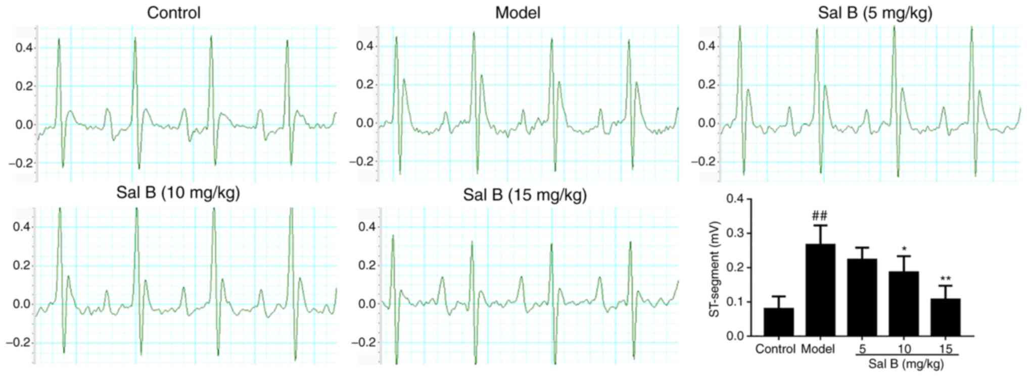
Sal B on the 7th day. Control rats exhibited a normal ECG pattern
with regular sinus rhythm (Fig.
1). However, an irregular sinus rhythm with significant
elevation of the ST-segment was observed in the ECGs of ISO-induced
model rats compared with those of control rats. Furthermore, 10 and
15 mg/kg Sal B treatment markedly reversed these ISO-induced
alterations (Fig. 1).
Pathological observation
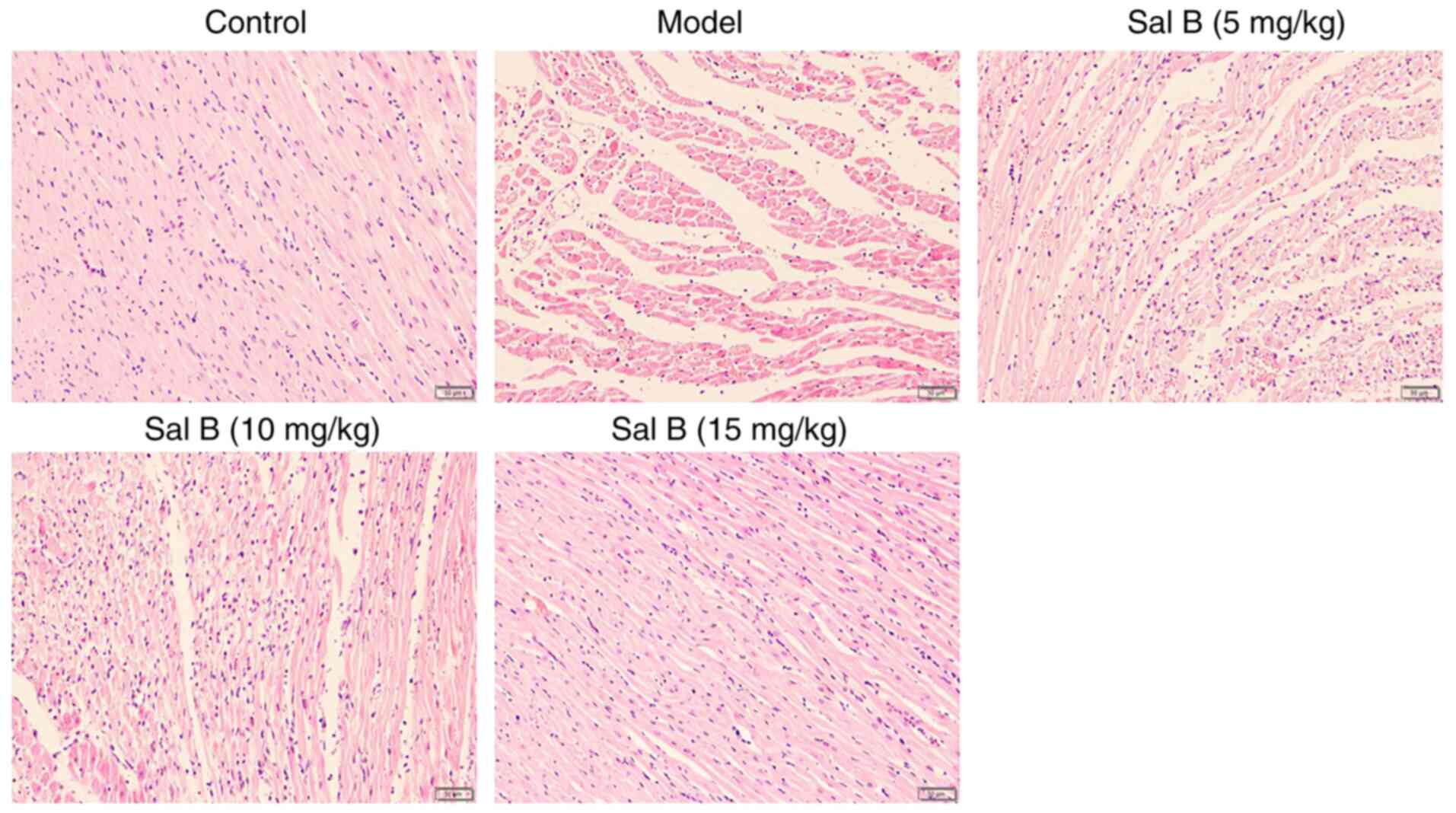
The results of H&E staining revealed that the
hearts of rats in the control group maintained a normal form and
function (Fig. 2). However, the
hearts of rats in the model group exhibited obvious rupture and
lysis of myocardial fibers, accompanied by marked inflammatory cell
infiltration. Notably, treatment with Sal B alleviated these
pathological observations (Fig.
2).
Sal B reduces the levels of CK-MB, GOT
and IL-1β in the serum of rats with myocardial ischemia
Elevation in the serum levels of cardiac markers,
such as CK-MB and GOT, is an important basis for the diagnosis of
acute myocardial ischemia (27).
To evaluate the effects of Sal B on myocardial ischemic injury, the
serum levels of CK-MB, GOT and IL-1β were determined. As shown in
Fig. 3, intraperitoneal injection
of ISO significantly increased the levels of CK-MB, GOT and IL-1β
compared with those in the control group; treatment with Sal B (5,
10 and 15 mg/kg) significantly reversed the levels of these
factors.
Effects of Sal B on the mRNA
expression levels of NLRP3, ASC, caspase-1 and IL-1β in rats with
ISO-induced myocardial ischemia
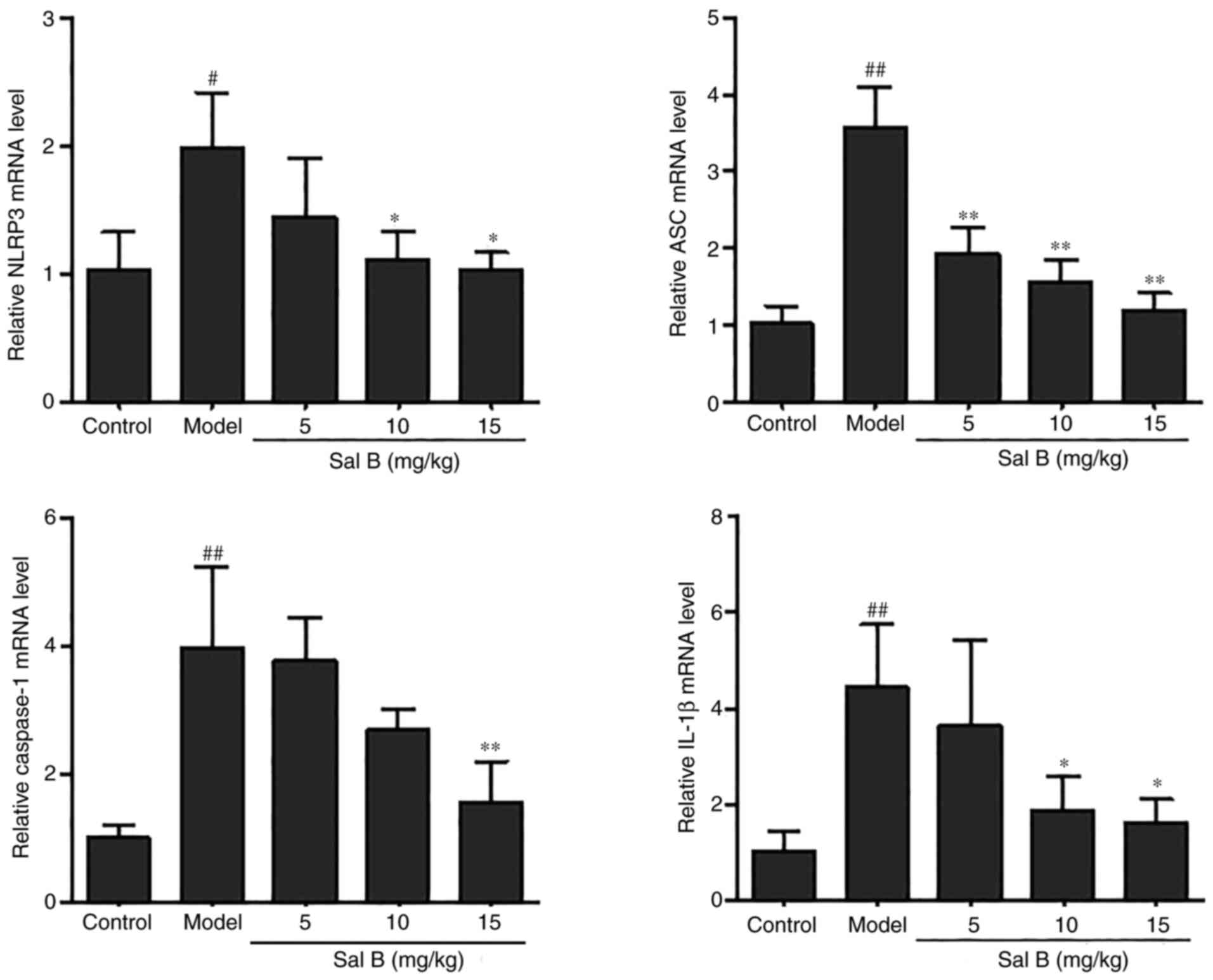
To investigate whether Sal B could inhibit the
expression of the NLRP3 inflammasome in rats with ISO-induced
myocardial ischemia, RT-qPCR analysis was conducted to determine
the mRNA expression levels of the related genes. ISO stimulation
significantly increased the mRNA expression levels of the NLRP3
inflammasome components compared with those in the control group
(Fig. 4). However, the expression
levels of NLRP3, ASC, caspase-1 and IL-1β in the Sal B-treated
group (15 mg/kg) were significantly reduced compared with those in
the model group (Fig. 4).
Sal B treatment promotes
SIRT1-mediated mitophagy
The contraction and functioning of the heart are
mainly dependent on mitochondria to provide energy. A previous
study demonstrated that myocardial ischemic injury is often
accompanied by mitochondrial dysfunction (28). Mitophagy is considered an effective
process to remove damaged mitochondria and maintain normal
mitochondrial function (29). To
investigate the effect of Sal B on mitophagy during myocardial
ischemic injury in cardiomyocytes, the expression levels of the
proteins SIRT1, PINK1 and Parkin and were detected in H9C2 cells
(Fig. 5). The differences in the
protein expression levels of SIRT1 and Parkin in the control and
LPS + ATP groups were not statistically significant; however, the
expression levels of SIRT1 and Parkin were significantly decreased
in the LPS + ATP group compared with those in the Sal B-treated
groups (5 and 25 µM). Furthermore, LPS + ATP stimulation
significantly increased the expression levels of PINK1 compared
with the control group; however, Sal B treatment (25 µM)
significantly reduced the expression levels. In addition, the
expression levels of autophagy-related proteins P62, LC3 and
Beclin-1 were evaluated. Notably, LPS + ATP stimulation
significantly increased the expression level of autophagy substrate
P62 compared with the control group; however, Sal B treatment (25
µM) significantly reduced the expression levels. Furthermore, LPS +
ATP significantly upregulated the expression levels of cellular
autophagy markers Beclin-1 and LC3-II compared with those in the
control group. The expression levels of Beclin-1 following Sal B
treatment (25 µM) and LC3-II following Sal B treatment (5 and 25
µM) were significantly increased compared with those in LPS + ATP
group, thus suggesting that Sal B (25 µM) may mediate the induction
of mitophagy and autophagy in myocardial ischemic injury.
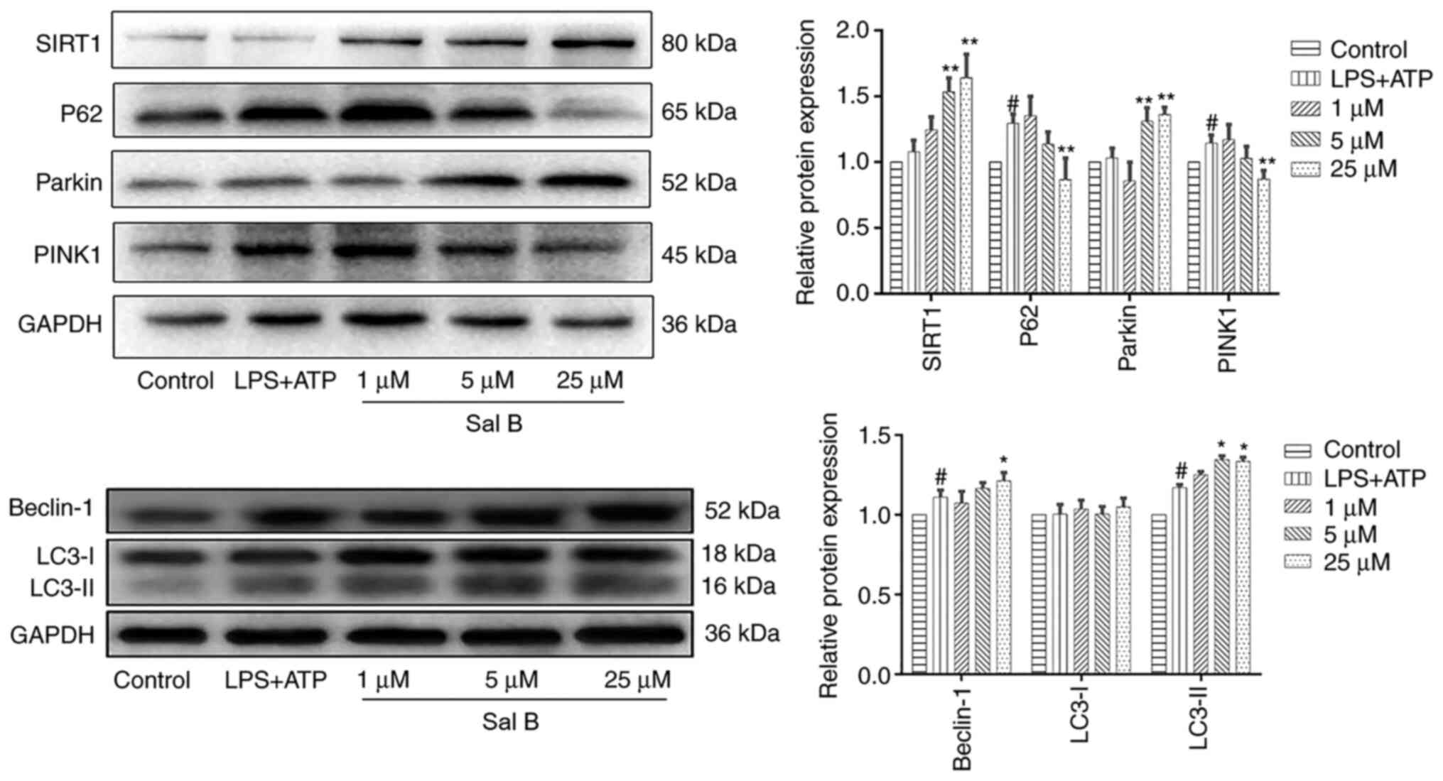 | Figure 5.Sal B treatment significantly
promotes mitophagy in H9C2 cells. Western blot analysis was
performed to determine the protein expression levels of SIRT1, P62,
Parkin, PINK1, Beclin-1 and LC3 in each group. The protein
expression levels were normalized to those of GAPDH. Data are
presented as the mean ± standard deviation from three independent
experiments. #P<0.05 vs. the control group;
*P<0.05, **P<0.01 vs. the LPS + ATP group. ATP, adenosine
triphosphate; LC3, microtubule-associated protein 1A/1B-light chain
3; LPS, lipopolysaccharide; PINK1, PTEN-induced kinase 1; Sal B,
salvianolic acid B; SIRT1, sirtuin 1. |
Sal B inhibits LPS + ATP-induced ROS
production
Excessive ROS production has been associated with
impaired mitochondrial function and increased H9C2 cell apoptotic
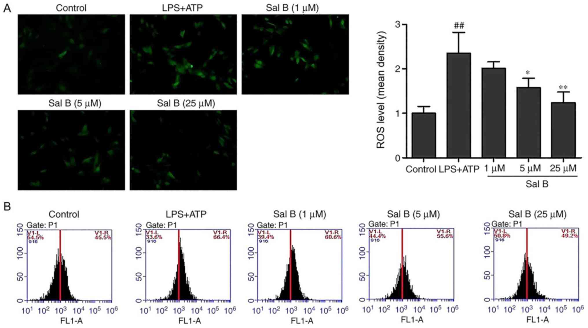
rate (30). As shown in the
fluorescence images in Fig. 6, LPS
+ ATP stimulation significantly increased the fluorescence
intensity compared with the control group, indicating the
production of ROS. Conversely, pretreatment with Sal B attenuated
the DCF fluorescence induced by LPS + ATP. Consistent with the
fluorescence results, flow cytometric analysis demonstrated that
the levels of intracellular ROS production in the LPS + ATP group
were notably increased compared with those in the control group,
whereas pretreatment with Sal B reversed this effect (Fig. 6).
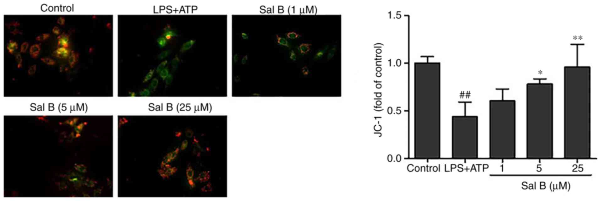
Effects of Sal B on MMP
A previous study reported that decreased MMP is
closely associated with mitochondrial dysfunction (31). To investigate whether Sal B could
inhibit mitochondrial dysfunction by increasing MMP, a JC-1 kit was
used. The JC-1 red/green fluorescence ratio was used as a parameter
to estimate changes in MMP. The results revealed that the MMP in
H9C2 cells was significantly reduced by LPS + ATP stimulation
compared with in the control group, whereas Sal B pre-treatment (5
and 25 µM) significantly increased its potential (Fig. 7).
Effects of Sal B on activation of the
NLRP3 inflammasome
A previous study suggested that the occurrence of
mitophagy may lead to degradation of the NLRP3 inflammasome, which
in turns prevents the activation of NLRP3 inflammasome signaling
and reduces the secretion of mature IL-1β (32). To explore whether Sal B treatment
could attenuate the expression levels of the NLRP3 inflammasome
components, the protein expression levels of NLRP3, caspase-1 and
IL-1β were evaluated. The results demonstrated that the protein
expression levels of NLRP3, caspase-1 and IL-1β were significantly
increased in H9C2 cells following LPS + ATP stimulation; however,
pre-treatment with Sal B (25 µM) significantly reduced the
expression levels of these proteins (Fig. 8).
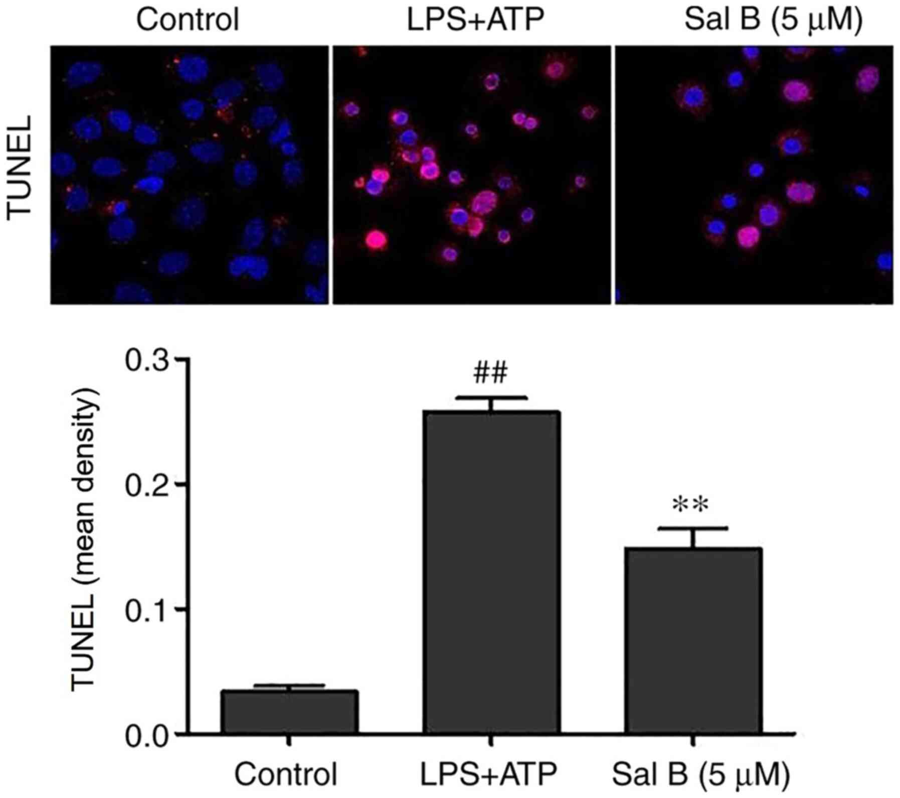
Effects of Sal B on the apoptosis of
H9C2 cells
Assembly of the NLRP3 inflammasome may activate
caspase-1 and promote apoptosis (33). As shown in Fig. 9, LPS + ATP stimulation
significantly promoted apoptosis compared with in the control
group, whereas Sal B pre-treatment (5 µM) significantly reversed
this effect.
Discussion
In the present study, Sal B markedly attenuated
ISO-induced acute myocardial ischemic injury in rats and reduced
the serum levels of several cytokines, including CK-MB, GOT and
IL-1β. Furthermore, to the best of our knowledge, the present study
was the first to demonstrate that Sal B treatment may be able to
maintain mitochondrial function, promote SIRT1-mediated mitophagy
and inhibit activation of the NLRP3 inflammasome. However, the
present study did not provide much information on cardiac pathology
and function; we aim to assess these further in future
experiments.
It is widely accepted that myocardial ischemia is
often accompanied by severe inflammatory response (34,35).
As the core target of inflammatory responses, the NLRP3
inflammasome has been considered the key regulator of numerous
inflammatory diseases (36). LPS +
ATP is a classical activator of the NLRP3 inflammasome; it is
widely used in studies of NLRP3 inflammasome-related diseases,
including myocardial ischemia (37,38).
Activation of the classic NLRP3 inflammasome requires two steps:
Firstly, microorganisms or other inflammatory factors bind to
Toll-like receptor 4 (TLR4) and activate the TLR4/NF-κB signaling
pathway to induce the expression of NLRP3, pro-caspase-1 and
pro-IL-1β (priming phase). Secondly, extracellular ATP or other
stimuli, such as lysosome destabilization and mitochondrial
dysfunction (39), directly
stimulate the activation of caspase-1, thereby leading to the
release of IL-1β and IL-18 (triggering phase). Activation of the
NLRP3 inflammasome in cardiomyocytes requires the involvement of
both priming and triggering phases (40). A previous study in our laboratory
reported that Sal B exerted its cardioprotective effects by
inhibiting the TLR4/NF-κB/NLRP3 signaling pathway (41), and myeloid differential protein-2
was considered the potential target. However, owing to the
multi-target characteristics of natural products, it was
hypothesized that Sal B may inhibit the NLRP3 inflammasome through
other mechanisms. Therefore, the present study explored whether Sal
B could inhibit the triggering phase of the NLRP3 inflammasome,
thereby attenuating myocardial ischemic injury.
The contraction and functioning of the heart are
mainly dependent on mitochondria to provide energy. Emerging
evidence has suggested that the accumulation of dysfunctional
mitochondria may result in increased intracellular ROS, thus
leading to cardiac dysfunction (42). Mitophagy is an effective process
that can remove damaged mitochondria and maintain mitochondrial
function (43). The association
between SIRT1 and mitophagy has also been previously reported. For
example, it has been reported that the SIRT1-PINK1-Parkin axis may
participate in resveratrol-induced mitophagy and cardioprotection
(44). Furthermore, it has been
revealed that promoting mitophagy may negatively regulate
activation of the NLRP3 inflammasome, which may exert significant
protective effects against myocardial ischemia (45,46).
In conclusion, the present study demonstrated that Sal B could
significantly inhibit intracellular ROS production, increase the
mitochondrial membrane potential and regulate the protein
expression levels of SIRT1, Parkin and PINK1 in myocardial
ischemia, which in turn could mediate the promotion of mitophagy
and inactivation of the NLRP3 inflammasome. Therefore, Sal B, as a
natural product with little toxicity, may provide a novel
therapeutic strategy for the treatment of myocardial ischemia.
Acknowledgements
Not applicable.
Funding
The present study was supported by The Jiangsu
Administration of Traditional Chinese Medicine (grant no.
ZD301701).
Availability of data and materials
The datasets used and/or analyzed during the current
study are available from the corresponding author on reasonable
request.
Authors' contributions
YH and XW contributed equally to this work. LX
designed this project. YH, XW, YP and QL performed the experiments
and analyzed the data. YH and XW contributed to the writing of the
manuscript. All authors contributed to the revision of this
manuscript, and read and approved the final manuscript.
Ethics approval and consent to
participate
The protocol was approved by the Animal Ethics
Committee of Nanjing University of Chinese Medicine (approval no.
201906A036).
Patient consent for publication
Not applicable.
Competing interests
The authors declare that they have no competing
interests.
References
|
1
|
Chen WW, Gao RL, Liu LS, Zhu ML, Wang W,
Wang YJ, Wu ZS, Li HJ, Gu DF, Yang YJ, et al: China cardiovascular
diseases report 2015: A summary. J Geriatr Cardiol. 14:1–10.
2017.PubMed/NCBI
|
|
2
|
Kang PF, Wu WJ, Tang Y, Xuan L, Guan SD,
Tang B, Zhang H, Gao Q and Wang HJ: Activation of ALDH2 with low
concentration of ethanol attenuates myocardial ischemia/reperfusion
injury in diabetes rat model. Oxid Med Cell Longev.
2016:61905042016. View Article : Google Scholar : PubMed/NCBI
|
|
3
|
Cao DJ, Schiattarella GG, Villalobos E,
Jiang N, May HI, Li T, Chen ZJ, Gillette TG and Hill JA: Cytosolic
DNA sensing promotes macrophage transformation and governs
myocardial ischemic injury. Circulation. 137:2613–2634. 2018.
View Article : Google Scholar : PubMed/NCBI
|
|
4
|
Jiang S, Liu Y, Wang J, Zhang Y, Rui Y,
Zhang Y and Li T: Cardioprotective effects of monocyte locomotion
inhibitory factor on myocardial ischemic injury by targeting
vimentin. Life Sci. 167:85–91. 2016. View Article : Google Scholar : PubMed/NCBI
|
|
5
|
Zhu L, Wei T, Gao J, Chang X, He H, Luo F,
Zhou R, Ma C, Liu Y and Yan T: The cardioprotective effect of
salidroside against myocardial ischemia reperfusion injury in rats
by inhibiting apoptosis and inflammation. Apoptosis. 20:1433–1443.
2015. View Article : Google Scholar : PubMed/NCBI
|
|
6
|
Marchetti C, Toldo S, Chojnacki J,
Mezzaroma E, Liu K, Salloum FN, Nordio A, Carbone S, Mauro AG, Das
A, et al: Pharmacologic inhibition of the NLRP3 inflammasome
preserves cardiac function after ischemic and nonischemic injury in
the mouse. J Cardiovasc Pharmacol. 66:1–8. 2015. View Article : Google Scholar : PubMed/NCBI
|
|
7
|
Swanson KV, Deng M and Ting JP: The NLRP3
inflammasome: Molecular activation and regulation to therapeutics.
Nat Rev Immunol. 19:477–489. 2019. View Article : Google Scholar : PubMed/NCBI
|
|
8
|
He Q, Li Z, Meng C, Wu J, Zhao Y and Zhao
J: Parkin-dependent mitophagy is required for the inhibition of
ATF4 on NLRP3 inflammasome activation in cerebral
ischemia-reperfusion injury in rats. Cells. 82019
|
|
9
|
Wan Z, Fan Y, Liu X, Xue J, Han Z, Zhu C
and Wang X: NLRP3 inflammasome promotes diabetes-induced
endothelial inflammation and atherosclerosis. Diabetes Metab Syndr
Obes. 12:1931–1942. 2019. View Article : Google Scholar : PubMed/NCBI
|
|
10
|
Mao L, Kitani A, Strober W and Fuss IJ:
The role of NLRP3 and IL-1β in the pathogenesis of inflammatory
bowel disease. Front Immunol. 9:25662018. View Article : Google Scholar : PubMed/NCBI
|
|
11
|
Toldo S, Marchetti C, Mauro AG, Chojnacki
J, Mezzaroma E, Carbone S, Zhang S, Van Tassell B, Salloum FN and
Abbate A: Inhibition of the NLRP3 inflammasome limits the
inflammatory injury following myocardial ischemia-reperfusion in
the mouse. Int J Cardiol. 209:215–220. 2016. View Article : Google Scholar : PubMed/NCBI
|
|
12
|
Han Y, Luo H, Wang H, Cai J and Zhang Y:
SIRT1 induces resistance to apoptosis in human granulosa cells by
activating the ERK pathway and inhibiting NF-κB signaling with
anti-inflammatory functions. Apoptosis. 22:1260–1272. 2017.
View Article : Google Scholar : PubMed/NCBI
|
|
13
|
Wong WT, Li LH, Rao YK, Yang SP, Cheng SM,
Lin WY, Cheng CC, Chen A and Hua KF: Repositioning of the
beta-blocker carvedilol as a novel autophagy inducer that inhibits
the NLRP3 inflammasome. Front Immunol. 9:19202018. View Article : Google Scholar : PubMed/NCBI
|
|
14
|
Akkafa F, Halil Altiparmak I, Erkus ME,
Aksoy N, Kaya C, Ozer A, Sezen H, Oztuzcu S, Koyuncu I and Umurhan
B: Reduced SIRT1 expression correlates with enhanced oxidative
stress in compensated and decompensated heart failure. Redox Biol.
6:169–173. 2015. View Article : Google Scholar : PubMed/NCBI
|
|
15
|
Wang L, Ma R, Liu C, Liu H, Zhu R, Guo S,
Tang M, Li Y, Niu J, Fu M, et al: Salvia miltiorrhiza: A
potential red light to the development of cardiovascular diseases.
Curr Pharm Des. 23:1077–1097. 2017. View Article : Google Scholar : PubMed/NCBI
|
|
16
|
Han F, Xing RH, Chen LQ, Chen L, Xiong W,
Yang M and Zhao ZD: Research progress of anti-drug resistance in
traditional Chinese medicine. Zhongguo Zhong Yao Za Zhi.
41:813–817. 2016.(In Chinese). PubMed/NCBI
|
|
17
|
Gong L, Di C, Xia X, Wang J, Chen G, Shi
J, Chen P, Xu H and Zhang W: AKT/mTOR signaling pathway is involved
in salvianolic acid B-induced autophagy and apoptosis in
hepatocellular carcinoma cells. Int J Oncol. 49:2538–2548. 2016.
View Article : Google Scholar : PubMed/NCBI
|
|
18
|
Lou Y, Wang C, Zheng W, Tang Q, Chen Y,
Zhang X, Guo X and Wang J: Salvianolic acid B inhibits
IL-1β-induced inflammatory cytokine production in human
osteoarthritis chondrocytes and has a protective effect in a mouse
osteoarthritis model. Int Immunopharmacol. 46:31–37. 2017.
View Article : Google Scholar : PubMed/NCBI
|
|
19
|
Liu X, Xavier C, Jann J and Wu H:
Salvianolic acid B (Sal B) protects retinal pigment epithelial
cells from oxidative stress-induced cell death by activating
glutaredoxin 1 (Grx1). Int J Mol Sci. 17:18352016. View Article : Google Scholar
|
|
20
|
Li D, Wang J, Hou J, Fu J, Liu J and Lin
R: Salvianolic acid B induced upregulation of miR-30a protects
cardiac myocytes from ischemia/reperfusion injury. BMC Complement
Altern Med. 16:3362016. View Article : Google Scholar : PubMed/NCBI
|
|
21
|
Dhivya V, Priya LB, Chirayil HT,
Sathiskumar S, Huang CY and Padma VV: Piperine modulates
isoproterenol induced myocardial ischemia through antioxidant and
anti-dyslipidemic effect in male Wistar rats. Biomed Pharmacother.
87:705–713. 2017. View Article : Google Scholar : PubMed/NCBI
|
|
22
|
Ke Z, Wang G, Yang L, Qiu H, Wu H, Du M,
Chen J, Song J, Jia X and Feng L: Crude terpene glycoside component
from Radix paeoniae rubra protects against isoproterenol-induced
myocardial ischemic injury via activation of the PI3K/AKT/mTOR
signaling pathway. J Ethnopharmacol. 206:160–169. 2017. View Article : Google Scholar : PubMed/NCBI
|
|
23
|
Livak KJ and Schmittgen TD: Analysis of
relative gene expression data using real-time quantitative PCR and
the 2(-Delta Delta C(T)) Method. Methods. 25:402–408. 2001.
View Article : Google Scholar : PubMed/NCBI
|
|
24
|
Huang X, Zuo L, Lv Y, Chen C, Yang Y, Xin
H, Li Y and Qian Y: Asiatic acid attenuates myocardial
ischemia/reperfusion injury via Akt/GSK-3β/HIF-1α signaling in rat
H9c2 cardiomyocytes. Molecules. 21:212016. View Article : Google Scholar
|
|
25
|
Li P, Lin N, Guo M, Huang H, Yu T and
Zhang L: REDD1 knockdown protects H9c2 cells against myocardial
ischemia/reperfusion injury through Akt/mTORC1/Nrf2
pathway-ameliorated oxidative stress: An in vitro study. Biochem
Biophys Res Commun. 519:179–185. 2019. View Article : Google Scholar : PubMed/NCBI
|
|
26
|
Zou J, Zhang Y, Sun J, Wang X, Tu H, Geng
S, Liu R, Chen Y and Bi Z: Deoxyelephantopin induces reactive
oxygen species-mediated apoptosis and autophagy in human
osteosarcoma cells. Cell Physiol Biochem. 42:1812–1821. 2017.
View Article : Google Scholar : PubMed/NCBI
|
|
27
|
Zhang X, Du Q, Yang Y, Wang J, Dou S, Liu
C and Duan J: The protective effect of Luteolin on myocardial
ischemia/reperfusion (I/R) injury through TLR4/NF-κB/NLRP3
inflammasome pathway. Biomed Pharmacother. 91:1042–1052. 2017.
View Article : Google Scholar : PubMed/NCBI
|
|
28
|
Xue W, Wang X, Tang H, Sun F, Zhu H, Huang
D and Dong L: Vitexin attenuates myocardial ischemia/reperfusion
injury in rats by regulating mitochondrial dysfunction induced by
mitochondrial dynamics imbalance. Biomed Pharmacother.
124:1098492020. View Article : Google Scholar : PubMed/NCBI
|
|
29
|
Youle RJ and Narendra DP: Mechanisms of
mitophagy. Nat Rev Mol Cell Biol. 12:9–14. 2011. View Article : Google Scholar : PubMed/NCBI
|
|
30
|
Wang M, Wang R, Xie X, Sun G and Sun X:
Araloside C protects H9c2 cardiomyoblasts against oxidative stress
via the modulation of mitochondrial function. Biomed Pharmacother.
117:1091432019. View Article : Google Scholar : PubMed/NCBI
|
|
31
|
Wu C, Zhao W, Yu J, Li S, Lin L and Chen
X: Induction of ferroptosis and mitochondrial dysfunction by
oxidative stress in PC12 cells. Sci Rep. 8:5742018. View Article : Google Scholar : PubMed/NCBI
|
|
32
|
Kim MJ, Yoon JH and Ryu JH: Mitophagy: A
balance regulator of NLRP3 inflammasome activation. BMB Rep.
49:529–535. 2016. View Article : Google Scholar : PubMed/NCBI
|
|
33
|
He Y, Hara H and Núñez G: Mechanism and
regulation of NLRP3 inflammasome activation. Trends Biochem Sci.
41:1012–1021. 2016. View Article : Google Scholar : PubMed/NCBI
|
|
34
|
Hammadah M, Sullivan S, Pearce B, Al Mheid
I, Wilmot K, Ramadan R, Tahhan AS, O'Neal WT, Obideen M, Alkhoder
A, et al: Inflammatory response to mental stress and mental stress
induced myocardial ischemia. Brain Behav Immun. 68:90–97. 2018.
View Article : Google Scholar : PubMed/NCBI
|
|
35
|
Hansson GK: Inflammation, atherosclerosis,
and coronary artery disease. N Engl J Med. 352:1685–1695. 2005.
View Article : Google Scholar : PubMed/NCBI
|
|
36
|
Mangan MSJ, Olhava EJ, Roush WR, Seidel
HM, Glick GD and Latz E: Targeting the NLRP3 inflammasome in
inflammatory diseases. Nat Rev Drug Discov. 17:588–606. 2018.
View Article : Google Scholar : PubMed/NCBI
|
|
37
|
Wang DS, Yan LY, Yang DZ, Lyu Y, Fang LH,
Wang SB and Du GH: Formononetin ameliorates myocardial
ischemia/reperfusion injury in rats by suppressing the
ROS-TXNIP-NLRP3 pathway. Biochem Biophys Res Commun. 525:759–766.
2020. View Article : Google Scholar : PubMed/NCBI
|
|
38
|
Yuan X, Juan Z, Zhang R, Sun X, Yan R, Yue
F, Huang Y, Yu J and Xia X: Clemastine fumarate protects against
myocardial ischemia reperfusion injury by activating the
TLR4/PI3K/Akt signaling pathway. Front Pharmacol. 11:282020.
View Article : Google Scholar : PubMed/NCBI
|
|
39
|
Gong Z, Pan J, Shen Q, Li M and Peng Y:
Mitochondrial dysfunction induces NLRP3 inflammasome activation
during cerebral ischemia/reperfusion injury. J Neuroinflammation.
15:2422018. View Article : Google Scholar : PubMed/NCBI
|
|
40
|
Latz E, Xiao TS and Stutz A: Activation
and regulation of the inflammasomes. Nat Rev Immunol. 13:397–411.
2013. View Article : Google Scholar : PubMed/NCBI
|
|
41
|
Hu Y, Li Q, Pan Y and Xu L: Sal B
alleviates myocardial ischemic injury by inhibiting TLR4 and the
priming phase of NLRP3 inflammasome. Molecules. 242019.
|
|
42
|
Zhu W, Liu F, Wang L, Yang B, Bai Y, Huang
Y, Li Y, Li W, Yuan Y, Chen C, et al: pPolyHb protects myocardial
H9C2 cells against ischemia-reperfusion injury by regulating the
Pink1-Parkin-mediated mitochondrial autophagy pathway. Artif Cells
Nanomed Biotechnol. 47:1248–1255. 2019. View Article : Google Scholar : PubMed/NCBI
|
|
43
|
Villanueva Paz M, Cotán D, Garrido-Maraver
J, Cordero MD, Oropesa-Ávila M, de La Mata M, Delgado Pavón A, de
Lavera I, Alcocer-Gómez E and Sánchez-Alcázar JA: Targeting
autophagy and mitophagy for mitochondrial diseases treatment.
Expert Opin Ther Targets. 20:487–500. 2016. View Article : Google Scholar : PubMed/NCBI
|
|
44
|
Das S, Mitrovsky G, Vasanthi HR and Das
DK: Antiaging properties of a grape-derived antioxidant are
regulated by mitochondrial balance of fusion and fission leading to
mitophagy triggered by a signaling network of
Sirt1-Sirt3-Foxo3-PINK1-PARKIN. Oxid Med Cell Longev.
2014:3451052014. View Article : Google Scholar : PubMed/NCBI
|
|
45
|
Mai CT, Wu MM, Wang CL, Su ZR, Cheng YY
and Zhang XJ: Palmatine attenuated dextran sulfate sodium
(DSS)-induced colitis via promoting mitophagy-mediated NLRP3
inflammasome inactivation. Mol Immunol. 105:76–85. 2019. View Article : Google Scholar : PubMed/NCBI
|
|
46
|
Cao S, Shrestha S, Li J, Yu X, Chen J, Yan
F, Ying G, Gu C, Wang L and Chen G: Melatonin-mediated mitophagy
protects against early brain injury after subarachnoid hemorrhage
through inhibition of NLRP3 inflammasome activation. Sci Rep.
7:24172017. View Article : Google Scholar : PubMed/NCBI
|


