Introduction
Human hepatocellular carcinoma (HCC) is a common
malignancy, with high incidence and mortality rates worldwide,
particularly in East Asia (1).
Hepatic surgery is the normal treatment and has undergone
improvement; however, recurrence and metastasis rates remain high
(2,3). Therefore, it is crucial to identify
and establish effective novel approaches to treat HCC. In the past
two decades, immunotherapy has proven to be a treatment strategy,
and active immunotherapy approaches using specific vaccines appear
to have potential in the treatment of HCC patients.
In recent years, a new family of tumor-specific
antigens has been identified, known as cancer/testis antigens
(CTA). CTA is a family that includes various types of
tumor-specific antigens primarily expressed in primitive germ
cells, spermatogonia and certain human tumors. The malignant
transformation of these antigens is frequently associated with the
activation or depression of genes, resulting in the expression of
the antigens. More than 50 types of CTA have been identified thus
far, including MAGE, GAGE, PAGE, NY-ESO-1, SSX, SPANX and XAGE
(4–8).
XAGE-1 was originally identified by database mining
of expressed sequence tags and belongs to a new family of CTA with
an expression pattern that is limited to the germ cells of the
testis and a variety of neoplastic tissues with immunogenicity
(9,10), but is abundantly expressed in
breast, prostate and lung cancer as well as in Ewing’s sarcomas and
rhabdomyosarcomas (11–14). The XAGE-1 gene lies on the X
chromosome and its encoded proteins have strong homology with
members of the GAGE/PAGE family in the COOH terminus. XAGE-1
expression in testis and cancer tissues is regulated by methylation
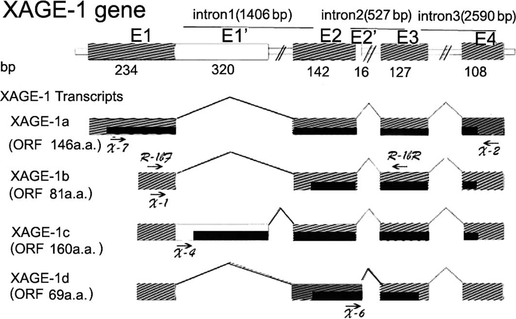
of the CpG island in the gene promoter (15). There are 4 transcript variants of
XAGE-1, i.e., XAGE-1a, XAGE-1b, XAGE-1c and XAGE-1d (10,13,16),
with XAGE-1b and XAGE-1d being specifically expressed in lung and
prostate cancer. These variants are immunogenic and can therefore
be used as immunotherapy targets. However, only limited research
haa been performed on XAGE-1 in HCC. The present study showed that
XAGE-1b was expressed strongly with a high frequency in HCC, while
XAGE-1d relatively less strongly with a lower frequency.
Additionally, more information is provided on this cancer antigen
family and its potential use in the diagnosis of or immunotherapy
for HCC.
Materials and methods
Patients and specimens
A total of 96 HCCs and corresponding adjacent
non-cancerous liver tissues from patients at Peking University
People’s Hospital, China, were surgically resected and dissected
into sections of ~0.3 × 0.3 × 0.3 cm and immediately stored in
liquid nitrogen. Written informed consent was obtained from each
patient for the use of the specimens and their clinicopathological
characteristics. The study was approved by the Ethics Committee of
Peking University People’s Hospital. HCC diagnosis was confirmed by
pathological examination and the TNM stage was determined on the
basis of the criteria issued by UICC in 2002.
Total RNA isolation and cDNA
synthesis
Total RNA was isolated from the frozen tumors and
adjacent non-cancerous liver specimens using RNeasy mini kit
(Qiagen, Hilden, Germany). A total of 2 μg was reverse-transcribed
into cDNA using M-MLV reverse transcriptase (Promega, Madison, WI,
USA). In addition to 200 units M-MLV, the reaction mixture
consisted of 1 μl oligo (dT)15 as a primer (Promega),
0.5 units RNase ribonuclease inhibitor (Promega), 4 μl of RT buffer
(250 mM Tris-HCl, pH 8.3, 375 mM KCl, 15 mM MgCl2 and 50
mM DTT) and 1 μl of 10 mM dNTPs (Phamcia), with DEPC-treated water
added to a final volume of 20 μl. The mixture was incubated for 5
min at 70°C and 5 min on ice, 60 min at 42°C and 5 min at 95°C. The
cDNAs were tested for integrity by amplifying the GAPDH gene.
Reverse transcription-polymerase chain
reaction (RT-PCR) for XAGE-1 gene
A schematic representation of the structure of the
XAGE-1 gene and transcripts, including locations of primers used in
this study, is shown in Fig. 1.
Primer pair sequences used for RT-PCR are listed in Table I, with X-7 and X-2 for XAGE-1a, X-1
and X-2 for the common region of XAGE-1b, X-4 and X-2 for XAGE-1c
and X-6 and X-2 for XAGE-1d. The amplification program for XAGE-1
transcript variants was 1 min at 94°C, 1 min at 60°C and 1 min at
72°C for 30 cycles after 10 min at 94°C. The amplification program
for GAPDH was 30 sec at 94°C, 45 sec at 57°C and 45 sec at 72°C for
30 cycles after 10 min at 94°C. The cycles were followed by a
10-min elongation step at 72°C. The PCR products were analyzed on
1.6% agarose gel electrophoresis. The positive PCR products were
then recovered and sequenced. Data were analyzed by comparison with
the respective sequences in GeneBank.
 | Table IPCR primers used in this study,
including GAPDH and XAGE-1. |
Table I
PCR primers used in this study,
including GAPDH and XAGE-1.
| Primer | Sequence | Notes |
|---|
| GAPDHF |
5′-GAAGGTGAAGGTCGGAGTC-3′ | Forward, sense |
| GAPDHR |
5′-GAAGATGGTGATGGGATTTC-3′ | Reverse,
anti-sense |
| X-7 |
5′-ACCTCAGTGCGCATGTTCAC-3′ | Forward, sense |
| X-1 |
5′-TTTCTCCGCTACTGAGACAC-3′ | Forward, sense |
| X-4 |
5′-CTGGGAGTTGAAGTGTGAGT-3′ | Forward, sense |
| X-2 |
5′-CAGGTGCTGGGAAGGGAAAT-3′ | Forward, sense |
| X-6 |
5′-CAGCTTGCGTTGTTTCAGCT-3′ | Reverse,
anti-sense |
| R-1bF |
5′-TACTGAGACACGGCGGAC-3′ | Forward, sense |
| R-1bR |
5′-TTCCATGTCGCGCACTG-3′ | Reverse,
anti-sense |
Real-time reverse
transcription-polymerase chain reaction (RT-PCR) for GAPDH and
XAGE-1
Sequences of gene-specific primers for XAGE-1b and
XAGE-1d GAPDH are shown in Table I.
Relative quantification using real-time RT-PCR was performed using
SYBR-Green PCR Mastermix (Applied Biosystems, Foster City, CA, USA)
in accordance with the manufacturer’s instructions, and the ABI
PRISM 7000 Sequence Detection System (Applied Biosystems).
Amplification was performed for 42 cycles with 1 μl cDNA
(corresponding to 60 ng total RNA) solution extracted from tumor
and non-cancerous specimens with 10 μl PCR SYBR-Green Master Mix
(Applied Biosystems) and 1 μl 750 nM forward and reverse primers in
a total volume of 20 μl. The amplification conditions were: 95°C
for 15 sec and 60°C for 60 sec.
Immunohistochemistry
Paraffin-embedded tumors and the adjacent
non-cancerous tissues of 4 μm were mounted on glass slides and
deparaffinized with xylene and ethanol. For antigen retrieval, the
tissues were water-bath heated in an antigen retrieval buffer [10
nM citrate buffer (pH 6.0)] for 10 min. Endogenous peroxidase was
inactivated with 0.3% H2O2 for 10 min.
Following pre-incubation with serum-free blocking solution, XAGE-1
mAb (Santa Cruz) was added at a concentration of 2 μg/ml and
incubated at 4°C overnight. Following PBS washing,
biotin-conjugated goat anti-mouse IgG and horseradish
peroxidase-conjugated avidin (Santa Cruz) were applied,
respectively, and incubated for 30 min at room temperature for each
step. The specimens were then visualized using
3,3′-diaminobenzidine (DAB) in H2O2 and
counterstained with hematoxylin solution.
Statistical analysis
Associations between the XAGE-1 expression and the
clinicopathological characteristics were analyzed using the
Chi-square or Fisher’s exact test, according to the test condition.
The survival probabilities were estimated using the Kaplan-Meier
method and were compared using the log-rank test. Overall survival
time was defined as the time from the date of surgery to the date
the patient succumbed to any cause. Patients who were alive at the
date of the last follow-up were censored on that date plus 1 day.
Confidence intervals of 95% were used throughout the analysis.
Statistical significance was defined as P<0.05. The statistical
tests were performed using the Statistical Package, SPSS 13.0 for
Windows (SPS Inc., Chicago, IL, USA).
Results
XAGE-1 mRNA expression in hepatocellular
carcinoma
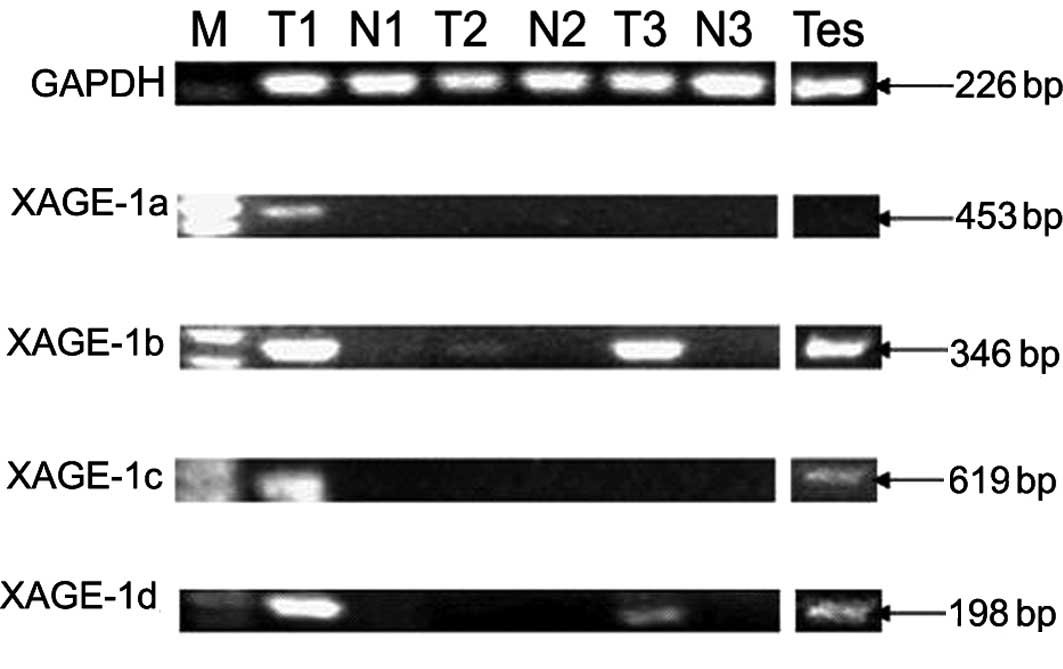
Expression of the four XAGE-1 transcript variants
was investigated in 96 HCC and adjacent non-cancerous liver tissues
by conventional 30-cycle RT-PCR using specific primer pairs
(Fig. 1, Table I). The PCR product was analyzed in
1.6% agarose gel and sequenced. Fig.
2 shows a representative RT-PCR and Table II summarizes the results. The
expression of XAGE-1b, XAGE-1d and XAGE-1c mRNA, but not XAGE-1a,
was observed, with the exception of 2 cases. Histologically,
XAGE-1b mRNA expression was observed in 40 of 96 (41.7%) HCC
patients. By contrast, XAGE-1d mRNA expression was observed in 25
(15.6%) HCC specimens and XAGE-1c mRNA in 15 (26.0%) HCC specimens.
XAGE-1a mRNA expression was observed in only 2 cases of HCC
tissues. XAGE-1c and XAGE-1d mRNA expression, except for 1 case,
was associated with XAGE-1b mRNA expression. No XAGE-1 mRNA
expression was observed in the 96 adjacent normal liver
tissues.
| Table IICorrelation between XAGE-1b, the
dominant type, XAGE-1c and XAGE-1d mRNA expression and
clinicopathological characteristics in HCC. |
Table II
Correlation between XAGE-1b, the
dominant type, XAGE-1c and XAGE-1d mRNA expression and
clinicopathological characteristics in HCC.
| XAGE-1b-positive | XAGE-1b-negative | P-value | One positive | Double positive | Triple positive | Tetra positive |
|---|
| Cases | 40 (41.7%) | 56 (58.3%) | | 16 | 9 | 14 | 1 |
|
| Age (mean ± SD) | 54.3±12.7 | 51.3±11.1 | 0.97 | 16 | 9 | 14 | 46 |
| ≤50 years | 18 | 25 | | 8 | 5 | 4 | 0 |
| >50 years | 22 | 31 | | 8 | 4 | 10 | 1 |
| Gender | | | 1.00 | | | | |
| Male | 30 | 42 | | 9 | 9 | 11 | 1 |
| Female | 10 | 14 | | 7 | 0 | 3 | 0 |
| HBV | | | 0.53 | | | | |
| Positive | 30 | 45 | | 13 | 7 | 10 | 0 |
| Negative | 10 | 11 | | 3 | 2 | 4 | 1 |
| AFP level | | | 0.29 | | | | |
| ≤20 μg/l | 16 | 28 | | 8 | 2 | 5 | 1 |
| >20 μg/l | 24 | 27 | | 8 | 7 | 9 | 0 |
| Tumor size | | | 0.70 | | | | |
| ≤5 cm | 22 | 33 | | 10 | 2 | 9 | 1 |
| >5 cm | 18 | 23 | | 6 | 7 | 5 | 0 |
| TNM stage | | | 0.88 | | | | |
| I/II | 27 | 37 | | 9 | 5 | 12 | 1 |
| III/IV | 13 | 19 | | 7 | 4 | 2 | 0 |
|
Differentiation | | | 0.20 | | | | |
| Well/moderate | 32 | 50 | | 15 | 6 | 10 | 1 |
| Poor | 8 | 6 | | 1 | 3 | 4 | 0 |
Quantitative real-time RT-PCR analysis of
XAGE-1b and XAGE-1d mRNA expression in HCC, adjacent normal liver
tissues and testis tissues
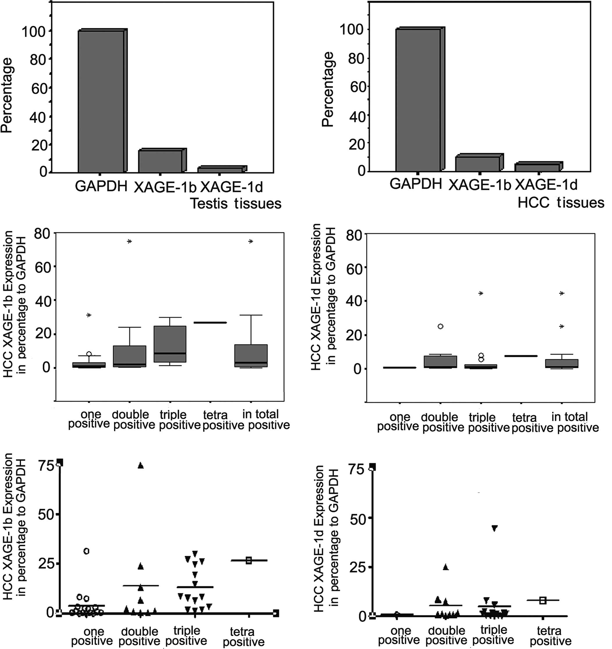
XAGE-1b and XAGE-1d mRNA expression was
quantitatively analyzed by real-time RT-PCR using SYBR-Green PCR
Mastermix with the procedure running on the ABI PRISM 7000 Sequence
Detection System and specific primers shown in Fig. 1 and Table I. As shown in Fig. 3, compared to GAPDH expression, the
XAGE-1b and XAGE-1d mRNA copy numbers ranged from 0.02 to 74.74%
and 0.001 to 44.44%, respectively. Additionally, the mean values
were 9.73 and 4.81%, respectively, and standard deviation was 14.48
and 9.85%, respectively, in the 40 and 25 HCC specimens found to be
positive for XAGE-1b and XAGE-1d mRNA expression by conventional
RT-PCR. Compared to GAPDH expression, XAGE-1b and XAGE-1d mRNA copy
numbers were 15.86 and 3.51%, respectively, in testis tissues.
XAGE-1 isoform expression in HCC cancer
by immunohistochemistry
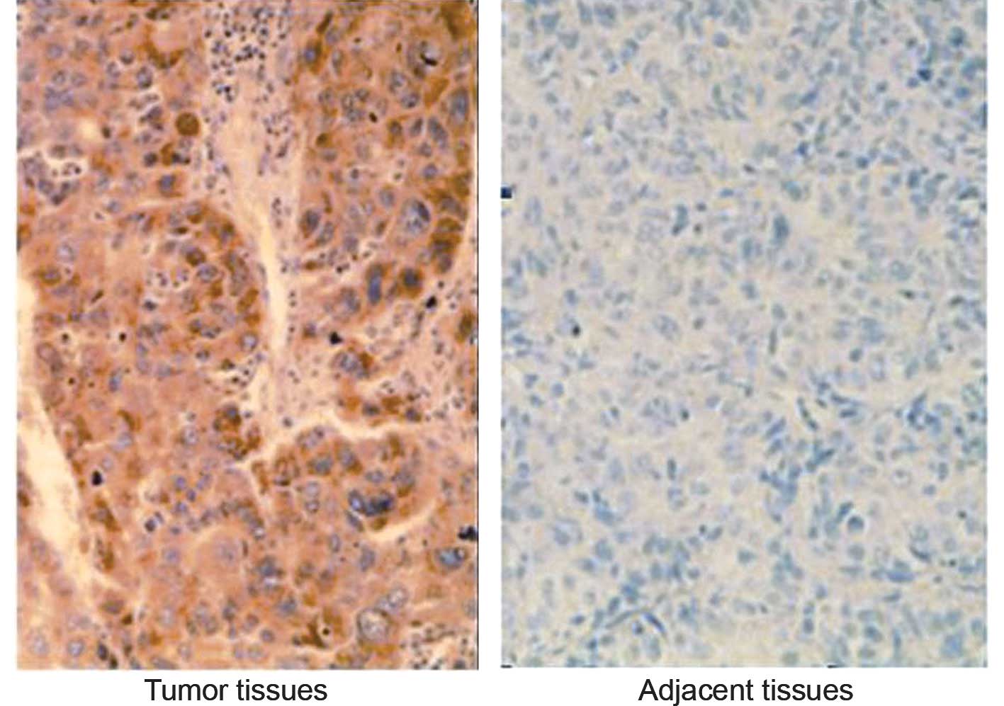
XAGE-1 protein expression was examined in 96 HCC
specimens by immunohistochemistry using a XAGE-1 mAb that reacted
against XAGE-1 isoforms, including XAGE-1b, XAGE-1c and XAGE-1d.
Cases with staining in >10% of the cells were considered to be
positive. Positive staining was observed in 39 of 40 (97.5%) of the
XAGE-1b mRNA-positive specimens, while no positive staining was
noted in mRNA-negative specimens (Fig.
4). No positive staining was observed in the adjacent
non-cancerous, relatively normal liver tissues examined.
Correlation between XAGE-1b and
clinicopathological characteristics
The various clinicopathological characteristics of
the patients and their tumors were compared according to the
XAGE-1b mRNA expression (Table
II). No significant association was found between age, gender,
HBV infection, tumor size, AFP level, TNM stage or differentiation.
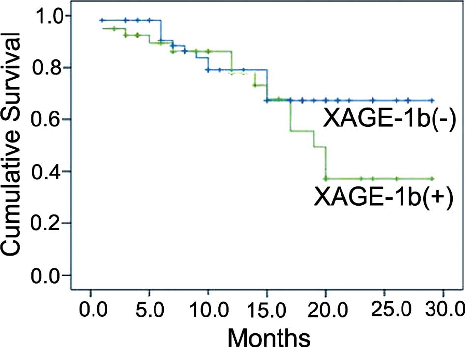
The effect of XAGE-1b mRNA expression on survival was examined.
Adequate clinical follow-up information was available for the 96
cases. The mean follow-up of the 96 cases was 12.8 months, (range
1–29). A total of 68 patients (70.8%) survived, but 28 patients
(29.2%) succumbed during the follow-up period. The 2-year survival
rate of the patients with a positive and negative XAGE-1b
expression was 37.1 and 67.3%, respectively, and the former group
exhibited a relatively shorter 2-year survival rate (P=0.045). The
Kaplan-Meier survival curves showed that patients with a positive
XAGE-1b expression did not have a significant survival difference
compared to patients with a negative XAGE-1b mRNA expression
(P=0.182) (Fig. 5).
Discussion
The present investigation showed that XAGE-1
expression occurred in tumor samples from HCC patients,
demonstrating a relatively high frequency expression of XAGE-1b and
XAGE-1d (41.7 and 26.0%, respectively). Additionally, the XAGE-1
protein was detected in 39 (40.6%) HCC tumor tissues, rendering
them ideal candidate antigens for antigen-specific HCC
immunotherapy. Moreover, our study qualitatively showed that
XAGE-1b and XAGE-1d exhibited a higher expression in certain HCC
cases compared to testis tissues. Concomitantly, we found no
relationship between the expression of XAGE-1b and the clinical
parameters. Furthermore, the HCC patient groups with a positive
XAGE-1 mRNA expression had a relatively shorter 2-year survival
rate compared to the negative group.
Studies have shown that XAGE-1 was highly expressed
in numerous malignancies. Using automated high-throughput filter
immunoscreening or cDNA phage surface display, XAGE-1 antigen was
identified in prostate cancer, particularly in the serum of
prostate cancer and lung adenocarcinoma patients (17–19).
The molecular mechanism regulating the expression of this CTA gene
may be epigenetic modulation of the gene promoter, histone
deacetylase and DNA methyltransferase inhibitors (20). Epigenetic modulation of this antigen
gene may be found in tumorigenic human mesenchymal stem cells that
can potentially be utilized in cancer therapy (21).
Furthermore, the dominant XAGE-1 isoform, XAGE-1b,
is known to be able to stimulate the immune response of patients
suffering from non-small cell lung or prostate cancer (22–24).
In addition, antibody responses to recombinant L552S protein, an
alternatively spliced isoform of XAGE-1, were observed in the
pleural effusion fluids of lung cancer patients (25). The XAGE-1b protein can be processed,
is present in the HLA-I molecule and stimulates the autologous
CD4+ T-lymphocyte response (26,27).
Patients with lung adenocarcinoma expressing both XAGE-1b and HLA
class I antigens were associated with prolonged survival time
(28).
Full-length XAGE-1b protein-pulsed dendritic cells
induce a specific cytotoxic T-lymphocyte response in vitro
that suggests a potential role of immunotherapy in various types of
cancer (29). Therefore, XAGE-1b is
a type of highly and specifically expressed XAGE-1 antigen in
tumors that can be used as targets for immunotherapy. However,
XAGE-1b expression and its correlations with the
clinicopathological characteristics have yet to be adequately
analyzed in human HCC cancer tissues. To the best of our knowledge,
this is the first study regarding the relationship between XAGE-1
expression and clinical parameters in a larger sample of patients
with HCC. Of note is that this study did not demonstrate
statistical significance of the survival difference on the basis of
XAGE-1b expression, which has been shown to be of prognostic
significance in lung cancer, as reported by Kikuchi et al
(28). This result may be due to
the fact that our study did not simultaneously evaluate the MHC-I
expression status.
Further prospective studies with a larger number of
cases in a wide range of clinical settings are required to evaluate
XAGE-1b expression in order for the unique role of XAGE-1b in HCC
development and progression to be determined. Moreover, humoral
immunity and autologous T-lymphocyte responses induced by XAGE-1b
should also be assayed. The results suggest that potential
strategies to reduce or antagonise the XAGE-1b expression may
become a valuable therapeutic approach in the treatment of HCC
patients. Furthermore, due to its highly specific expression and
immunogenic effects, XAGE-1b may be used as a vaccine applicable in
the immunotherapy of HCC. However, further investigations regarding
the application of XAGE-1b are crucial.
References
|
1
|
Bruix J, Boix L and Sala M: Focus on
hepatocellular carcinoma. Cancer Cell. 5:215–219. 2004. View Article : Google Scholar
|
|
2
|
Tang ZY: Hepatocellular carcinoma – cause,
treatment and metastasis. World J Gastroenterol. 7:445–454.
2001.
|
|
3
|
Kim W, Gores G, Benson JT, Therneau TM and
Melton LJ III: Mortality and hospital utilization for
hepatocellular carcinoma in the United States. Gastroenterology.
129:486–493. 2005. View Article : Google Scholar : PubMed/NCBI
|
|
4
|
Chen CH, Chen GL, Lee HS and Huang GT:
Expressions of cancer-testis antigens in human hepatocellular
carcinomas. Cancer Lett. 164:189–195. 2001. View Article : Google Scholar : PubMed/NCBI
|
|
5
|
Chomez P, De Backer O and Bertrand M: An
overview of the MAGE gene family with the identification of all
human members of the family. Cancer Res. 61:5544–5551.
2001.PubMed/NCBI
|
|
6
|
Luo G, Huang S and Xie X: Expression of
cancer-testis genes in human hepatocellular carcinomas. Cancer
Immunity. 2:11–21. 2002.PubMed/NCBI
|
|
7
|
Peng JR, Chen HS and Mou DC: Expression of
cancer/testis (CT) antigens in Chinese hepatocellular carcinoma and
its correlation with clinical parameters. Cancer Lett. 219:223–232.
2005. View Article : Google Scholar : PubMed/NCBI
|
|
8
|
Tajima K, Obata Y and Tamaki H: Expression
of cancer/testis (CT) antigens in lung cancer. Lung Cancer.
42:23–33. 2003. View Article : Google Scholar
|
|
9
|
Caballero OL and Chen YT: Cancer/testis
(CT) antigens: potential targets for immunotherapy. Cancer Sci.
100:2014–2021. 2009. View Article : Google Scholar : PubMed/NCBI
|
|
10
|
Brinkmann U, Vasmatzis G and Lee B: Novel
genes in the PAGE and GAGE family of tumor antigens found by
homology walking in the dbEST database. Cancer Res. 59:1445–1448.
1999.PubMed/NCBI
|
|
11
|
Liu XF, Helman LJ and Yeung C: XAGE-1, a
new gene that is frequently expressed in Ewing’s sarcoma. Cancer
Res. 60:4752–4755. 2000.PubMed/NCBI
|
|
12
|
Zendman AJ, van Kraats AA and den
Hollander AI: Characterization of XAGE-1b, a short major transcript
of cancer/testis-associated gene XAGE-1, induced in melanoma
metastasis. Int J Cancer. 97:195–204. 2002. View Article : Google Scholar : PubMed/NCBI
|
|
13
|
Zendman AJ, van Kraats AA and Weidle UH:
The XAGE family of cancer/testis-associated genes: alignment and
expression profile in normal tissues, melanoma lesions and Ewing’s
sarcoma. Int J Cancer. 99:361–369. 2002.PubMed/NCBI
|
|
14
|
Egland KA, Kumar V and Duray P:
Characterization of overlapping XAGE-1 transcripts encoding a
cancer testis antigen expressed in lung, breast, and other types of
cancers. Mol Cancer Ther. 1:441–450. 2002.PubMed/NCBI
|
|
15
|
Jun HL, Sang PK and Edward G: Activation
of human cancer/testis antigen gene, XAGE-1, in tumor cells is
correlated with CpG island hypomethylation. Int J Cancer.
116:200–206. 2005. View Article : Google Scholar : PubMed/NCBI
|
|
16
|
Sato S, Noguchi Y and Ohara N:
Identification of XAGE-1 isoforms: predominant expression of
XAGE-1b in testis and tumors. Cancer Immunity. 7:5–12.
2007.PubMed/NCBI
|
|
17
|
Alsoe L, Stacy JE and Fossa A:
Identification of prostate cancer antigens by automated
high-throughput filter immunoscreening. J Immunol Methods.
330:12–23. 2008. View Article : Google Scholar : PubMed/NCBI
|
|
18
|
Fossa A, Alsoe L and Crameri R:
Serological cloning of cancer/testis antigens expressed in prostate
cancer using cDNA phage surface display. Cancer Immunol Immunother.
53:431–438. 2004. View Article : Google Scholar : PubMed/NCBI
|
|
19
|
Ali Eldib AM, Ono T and Shimono M:
Immunoscreening of a cDNA library from a lung cancer cell line
using autologous patient serum: identification of XAGE-1b as a
dominant antigen and its immunogenicity in lung adenocarcinoma. Int
J Cancer. 108:558–563. 2004.PubMed/NCBI
|
|
20
|
James SR, Link PA and Karpf AR: Epigenetic
regulation of X-linked cancer/germline antigen genes by DNMT1 and
DNMT3b. Oncogene. 25:6975–6985. 2006. View Article : Google Scholar : PubMed/NCBI
|
|
21
|
Gjerstorff M, Burns JS and Nielsen O:
Epigenetic modulation of cancer-germline antigen gene expression in
tumorigenic human mesenchymal stem cells: implications for cancer
therapy. Am J Pathol. 175:314–323. 2009. View Article : Google Scholar : PubMed/NCBI
|
|
22
|
Koizumi F, Noguchi Y and Saika T: XAGE-1
mRNA expression in prostate cancer and antibody response in
patients. Microbiol Immunol. 49:471–476. 2005. View Article : Google Scholar : PubMed/NCBI
|
|
23
|
Nakagawa K, Noguchi Y and Uenaka A: XAGE-1
expression in non-small cell lung cancer and antibody response in
patients. Clin Cancer Res. 11:5496–5503. 2005. View Article : Google Scholar : PubMed/NCBI
|
|
24
|
Watanabe Y and LePage S: Characterization
of preexisting humoral immunity specific for two cancer-testis
antigens overexpressed at the mRNA level in non-small cell lung
cancer. Cancer Immunity. 6:3–11. 2006.PubMed/NCBI
|
|
25
|
Wang T, Fan L and Watanabe Y: L552S, an
alternatively spliced isoform of XAGE-1, is overexpressed in lung
adenocarcinoma. Oncogene. 20:7699–7709. 2001. View Article : Google Scholar : PubMed/NCBI
|
|
26
|
Morishita Y, Uenaka A and Kaya S:
HLA-DRB1*0410-restricted recognition of XAGE-1b 37-48
peptide by CD4 T cells. Microbiol Immunol. 51:755–762. 2007.
|
|
27
|
Shimono M, Uenaka A and Noguchi Y:
Identification of DR9-restricted XAGE antigen on lung
adenocarcinoma recognized by autologous CD4 T-cells. Int J Oncol.
30:835–840. 2007.PubMed/NCBI
|
|
28
|
Kikuchi E, Yamazaki K and Nakayama E:
Prolonged survival of patients with lung adenocarcinoma expressing
XAGE-1b and HLA class I antigens. Cancer Immunity. 8:13–18.
2008.PubMed/NCBI
|
|
29
|
Zhou Q, Guo AL and Xu CR: A dendritic
cell-based tumour vaccine for lung cancer: full-length XAGE-1b
protein-pulsed dendritic cells induce specific cytotoxic T
lymphocytes in vitro. Clin Exp Immunol. 153:392–400. 2008.
View Article : Google Scholar : PubMed/NCBI
|