Introduction
Extraskeletal osteosarcoma (ESOS) is a rare
malignancy that accounts for approximately 1% of all soft tissue
sarcomas and for 2–4% of all osteosarcomas (1–3). ESOS
usually occurs in the deep soft tissue of the extremities of adults
(4). It typically arises in the
deep soft tissue of the thigh. Other less frequent sites include
the buttock, shoulder, trunk and retroperitoneum. Approximately 24%
of cases have been associated with previous radiotherapy or trauma
(5). In contrast to primary
osteosarcoma of the bone, this variant typically develops after the
fifth decade of life, and the prognosis is uniformly poor (5,6). The
present report documented the clinicopathological findings in a
patient who had an ESOS arising from the subcutaneous tissue of the
upper arm and reviews previous cases of subcutaneous ESOS.
Case report
A 79-year-old male was referred to the Mie
University Hospital due to an enlarged, slightly painful mass in
the left upper arm. The patient first noted the mass 3 years prior
to presentation. No history of trauma or therapy had previously
occurred at this site. Moreover, the patient had experienced no
recent health problems and recalled no family history of cancer. A
physical examination confirmed the presence of a lobulated hard
mass with a diameter of 4 cm at the lateral side of the left upper
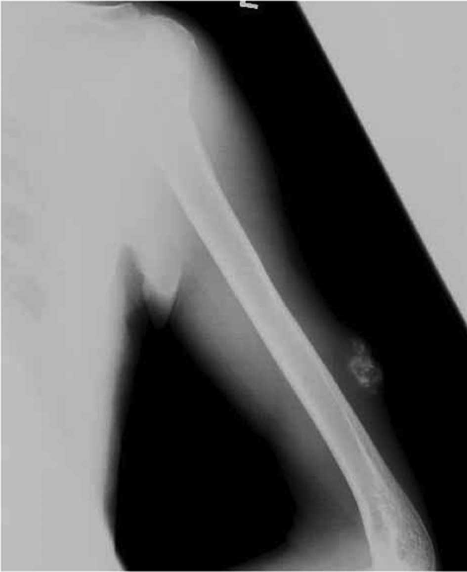
arm. Radiographs of the left upper arm revealed a mass with
ossification which appeared to be separated from the humerus
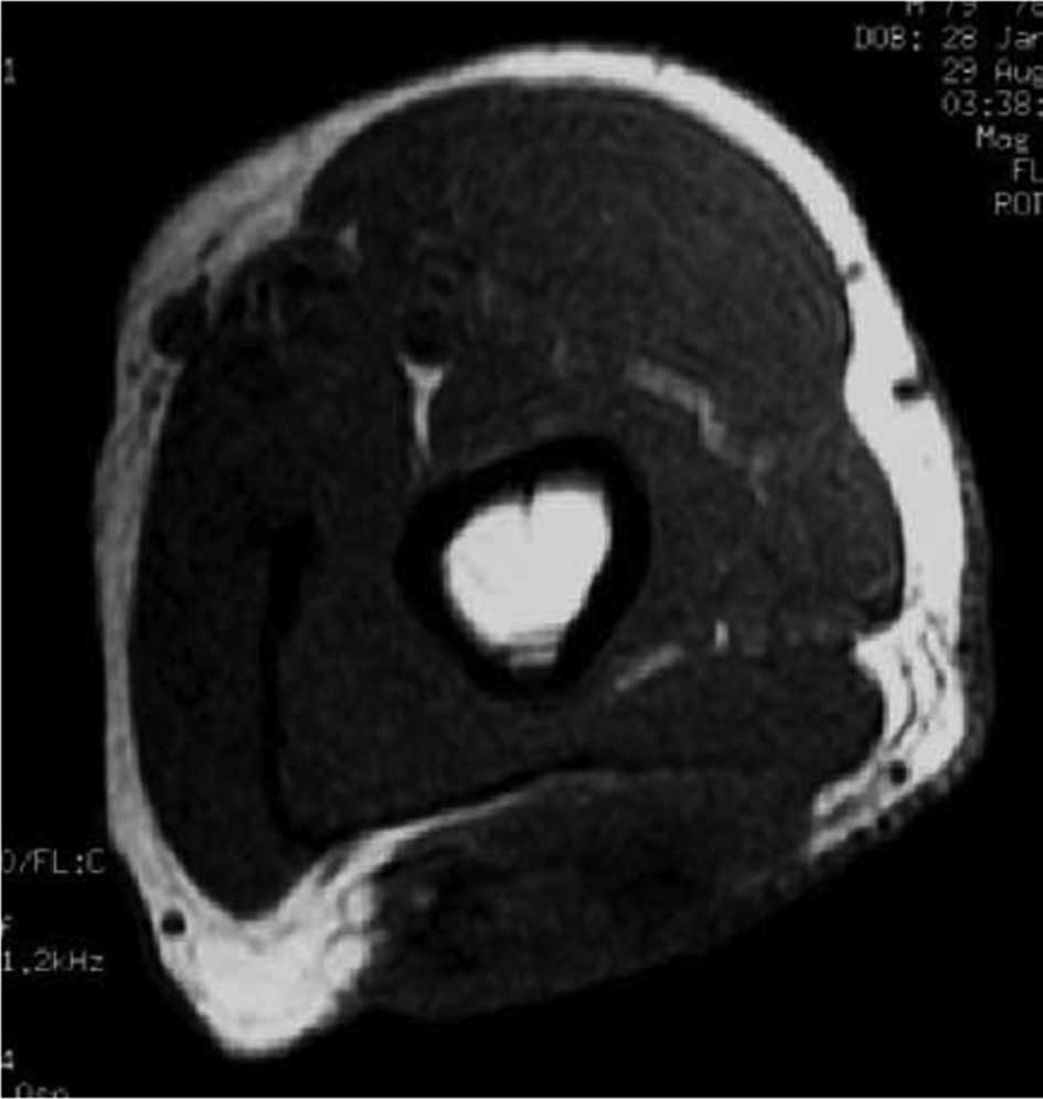
(Fig. 1). Magnetic resonance images
(MRI) showed a soft tissue mass above the fascia of the triceps
with a low signal intensity on T1-weighted images and a
heterogeneous signal intensity on T2-weighted images (Fig. 2). Computed tomography (CT) of the
chest did not demonstrate any pulmonary masses. All routine
laboratory data were normal. The long clinical course from the
first awareness of the tumor and the clinical findings, suggested
that the tumor was benign or a malignant calcifying epithelioma
with ossification. The patient was treated with wide resection and
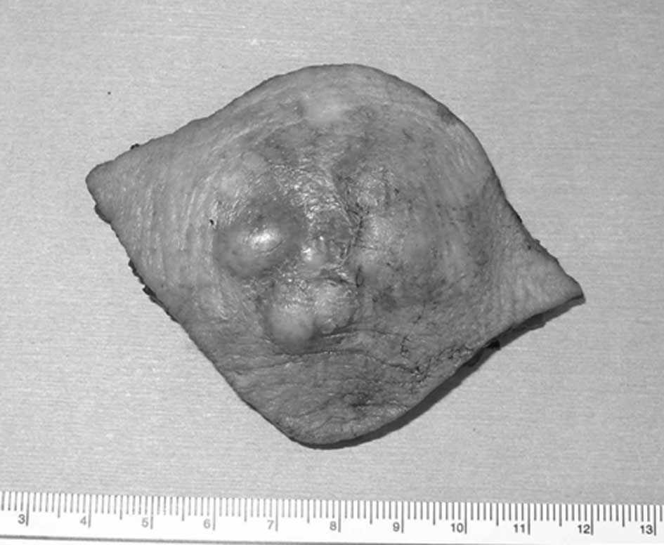
a skin graft from an inguinal lesion. Gross sectioning of the
specimen showed a 4×4×2.5 cm firm and solid mass in the deep dermis
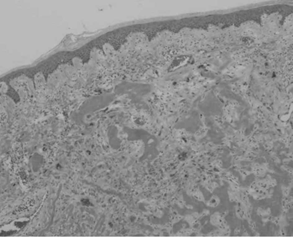
and subcutaneous tissue (Fig. 3). A
microscopic examination showed the presence of numerous spindle and
atypical cells often exhibiting pronounced nuclear atypia or
multinucleated giant cells with bone and osteoid formation. A high
mitotic activity with numerous atypical mitoses was noted (Fig. 4). These findings led to the
conclusion that this soft tissue tumor was a subcutaneous ESOS. The
patient had no evidence of local recurrence and distant metastasis
1 year following resection.
Discussion
Since ESOS was initially described by Wilson in
1941, approximately 300 cases have been reported thus far (5,6). ESOS
is defined as a malignant mesenchymal neoplasm composed of cells
producing osteoid, bone and/or chondroid material, with no
attachment to bone or periosteum (7). It occurs most often in the deep soft
tissues of the extremities of adults, at an average age of 50
years. Clinically, ESOS usually carries an extremely poor
prognosis. The 5-year survival rate ranges from 25 to 37% for ESOS
as previously reported (3,4). The tumor size appears to be the only
reliable prognostic variable in that tumors greater than 5 cm have
a poor prognosis (3).
Subcutaneous ESOS was rarely reported. Only eight
cases, including the present one, were found in the literature
(6,8–13)
(Table I). Patients in those
studies included 4 males and 4 females, ranging in age from 25 to
79 years. Lesions were located in the buttock in 2 cases and in the
scalp, forehead, jaw, abdominal wall, lower leg and upper arm each
in 1 case. In general, ESOS develops in the lower extremities, with
the thigh being involved most; however, these 8 cases developed
ESOS in various anatomical sites. The tumor size was less than 5 cm
in all but 2 cases. A surgical resection was performed in all
cases. The consequent surgical margin was wide in 7 patients,
including an additional wide resection in 2 cases and an
intralesional margin in 1 case. A total of three patients received
chemotherapy. A wide surgical resection was performed in the
present case. No chemotherapy was administered as a result of the
advanced age of the patient. A wide margin is generally recommended
for ESOS, as for other high-grade sarcomas (14). Lee et al reported that
recurrence is common in ESOS and usually occurs in more than half
of the patients (3). However, a
wide (or radical) resection should decrease the recurrence of
ESOS.
 | Table IReview of the literature describing
cases of primary subcutaneous extraskeletal osteosarcoma. |
Table I
Review of the literature describing
cases of primary subcutaneous extraskeletal osteosarcoma.
| Author | Age/gender | Location | Size (cm) | Therapy | Outcome |
|---|
| Fang et al
(8) | 59/female | Abdominal wall | 1.0 | Surgery | CDF, 16 years |
| Yamakage et al
(9) | 62/male | Forehead | 3.0 | Surgery | DOD, 2 months (brain
metastasis) |
| Dubec et al
(10) | 75/female | Lower leg | 15.0 | Surgery | CDF, 12 months |
| Pillay et al
(11) | 56/male | Scalp | 10.0 | Surgery and
chemotherapy | Unknown |
| Oonuma et al
(6) | 55/female | Buttock | 1.0 | Surgery and
chemotherapy | CDF, 48 months |
| Matsumoto et
al (12) | 68/ female | Buttock | 1.0 | Surgery | CDF, 16 months |
| Hatano et al
(13) | 25/male | Jaw | 1.5 | Surgery and
chemotherapy | CDF, 16 months |
| Nakamura et al
(Present study) | 79/male | Upper arm | 4.0 | Surgery | CDF, 12 months |
The role of adjuvant chemotherapy in ESOS is
unclear. A recent series (14,15)
found that the 5-year survival rate of patients with ESOS receiving
chemotherapy showed an obvious improvement in comparison to what
was described in previous reports (3,4). The
two most recent reports found that the 5-year survival rate of
patients with chemotherapy was approximately 70% (14,15).
Although adjuvant therapy for ESOS remains controversial,
chemotherapy may be useful in an aggressive multimodality approach
to this tumor.
The 5-year survival rates associated with ESOS are
relatively poor. However, 7 of the 8 cases of subcutaneous ESOS
were continuously disease-free. The prognostic significance of the
tumor location with respect to its relationship to the superficial
fascia of the extremity or trunk was incorporated into the staging
system of soft tissue sarcoma in 1998 (16). Although only 9 patients with primary
subcutaneous ESOS were previously reported in the literature, these
reports may indicate that subcutaneous ESOS has a more favorable
prognosis than their more deeply situated counterparts.
References
|
1
|
Allan CJ and Soule EH: Osteogenic sarcoma
of the somatic soft tissue. Clinicopathologic study of 26 cases and
review of literature. Cancer. 27:1121–1133. 1971. View Article : Google Scholar : PubMed/NCBI
|
|
2
|
McCarter MD, Lewis JJ, Antonescu CR and
Brennan MF: Extraskeletal osteosarcoma: analysis of outcome of a
rare neoplasm. Sarcoma. 4:119–123. 2000. View Article : Google Scholar : PubMed/NCBI
|
|
3
|
Lee JY, Fetsch JF, Wasdhal DA, Lee BP,
Pritchard DJ and Nascimento AG: A review of 40 patients with
extraskeletal osteosarcoma. Cancer. 76:2253–2259. 1995. View Article : Google Scholar : PubMed/NCBI
|
|
4
|
Kransdorf MJ and Meis JM: From the
archives of the AFIP. Extraskeletal osseous and cartilaginous
tumors of the extremities. Radiographics. 13:853–884. 1993.
View Article : Google Scholar : PubMed/NCBI
|
|
5
|
Wilson H: Extraskeletal ossifying tumors.
Ann Surg. 113:95–112. 1941. View Article : Google Scholar : PubMed/NCBI
|
|
6
|
Oonuma M, Hatori M, Hosaka M and Kokubun
S: Extraskeletal osteosarcoma arising in the buttock. Upsala J Med
Sci. 106:211–215. 2001. View Article : Google Scholar : PubMed/NCBI
|
|
7
|
Enzinger FM and Weis SW: Osseous soft
tissue tumors. Soft Tissue Tumors. Enzinger FM and Weiss SW: CV
Mosby Company; St Louis: pp. 1389–1417. 2001
|
|
8
|
Fang Z, Yokoyama R, Murai K, Beppu Y and
Fukuma H: Extraskeletal osteosarcoma: a clinicopathologic study of
four cases. Jpn J Clin Oncol. 25:55–60. 1995.PubMed/NCBI
|
|
9
|
Yamakage A, Kohnoike N, Shiraishi M, et
al: A case of extraskeletal osteosarcoma, which has been suspected
to be malignant fibrous histiocytoma. Skin Cancer (in Japanese).
11:161–166. 1996.
|
|
10
|
Dubec JJ, Munk PL, O’Connell JX, et al:
Soft tissue osteosarcoma with telangiectatic features: MR imaging
findings in two cases. Skeletal Radiol. 26:732–736. 1997.
View Article : Google Scholar : PubMed/NCBI
|
|
11
|
Pillay P, Simango S and Govender D:
Extraskeletal osteosarcoma of the scalp. Pathology. 32:154–157.
2000. View Article : Google Scholar
|
|
12
|
Matsumoto K, Sakai S, Iijima M, Matsumoto
K, Saida T and Kanai S: Subcutaneous tumor on the left buttock. J
Jpn Dermatohistopathol Assoc (in Japanese). 19:18–21. 2003.
|
|
13
|
Hatano H, Morita T, Kobayashi H, Ito T,
Segawa H and Hasegawa S: Extraskeletal osteosarcoma of the jaw.
Skeletal Radiol. 34:171–175. 2005. View Article : Google Scholar : PubMed/NCBI
|
|
14
|
Torigoe T, Yazawa Y, Takagi T, Terakado A
and Kurosawa H: Extraskeletal osteosarcoma in Japan:
multiinstitutional study of 20 patients from the Japanese
Musculoskeletal Oncology Group. J Orthop Sci. 12:424–429. 2007.
View Article : Google Scholar : PubMed/NCBI
|
|
15
|
Markin HJ, Hornicek FJ, Rosenberg AE,
Harmon DC and Gebhardt MC: Survival data for 648 patients with
osteosarcoma treated at one institution. Clin Orthop. 429:286–291.
2004. View Article : Google Scholar : PubMed/NCBI
|
|
16
|
Fleming ID, Phillips JL, Menck HR, Murphy
GP and Winchester DP: The National Cancer Data Base report on
recent hospital cancer program progress toward complete American
Joint Committee on Cancer/TNM staging. Cancer. 80:2305–2310. 1997.
View Article : Google Scholar : PubMed/NCBI
|