Introduction
High-dose chemotherapy with autologous hematopoietic
stem cell transplantation has been the cornerstone of salvage
therapy for patients with relapsed or refractory lymphoid
malignancies. However, this method has limited applications.
Recently, alternative treatment strategies such as
immunotherapeutic interventions, which activate autologous effector
cells to identify and kill tumor targets have been explored.
Cytokine-induced killer (CIK) cell therapy, a novel
T-cell adoptive immunotherapy, was first introduced by Schmidt-Wolf
et al, in 1991, where CIK cells were developed by growing
peripheral blood mononuclear cells in the presence of interferon
(IFN)-γ, anti-CD3 mAb, and interleukin (IL)-2 (1). Since then, the development of CIK cell
adoptive immunotherapy for the treatment of malignant diseases has
received considerable attention. CIK cells are ex vivo
activated and expanded CD8+ natural killer T cells that
have been shown to have anti-tumor activity (2,3). CIK
cells are thought to have high cytotoxic activity against lymphoma
cells, while exhibiting little toxicity against a subset of normal
human hematopoietic precursor cells. Preclinical studies have shown
that the adoptive transfer of CIK cells significantly reduces tumor
burden and improves survival in hematological malignancies and
solid tumors in mouse models (4).
In clinical studies, autologous CIK cell therapy was found to
ameliorate the symptoms of patients with primary hepatocellular
carcinoma, rhabdomyosarcoma and renal cancer, and no severe side
effects were found (5–7). CIK cells have been found to reduce
acquisition of homing molecules required for the entry of cells
into inflamed graft-versus-host disease (GVHD) target organs,
causing GVHD (8). CIK cells can
also be an alternative to bulk donor lymphocyte infusion (DLI)
(9).
In a previous study, the culture method for
cytokine-induced killer cells was investigated using CD3 mAb, IL-2,
IFN-γ and IL-1α in vitro (3). Results of that study showed that CIK
cells developed by PMBCs, collected from patients with refractory
lymphoma, possessed the ability to achieve a high proliferation
rate, and the immunophenotype of the cells indicated strong
antitumor activities.
In this study, CIK cells were developed under good
manufacturin practice (GMP) laboratory conditions and were
transfused back to the patients for treatment. The CIK cell count
and phenotype was investigated, and the clinical efficacy on
patients was also measured.
Materials and methods
Patients
A total of 8 male patients with a mean age of 41
years (range 22–65), who were pathologically diagnosed with
malignant lymphoma (Hodgkin's disease, 2 and non-Hodgkin's
lymphoma, 6) and admitted to the Beijing Military Hospital from
September 2006 to December 2009, were treated. The patients signed
an informed consent approved by the Beijing Ethics Commission.
Patients had involvement of at least 2–3 extranodal organs at
onset. ABVD, CHOP, MINI, ESHAP, DICE and VIP chemotherapy for 6–10
cycles were administered without remission (Table I).
 | Table IGeneral conditions of the
patients. |
Table I
General conditions of the
patients.
| Pt. no. | Age (years) | Gender | ECOG performance
status | Pathological
type | Stage | Extranodal organ
involvement | Tumor (>10
cm) | Chemotherapy (x no.
of cycles) | Other treatment | Response | Clinical course
(months) |
|---|
| 1 | 22 | M | 0 | HL MC | IVB | Lung, spleen,
marrow | 0 | ABVD x6 MOPP x3 | Interferon | NR | 18 |
| 2 | 26 | M | 1 | HL NS | IVB | Lung,
thoracic-abdominal wall, marrow | 0 | ABVD x4 MOPP x3 | Radiotherapy | NR | 16 |
| 3 | 36 | M | 1 | NHL DLBCL | IVA | Liver, spleen,
pancreas, marrow | 0 | CHOP x3 DICE x3 | MabThera | NR | 10 |
| 4 | 39 | M | 2 | NHL ALXL | IVB | Liver, spleen,
marrow | 0 | CHOP x3 CHOEP x3 DICE
x2 | Radiotherapy | NR | 12 |
| 5 | 41 | M | 1 | NHL SMZL | IVB | Liver, spleen,
pancreas, thoracic wall, marrow | 0 | CHOP x5 ESHAP x3 DICE
x3 | MabThera | NR | 35 |
| 6 | 45 | M | 0 | NHL DLBCL | IVB | Lung, liver | 0 | CHOP x3 ESHAP x2 DICE
x1 | Interferon | NR | 12 |
| 7 | 55 | M | 4 | NHL ALXL | IVB | Lung,
thoracic-abdominal wall, marrow | 1 | BCHOP x3 ESHAP x2
DICE x2 | Radiotherapy | NR | 13 |
| 8 | 65 | M | 3 | NHL MCL | IVA | Lung, pancreas,
liver, marrow | 0 | CHOP x3 DICE x3 | MabThera | NR | 12 |
Peripheral blood mononuclear cell
collection and separation
Peripheral blood (5–6 l) of each patient was
circulated, and 50–100 ml peripheral blood mononuclear cell (PBMCs)
concentrates were collected using a CS3000 Plus blood cell
separator. We purified the PBMC concentrates with equal amounts of
Ficoll-Paque Plus, centrifuged at 2,000 rpm for 20 min at room
temperature, and then mixed with sterile saline, centrifuged at
1,500 rpm for 5 min at 4°C.
CIK cell culture and count
Subject to GMP laboratory conditions,
0.6–1.2×10¹⁰ PBMCs were obtained from each patient and
among these 1–2×10⁸ CD3+CD56+ were
effector cells. PBMC concentrates were cultured in 10% AB
serum/RPMI-1640 with a cell concentration of 1–3×10⁶/ml
at 37°C and 5% CO2 in culture bags. Anti-CD3 mAb (100
ng/ml), recombinant human IL-1α (100 U/ml) and recombinant human
IFN-γ (1,000 U/ml) were added on day 1. Recombinant human IL-2 (300
U/ml) was added on day 2. Culture solution was changed every 3
days, and recombinant IL-2 and recombinant IFN-γ were added to
maintain its concentration. The cell survival rate was evaluated by
trypan blue staining on days 1, 4, 7, 10 and 13 of culture. In
accordance with the ideal conditions for adoptive cellular
immunotherapy, after 10–13 days, with cell viability >70%,
CD3+CD56+ NKT >30%, CD3+
CD8+ T cells >30%, bacterial and fungal cultures
negative, the cells were subsequently harvested.
CIK cell phenotype analysis
Phenotypic analysis of the CIK cells was achieved by
using fluorescein-labeled monoclonal antibodies to CD3+
and CD16+ and phycoerythrin-labeled antibodies to
CD56+, CD4+ and CD8+. Cells were
incubated with antibodies for 30 min at 4°C. The immunophenotype of
the PBMCs was also analyzed as a comparison.
CIK cell transfusion
The CIK cells were diluted with 100 ml saline and
transfused back to the patients, 1/3 to 4 fractions at a time, once
every 2–3 days. The cell count at each transfusion was
>109. Deproteinized nutrition cell extract of calf
blood and >2,000 ml fluid were administered following
transfusion.
CIK cell treatment
During every course of treatment, CIK cells were
infused 2–5 times. Each time the number of infused cells was not
<109, and the total number of cells per treatment was
>5×10⁹. Indomethacin (25 mg, oral) or phenergan (25
mg, intramuscular injection) was administered to the patients 30
min before treatment to prevent adverse reactions. Saline infusion
was administered 2 h prior to cell transfusion, to ensure smooth
fluid pipe flow. In the first year, the treatment of CIK cells
should be repeated every 3–6 months, while in the second year
treatment can be repeated every 6 months. After that, the CIK
therapy should be performed once every year.
Main outcome measures
According to the Cheson criteria, the response
criteria were: complete response (CR), partial response (PR), no
response (NR) and progression of disease (PD). Primary clinical
effect assessment included radiographic images (CT/ECT/PETCT);
secondary clinical effect evaluation included T-cell subsets,
biochemical, hemogram, bone marrow morphology test and clinical
symptoms; and safety assessment included side effects, mortality
rate and complications.
Statistical analysis
Data were analyzed using SPSS 13.0 software (SPSS,
Chicago, IL, USA). The results were expressed as mean ± SD. A
complete randomized analysis of variance was used to compare
differences among groups. P<0.05 was considered to be
statistically significant.
Results
CIK count
The total CIK cell count was 7–18×109
(median 12.7×10⁹). The absolute count was increased 44-
to 140-times (median 98) compared to the previous culture. The
survival rate evaluated by trypan blue staining was >90%
(Table II).
| Table IICD3+CD56+
effector cell count (x108) at different culture
times. |
Table II
CD3+CD56+
effector cell count (x108) at different culture
times.
| Sample no. | Day 1 | Day 4 | Day 7 | Day 10 | Day 13 |
|---|
| 1 | 1.5 | 4.5 | 7.3 | 45 | 180 |
| 2 | 1.2 | 3.7 | 6.9 | 35 | 130 |
| 3 | 1.8 | 3.4 | 5.1 | 38 | 160 |
| 4 | 1.0 | 2.8 | 4.6 | 32 | 140 |
| 5 | 1.4 | 2.9 | 5.6 | 36 | 130 |
| 6 | 1.6 | 2.6 | 4.3 | 30 | 70 |
| 7 | 1.2 | 2.8 | 4.6 | 33 | 120 |
| 8 | 1.2 | 1.9 | 3.8 | 28 | 90 |
| Average | 1.3±0.3 | 3.0±0.8 | 5.2±1.3 | 34.6±5.3 | 127.5±35.3 |
CIK phenotype
The average count of the CD3+,
CD4+, CD8+ and
CD3+CD56+ cells for the 8 samples was
increased to 90.2±1.6, 40.6±5.5, 52.8±4.9 and 33.1±4.0% on day 13
(Table II), compared to 50.9±3.5,
29.9±1.7, 41.3±3.2 and 1.6±0.2% before culture.
CD3+CD56+ cells consisted of 2% of the PBMCs
before culture, and the percentage increased to 8.4% on day 7,
reaching 40–50% on day 13. Considering the total proliferation rate
of CIK cells, the absolute number for this type of cell was
multiplied hundreds of times (Table
III).
| Table IIIImmunophenotype of the 8 samples
before and after culture. |
Table III
Immunophenotype of the 8 samples
before and after culture.
| Sample no. |
CD3+ |
CD4+ |
CD8+ |
CD3+CD56+ |
|---|
|
|
|
|
|
|---|
| Culture | Culture | Culture | Culture |
|---|
| Before | After | Before | After | Before | After | Before | After |
|---|
| 1 | 45.9 | 90.2 | 31.2 | 42.5 | 41.9 | 58.3 | 1.8 | 38.8 |
| 2 | 54.2 | 90.4 | 29.4 | 41.3 | 40.1 | 57.2 | 1.7 | 32.6 |
| 3 | 51.6 | 89.6 | 33.2 | 39.5 | 38.8 | 52.8 | 1.5 | 36.6 |
| 4 | 49.9 | 91.2 | 27.8 | 32.2 | 45.5 | 57.4 | 1.3 | 32.1 |
| 5 | 56.3 | 88.6 | 30.1 | 47.1 | 36.6 | 46.3 | 1.5 | 28.8 |
| 6 | 48.7 | 92.3 | 28.8 | 35.5 | 45.1 | 51.5 | 2.1 | 27.5 |
| 7 | 53.2 | 91.6 | 28.6 | 34.8 | 38.9 | 45.4 | 1.8 | 36.7 |
| 8 | 47.6 | 87.6 | 30.6 | 46.6 | 43.5 | 53.8 | 1.6 | 31.4 |
Measurement of the clinical effect
Radiographic images of the patients included
positron emission tomography-computed tomography (PET-CT), emission
computed tomography (ECT) or computerized tomography (CT) one month
before and after treatment. Metastasis in the liver, spleen,
pancreatic tail, abdominal aorta, and bone marrow of one case
(SMZL) was eradicated, and the other 2 cases (DLBCL, MC) also
showed measurable radiographic tumor reduction of the metastases in
the lung, liver, and spleen. Other cases also showed tumor
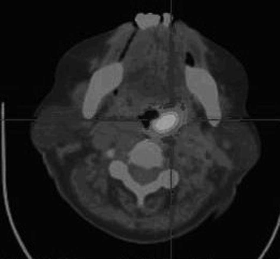
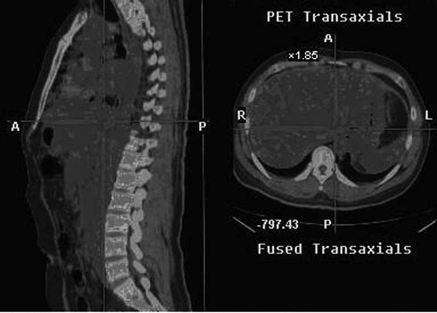
reduction (Fig. 1A, the
hypermetabolic area prior to CIK treatment as shown in PET-CT;
Fig. 1B, following CIK treatment,
the hypermetabolic area of the same patient showing measurable
radiographic tumor reduction) (Fig.
2A, liver metastasis prior to treatment as shown in PET-CT;
Fig. 2B, the metastasis of liver in
the same patient following treatment).
T-cell subsets
T-cell subsets were determined when the nucleated
cells of the patients were sampled 1 week prior to and after the
administration of CIK cell transfusion.
The ratio of CD3+, CD4+ and
CD8+ cells in the peripheral blood cells in the 8 male
patients improved significantly following CIK cell transfusion
(P<0.05) (Table IV).
| Table IVThe ratio of CD3+,
CD4+ and CD8+ cells in the peripheral blood
cells of the 8 patients. |
Table IV
The ratio of CD3+,
CD4+ and CD8+ cells in the peripheral blood
cells of the 8 patients.
|
CD3+ |
CD4+ |
CD8+ |
CD4+/CD8+ |
|---|
| Before
treatment | 61.8±5.6 | 28.1±5.2 | 30.0±9.5 | 0.8±0.5 |
| After
treatment | 69.8±6.2 | 32.1±5.6 | 32.5±11.5 | 1.0±0.4 |
Bone marrow examination
Patients with bone marrow infiltration had
reevaluation of the bone marrow after treatment. The lymphoma cells
in the bone marrow of 1 case (SMZL) achieved complete remission
compared to 16% before treatment. Other cases also showed reduced
levels of lymphoma cells in the bone marrow.
Results of biochemical and hemogram tests of the
patients before and after the treatment indicated that the CIK
therapy had little toxicity (Table
V).
| Table VGeneral conditions of the patients
after treatment. |
Table V
General conditions of the patients
after treatment.
| Pt. no. | Age (years) | Gender | ECOG Performance
status | Extra-organ
involvement | Tumor | CIK transfusion
times/cycle(cell count/cycle ×109) | Cell count
(×109) | Cycles | Response |
|---|
| 1 | 22 | M | 0 | None | 0 | 3 (6,8,4) | 18 | 1 | PR |
| 2 | 26 | M | 0 | Lung, bone
marrow
Thoracic-abdominal wall | 0 | 3 (5,5,3) | 13 | 1 | PR |
| 3 | 36 | M | 0 | Liver, bone
marrow | 0 | 3 (5,6,5) | 16 | 1 | PR |
| 4 | 39 | M | 0 | Liver,
spleen
Thoracic-abdominal wall | 0 | 3 (3,8,3) | 14 | 1 | PR |
| 5 | 41 | M | 0 | – | 0 | 4 (2,8,1,2) | 13 | 1 | CR |
| 6 | 45 | M | 0 | – | 0 | 3 (3,3,1) | 7 | 1 | PR |
| 7 | 55 | M | 2 | Bone
marrow
Thoracic-abdominal wall | 0 | 4 (3,3,4,2) | 12 | 1 | PR |
| 8 | 65 | M | 1 | Pancreas,
liver
Bone marrow | 0 | 2 (6,3) | 9 | 1 | PR |
Side effects associated with the CIK
treatment
During and after CIK cell transfusion, all of the 8
patients had varying degrees of fever, usually occurring 1–2 h
after the transfusion which lasted for ~8–15 h. The temperatures
were ~38°C. Some patients also developed sustainable low heat one
week after treatment. Analgesics were administered. The fever was
considered to be the result of metabolic syndrome. One patient
developed headache, nausea and vomiting while 2 patients exhibited
irritation, which disappeared after treatment withdrawal. None of
the patients experienced skin allergies, itching, tachycardia,
liver and kidney failure or other adverse reactions.
Complications and symptoms
No incidents of any significant complications
following CIK cell transfusion were noted. Most of the patients had
improved physical conditions, such as sleep, physical strength and
appetite. No delayed side effects occurred.
Discussion
Refractory lymphoma is generally defined as
treatment ineffective after at least 2 cycles of chemotherapy, or
relapse shortly after withdrawal. Although autologous hematopoietic
cell transplantation has proven to be an effective therapy for
patients with refractory and relapsed malignant lymphoma,
feasibility and affordability limit its widespread application.
Therefore, applicable and effective strategies should be developed.
A variety of treatment strategies have been explored, including
cytokines, activated NK cells and monoclonal antibodies.
In the present study, CIK cells were cultured using
PBMCs collected by a blood cell separator from 8 patients with
refractory lymphoma in vitro. We previously demonstrated
that CIK cells developed in this way possess the ability to achieve
a high proliferation rate, and the immunophenotype of the cells
indicates strong anti-tumor activities. The time course for the
addition of IFN-γ was found to be crucial in enhancing the
cytotoxic activity. The addition of IL-2 following that of IFN-γ
led to an increase in cytotoxicity. Furthermore, the combination of
IFN-γ and anti-CD3 enhances the proliferative effect of the
anti-CD3 treatment with an increase in cytotoxicity by IFN-γ.
This is the first study to evaluate the clinical
efficacy of CIK cell treatment on patients with refractory
lymphoma. The anti-tumor effect of CIK cells was shown mainly for
lymphoma cell lines and leukemic blasts either in vitro or
in vivo (2,10,11).
The mechanism of CIK anti-tumor activity mainly involves the
following procedures: CD3+CD56+ cells release
large amounts of cytotoxic granules that dissolve target cells. In
addition, CIK cells secrete a variety of cell-activating factors,
such as IFN-γ, TNF and IL-2, which not only directly inhibit tumor
growth, but also regulate the body's immune system response to the
indirect killing of tumor cells. CIK cells also induce tumor cell
apoptosis. Their cytotoxic effect is mediated by a
perforin/granzyme-dependent mechanism, and targeted tumor
recognition capacity appears to be partly mediated by NKG2D, an
activating receptor on NK cells, and the adhesion receptor
leukocyte function-associated antigen-1 (LFA-1) (12,13).
Recently, it has been reported that CIK cells use tumor necrosis
factor-related apoptosis-inducing ligand (TRAIL) to activate
caspase-3, which is responsible for apoptosis (6).
We assessed T-cell subsets before and after
treatment, which reflected the immune function of the patients. The
results showed that CIK cell therapy significantly improved
CD3+, CD4+, CD4+CD8+
T-cell counts, which is crucial for malignant lymphoma patients
whose immune functions are extremely low after routine treatment.
In addition, patients with loss of appetite and sleep, showed
relief of these symptoms following treatment, showing that patient
quality of life is significantly improved. Furthermore, no serious
side effects occurred after CIK treatment, indicating that CIK
therapy is safe for clinical application although larger population
cohorts should be investigated. To gain a better understanding of
the long-term efficacy of CIK therapy, follow-up of these patients
is required.
In conclusion, CIK cells are readily isolated and
expanded from peripheral blood mononuclear cells of patients with
malignant lymphoma. Due to the low level of side effects and
effective anti-tumor activity, CIK therapy may be a novel option in
the treatment of patients with refractory and relapsed malignant
lymphoma.
References
|
1
|
Schmidt-Wolf IG, Negrin RS, Kiem HP, et
al: Use of a SCID mouse/human lymphoma model to evaluate
cytokine-induced killer cells with potent antitumor cell activity.
Exp Med. 174:139–149. 1999. View Article : Google Scholar : PubMed/NCBI
|
|
2
|
Linn YC and Hui KM: Cytokine-induced
killer cells: NK-like T cells with cytotolytic specificity against
leukemia. Leuk Lymphoma. 44:1457–1462. 2003. View Article : Google Scholar : PubMed/NCBI
|
|
3
|
Leemhuis T, Wells S, Scheffold C, et al: A
phase I trial of autologous cytokine-induced killer cells for the
treatment of relapsed Hodgkin disease and non-Hodgkin lymphoma.
Biol Blood Marrow Transplant. 11:181–187. 2005. View Article : Google Scholar : PubMed/NCBI
|
|
4
|
Chan JK, Hamilton CA, Cheung MK, et al:
Enhanced killing of primary ovarian cancer by retargeting
autologous cytokine-induced killer cells with bispecific
antibodies: a preclinical study. Clin Cancer Res. 12:1859–1867.
2006. View Article : Google Scholar : PubMed/NCBI
|
|
5
|
Shi M, Zhang B, Tang ZR, et al: Autologous
cytokine-induced killer cell therapy in clinical trial phase I is
safe in patients with primary hepatocellular carcinoma. Clin Trial.
10:1146–1151. 2004.PubMed/NCBI
|
|
6
|
Kuci S, Rettinger E, Voss B, et al:
Efficient lysis of rhabdomyosarcoma cells by cytokine-induced
killer cells: implications for adoptive immunotherapy after
allogeneic stem cell transplantation. Haematologica. 95:1579–1586.
2010. View Article : Google Scholar : PubMed/NCBI
|
|
7
|
Sangiolo D, Mesiano G, Carnevale SF, et
al: Cytokine induced killer cells as adoptive immunotherapy
strategy to augment graft versus tumor after hematopoietic cell
transplantation. Expert Opin Biol Ther. 9:831–840. 2009. View Article : Google Scholar
|
|
8
|
Nishimura R, Baker J, Beilhack A, et al:
In vivo trafficking and survival of cytokine-induced killer
cells resulting in minimal GVHD with retention of anti-tumor
activity. Blood. 112:2563–2574. 2008. View Article : Google Scholar
|
|
9
|
Sangiolo D, Martinuzzi E, Todorovic M, et
al: Alloreactivity and anti-tumor activity segregate within two
distinct subsets of cytokine-induced killer (CIK) cells:
implications for their infusion across major HLA barriers. Inter
Immunol. 20:841–848. 2008. View Article : Google Scholar
|
|
10
|
Alvarnas JC, Linn YC, Hope EG, et al:
Expansion of cytotoxic CD3+CD56+ cells from
peripheral blood progenitor cells of patients undergoing autologous
hematopoietic cell transplantation. Biol Blood Marrow Transplant.
7:216–222. 2001.
|
|
11
|
Kornacker M, Moldenhauer G, Herbst M, et
al: Cytokine-induced killer cells against autologous CLL: direct
cytotoxic effects and induction of immune accessory molecules by
interferon-gamma. Int J Cancer. 119:1377–1382. 2006. View Article : Google Scholar
|
|
12
|
Mehta BA, Schmidt-Wolf IG, Weissman IL, et
al: Two pathways of exocytosis of cytoplasmic granule contents and
target cell killing by cytokine-induced
CD3+CD56+ killer cells. Blood. 86:3493–3499.
1995.PubMed/NCBI
|
|
13
|
Verneris MR, Karami M, Baker J, et al:
Role of NKG2D signaling in the cytotoxicity of activated and
expanded CD8+ T cells. Blood. 103:3065–3072. 2004.
View Article : Google Scholar : PubMed/NCBI
|