Introduction
Lifeguard (LFG), a member of a unique gene family
with high structural similarity (1), was isolated and identified as a
molecule that inhibits death mediated by Fas in tumor cells
(2). The anti-apoptotic role of LFG
was confirmed in LN-18 astrocytoma, and cervical carcinoma HeLa and
Jurkat T cell lines (3). However,
the exact role of LFG in apoptosis remains to be determined. While
it has been shown that LFG interacts with Bax and localizes in
cellular membranes, including the endoplasmic reticulum and plasma
membrane (4), it is well documented
that endogenous LFG localizes to lipid rafts (3). Its mode of action depends on
Akt/protein kinase B (PKB) signaling as dominant negative Akt/PKB
inhibits LFG activity, whereas the overexpression of constitutively
active Act/PKB leads to an increase in LFG activity (3). Previously, it was found that LFG
expression correlates with high tumor grades in primary breast
tumors and that the expression of LFG mRNA in breast cancer depends
on the activity of Akt/LEF-1 signaling (5,6).
Perifosine is an alkylphospholipid (APL), a novel
class of antitumor agents, structurally related to ether lipids
that interact with the cell membrane, thereby modulating
intracellular growth signal transduction pathways. Notably,
perifosine inhibits Akt/PKB activity, which is associated with
activation of the stress-activated protein kinase (SAPK)/JNK
pathway, without affecting PI3-K or PDK-1 activity (7). Perifosine was found to induce
apoptotic cell death in a variety of tumor cell lines and cause
inhibition of PC-3 prostate carcinoma cell growth. Perifosine
induces p21WAF1 expression in squamous carcinoma cells through a
p53-independent pathway, leading to the loss of cyclin-dependent
kinase activity and cell cycle arrest (7,8).
While it has been shown that the cellular uptake of
perifosine is required in its role as an anticancer drug, the
significance of lipid raft-mediated endocytosis has yet to be
determined. Various studies have shown that perifosine accumulates
in lipid rafts in a manner sensitive to raft disruption by
cholesterol depletion (9–11). Subsequently, it was suggested that
LFG, an anti-apoptotic protein with subcellular localization in
lipid rafts, may interfere with perifosine-induced apoptosis. Thus,
it was postulated that high expression rates of LFG confer
resistance to perifosine-induced apoptosis and that downregulation
of LFG expression sensitizes cell lines to this drug. Consequently,
LFG expression was reduced in selected cellular models by small
interfering (si)RNA transfection and the impact on
perifosine-induced apoptosis was measured.
Materials and methods
Cell lines and culture condition
Human breast carcinoma MCF7 and liposarcoma
carcinoma SW872 cell lines were obtained from the American Type
Culture Collection (Rockville, MD, USA) and grown in Dulbecco’s
modified Eagle’s medium (DMEM) (PAA, Cölbe, Germany) supplemented
with 10% fetal calf serum (Biochrom, Berlin, Germany) and 50 mg/ml
penicillin-streptomycin. Cultures were maintained at 37°C in a
humidified atmosphere with 5% CO2.
Caspase assay
Activation of caspase-3/7 was determined using the
Apo-One Homogeneous Caspase-3/7 assay (Promega, Madison, WI, USA)
according to the manufacturer’s instructions. Briefly, MCF-7 breast
cancer and SW872 sarcoma cancer cells were seeded
(1×10⁴/well) in a 96-well plate and infected with siRNA
LFG and control siRNA [107 VP (viral particles)/ml] for
48 h. After 48 h, the cells were incubated with either 25–50 ng/ml
of agonistic anti-Fas (clone CH11) or 2.5–5 μM of perifosine
(soluble in water at 10 mg/ml) for 4 h. Following treatment, the
cells were mixed with the same volume of Apo-One Homogeneous
Caspase-3/7 reagent and incubated at room temperature for 2 h.
Caspase-3/7 activation was estimated from sample fluorescence at
the excitation wavelength of 492 nm and the emission wavelength of
521 nm using the fluorescence plate reader Tecan GENios (Tecan
Schweiz AB, Zurich, Switzerland).
Small interfering RNA
MCF-7 and SW873 cells were transfected with siRNA
LFG-780 5′-gagcgggtgtatttacattg-3′ and siRNA LFG-650
5′-cctcctacccttccaatatgt-3 (designed by Sirion, Munich, Germany)
and the appropriate control vector. The algorithm used by Sirion
for the siRNA design was optimized for maximum gene specificity and
KD efficiency. Subsequent virus rescue and production were carried
out in HEK 293 cells. Virus purification was performed using the
ViraBind™ Adenovirus Miniprep kit (Cell Biolabs, Inc., USA). The
cells were seeded at 2×10⁴ cells/cm2 and
incubated at 37°C in a humidified atmosphere with 5% CO2
for 48 h before being analyzed.
Real-time polymerase chain reaction
(RT-PCR) analysis
Total RNA was extracted using the NucleoSpin RNAII
kit (MN Macherey-Nagel, Duren, Germany). RNA (1 μg) was then
reverse transcribed into cDNA and amplified using the iScript™ cDNA
kit (Bio-Rad Laboratories, Hercules, CA, USA). The reverse (R) and
forward (F) primers used were: LFG-F 5′-gactcatcctggccatcctcctac-3′
and LFG-R 5′-ggcgtcggtt acccatcagc-3′; and 18S-F
5′-gagcggtcggcgtcccccaacttc-3′ and 18S-R
5′-gcgcgtgcagccccggacatctaa-3′. PCR was carried out in 20-μl
samples with 5 ng cDNA and 10 pM of each forward and reverse primer
and the 2X SYBR-Green SensiMix DNA kit (Quantace, London, UK).
Relative gene expression was determined by normalization of the
fluorescence intensity to the expression of the 18S gene.
Amplification cycles were: 40 cycles at 94°C for 30 sec, 65°C for
30 sec and 72°C for 1 min.
Results
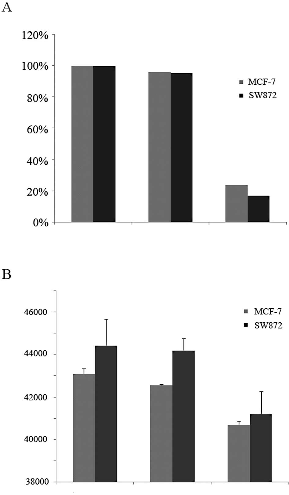
To investigate the ability of LFG expression to
suppress APL-induced apoptosis, two human solid tumor-derived cell
lines with high endogenous LFG expression were selected: breast
carcinoma MCF7 and liposarcoma SW872. Two siRNAs (650 and 780) were
designed and tested for their silencing activity in MCF-7 and SW872
cells. Ad-sh-LFG-650 significantly decreased LFG mRNA as shown by
semi-quantitative RT-PCR at 48 h following transfection (Fig. 1A).
We subsequently investigated whether downregulation
of LFG expression leads to increased rates of apoptosis in the
selected cell lines. The cells did not exhibit a significant
increase in cellular apoptosis over a 48-h period; the level of
apoptosis remained at an almost constant level comparable to the
control (Fig. 1B).
Since the LFG-mediated inhibition of Fas-induced
cell death is well documented in the literature, we assessed the
efficiency of LFG suppression on Fas-mediated apoptosis. MCF-7 and
SW872 cancer cells were infected with Ad-sh-LFG-650 and the control
siRNA (107 VP/ml) vector for 48 h followed by incubation
with 25–50 ng/ml of agonistic anti-Fas (clone CH11) for an
additional 4 h. While a dose-dependent increase in apoptosis in the
control cells indicated a general sensitivity of MCF-7 and SW872
cells to treatment with agonistic anti-Fas, we found that the cells
with a downregulated expression of LFG exhhibited significantly
increased rates of apoptosis (SW872: P=4.94581E-07, P=1.7476E-07;
MCF7: P=2.9744E-05, P=0.00282759) indicating an increase in
sensitivity to the treatment (Fig. 2A
and B).
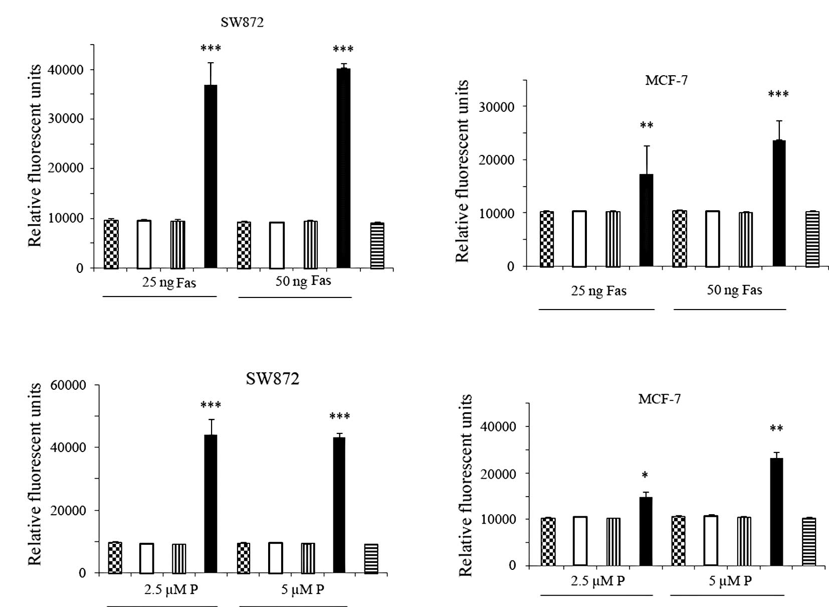 | Figure 2MCF-7 and SW872 cells were transfected
with LFG-specific siRNA (Ad-sh-LFG-650) for 48 h. Non-transfected
and Ad-sh-control vector-transfected cells were used as negative
controls. Following transfection, the cells were treated with
agonistic anti-Fas (clone CH11; 25–50 ng/ml) and perifosine (2.5–5
μM). Apoptosis was quantified by measuring the levels of active
caspase 3 using the Apo-One assay. (A) SW872 and (B) MCF-7 cells
with downregulated LFG expression showed a statistically
significant increase in apoptosis compared to the control when
treated with Fas (P=4.94581E-07, P=1.7476E-07 and P=2.9744E-05,
P=0.00282759, respectively). Transfected (C) SW872 and (D) MCF-7
cells showed a statistically significant increase in apoptosis
compared to the control when treated with perifosine (P)
(P=0.00029973, P=0.00027001 and P=0.04668954, P=0.00152829,
respectively). Data are the means ± SD of triplicate determinations
which were repeated in three separate experiments.
*p<0.05, **p<0.01,
***p<0.001 vs. the control. Checkered bar, control;
white bar, Ad-sh-conrol; vertical-striated bar, LFG vector; black
bar, Ad-sh-LFG-650; horizontal-striated bar, untreated control. |
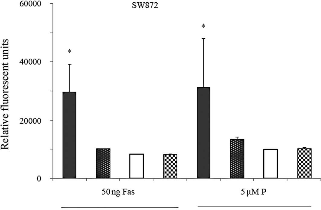
Involvement of LFG in resistance against
perifosine-mediated apoptosis was assessed to test the effect of
LFG downregulation in the cellular models. We transfected MCF-7 and
SW872 cells with the Ad-sh-LFG-650 vector 48 h prior to perifosine
treatment. Treatment of the transfected MCF-7 and SW872 cells with
2.5–5 μM perifosine for 4 h resulted in a significant increase in
cell death compared to the control cells (SW872: P=0.00029973,
P=0.00027001; MCF-7: P=0.04668954, P=0.00152829) (Fig. 2C and D).
To test for specificity of the observed effect, we
performed rescue experiments by first transfecting SW872 cells with
the Ad-sh-LFG-650 vector (downregulation) for 48 h followed by an
LFG-encoding expression vector for an additional 24 h. Following
the given time, cells were incubated with 50 ng/ml of agonistic
anti-Fas (clone CH11) and 5 μM of perifosine. Four hours after
treatment, apoptosis rates were found to be significantly decreased
in the LFG-transfected cells (Fig.
3).
Discussion
In the present study, we demonstrated that silencing
of LFG expression increased apoptosis in Fas- and
perifosine-treated MCF7 and SW872 cancer cells (Fig. 2). This is a significant finding as
clinical phase II studies on perifosine for treatment of inoperable
soft tissue sarcomas (12,13) and metastatic breast cancer patients
(14) are underway for the purpose
of identifying sensitive tumor populations.
Although our data clearly revealed that LFG was able
to reduce perifosine activity in the cell models, a number of
issues require elucidation. Investigation of the cellular uptake of
APL under given experimental conditions is warranted as uptake has
been shown to be crucial to its antitumoral activity (10). Although different types of
alkylphospholipids depend on the same modes of cellular uptake, it
appears that observed differences in kinetics and efficiencies may
depend on variations in the importance of singular pathways, e.g.,
raft-mediated endocytosis via ATP-dependent translocase activity
(15,16). This may explain differences in the
biological activity of modified alkylphospholipids as found in the
study by Mravljak et al (17) which confirms our results of reduced
sensitivity of MCF-7 cells to treatment with perifosine.
It is crucial to determine whether prolonged
treatment with perifosine induces any changes in the LFG expression
since perofisone is a known regulator of survival and proliferation
pathways, such as PI3K/Akt and ERK 1/2 (7,18,19).
Previously, we demonstrated that LFG is a new target gene of the
Akt/LEF-1 pathway (6) and
consequently a potential target of perifosine activity as well.
A number of studies aimed to sensitize cells to
undergo apoptosis by reducing the levels of anti-apoptotic
proteins, such as Bcl-2 and Bax-XL. In a large number of cell
culture studies, antisense oligonucleotides have been used to block
the expression of anti-apoptotic proteins, thereby sensitizing
cells to chemotherapy (20–24). The results of these studies showed
that altering the homeostasis of pro- and anti-apoptotic proteins
can facilitate cell death, suggesting a potential therapeutic
application of antisense oligonucleotides in the treatment of
cancer. This is the first study to report an enhanced sensitivity
to perifosine-mediated apoptosis following LFG downregulation in
carcinoma cells. Collectively, these data show that LFG may serve
as a target gene for developing new therapeutic strategies against
certain types of cancer.
Acknowledgements
This study was funded by the Claudia-von-Schilling
Breast Cancer Foundation and Niedersächsische Krebsgesellschaft. We
are most grateful to AEterna Zentaris Inc. for providing
perifosine. We also thank Dr Christine Radtke for the helpful
discussion and critical reading of the manuscript and Nikolas
Bautsch for the excellent technical assistance.
References
|
1
|
Hu L, Smith TF and Goldberger G: LFG: A
candidate apoptosis regulatory gene family. Apoptosis.
14:1255–1265. 2009. View Article : Google Scholar : PubMed/NCBI
|
|
2
|
Somia NV, Schmitt MJ, Vetter DE, et al:
LFG: an anti-apoptotic gene that provides protection from
Fas-mediated cell death. Proc Natl Acad Sci USA. 96:12667–12672.
1999. View Article : Google Scholar : PubMed/NCBI
|
|
3
|
Beier CP, Wischhusen J, Gleichmann M, et
al: FasL (CD95L/APO-1L) resistance of neurons mediated by
phosphatidylinositol 3-kinase-Akt/protein kinase B-dependent
expression of lifeguard/neuronal membrane protein 35. J Neurosci.
25:6765–6774. 2005. View Article : Google Scholar : PubMed/NCBI
|
|
4
|
Reimers K, Choi CY, Mau-Thek E, et al:
Sequence analysis shows that Lifeguard belongs to a new
evolutionarily conserved cytoprotective family. Int J Mol Med.
18:729–734. 2006.PubMed/NCBI
|
|
5
|
Bucan V, Reimers K, Choi CY, et al: The
anti-apoptotic protein lifeguard is expressed in breast cancer
cells and tissues. Cell Mol Biol Lett. 15:296–310. 2010. View Article : Google Scholar : PubMed/NCBI
|
|
6
|
Bucan V, Adili MY, Choi CY, et al:
Transactivation of lifeguard (LFG) by Akt-/LEF-1 pathway in MCF-7
and MDA-MB 231 human breast cancer cells. Apoptosis. 15:814–821.
2010. View Article : Google Scholar : PubMed/NCBI
|
|
7
|
Kondapaka SB, Singh SS, Dasmahapatra GP,
et al: Perifosine, a novel alkylphospholipid, inhibits protein
kinase B activation. Mol Cancer Ther. 2:1093–1103. 2003.PubMed/NCBI
|
|
8
|
Patel V, Lahusen T, Sy T, et al:
Perifosine, a novel alkylphospholipid, induces p21(WAF1) expression
in squamous carcinoma cells through a p53-independent pathway,
leading to loss in cyclin-dependent kinase activity and cell cycle
arrest. Cancer Res. 62:1401–1409. 2002.
|
|
9
|
Gajate C and Mollinedo F: Edelfosine and
perifosine induce selective apoptosis in multiple myeloma by
recruitment of death receptors and downstream signaling molecules
into lipid rafts. Blood. 109:711–719. 2007. View Article : Google Scholar
|
|
10
|
Mollinedo F, de la Iglesia-Vicente J,
Gajate C, et al: In vitro and in vivo selective antitumor activity
of Edelfosine against mantle cell lymphoma and chronic lymphocytic
leukemia involving lipid rafts. Clin Cancer Res. 16:2046–2054.
2010. View Article : Google Scholar : PubMed/NCBI
|
|
11
|
Van der Luit AH, Vink SR, Klarenbeek JB,
et al: A new class of anticancer alkylphospholipids uses lipid
rafts as membrane gateways to induce apoptosis in lymphoma cells.
Mol Cancer Ther. 6:2337–2345. 2007.PubMed/NCBI
|
|
12
|
Bailey HH, Mahoney MR, Ettinger DS, et al:
Phase II study of daily oral perifosine in patients with advanced
soft tissue sarcoma. Cancer. 107:2462–2467. 2006. View Article : Google Scholar : PubMed/NCBI
|
|
13
|
Knowling M, Blackstein M, Tozer R, et al:
A phase II study of perifosine (D-21226) in patients with
previously untreated metastatic or locally advanced soft tissue
sarcoma: A National Cancer Institute of Canada Clinical Trials
Group trial. Invest New Drugs. 24:435–439. 2006. View Article : Google Scholar
|
|
14
|
Leighl NB, Dent S, Clemons M, et al: A
Phase II study of perifosine in advanced or metastatic breast
cancer. Breast Cancer Res Treat. 108:87–92. 2008. View Article : Google Scholar : PubMed/NCBI
|
|
15
|
Munoz-Martinez F, Torres C, Castanys S, et
al: The anti-tumor alkylphospholipid perifosine is internalized by
an ATP-dependent translocase activity across the plasma membrane of
human KB carcinoma cells. Biochim Biophys Acta. 1778:530–540. 2008.
View Article : Google Scholar
|
|
16
|
Vink SR, van der Luit AH, Klarenbeek JB,
et al: Lipid rafts and metabolic energy differentially determine
uptake of anti-cancer alkylphospholipids in lymphoma versus
carcinoma cells. Biochem Pharmacol. 74:1456–1465. 2007. View Article : Google Scholar
|
|
17
|
Mravljak J, Zeisig R and Pecar S:
Synthesis and biological evaluation of spin-labeled
alkylphospholipid analogs. J Med Chem. 48:6393–6399. 2005.
View Article : Google Scholar : PubMed/NCBI
|
|
18
|
Ruiter GA, Zerp SF, Bartelink H, et al:
Anti-cancer alkyl-lysophospholipids inhibit the
phosphatidylinositol 3-kinase-Akt/PKB survival pathway. Anticancer
Drugs. 14:167–173. 2003. View Article : Google Scholar : PubMed/NCBI
|
|
19
|
Vink SR, van Blitterswijk WJ, Schellens
JH, et al: Rationale and clinical application of alkylphospholipid
analogues in combination with radiotherapy. Cancer Treat Rev.
33:191–202. 2007. View Article : Google Scholar : PubMed/NCBI
|
|
20
|
Duggan BJ, Maxwell P, Kelly JD, et al: The
effect of antisense Bcl-2 oligonucleotides on Bcl-2 protein
expression and apoptosis in human bladder transitional cell
carcinoma. J Urol. 166:1098–1105. 2001. View Article : Google Scholar : PubMed/NCBI
|
|
21
|
Li F, Srinivasan A, Wang Y, et al:
Cell-specific induction of apoptosis by microinjection of
cytochrome c. Bcl-xL has activity independent of cytochrome c
release. J Biol Chem. 272:30299–30305. 1997. View Article : Google Scholar : PubMed/NCBI
|
|
22
|
Strasberg RM, Zangemeister-Wittke U and
Rieber M: p53- independent induction of apoptosis in human melanoma
cells by a bcl-2/bcl-xL bispecific antisense oligonucleotide. Clin
Cancer Res. 7:1446–1451. 2001.PubMed/NCBI
|
|
23
|
Tortora G, Caputo R, Damiano V, et al:
Combined blockade of protein kinase A and bcl-2 by antisense
strategy induces apoptosis and inhibits tumor growth and
angiogenesis. Clin Cancer Res. 7:2537–2544. 2001.
|
|
24
|
Zangemeister-Wittke U, Leech SH, Olie RA,
et al: A novel bispecific antisense oligonucleotide inhibiting both
bcl-2 and bcl-xL expression efficiently induces apoptosis in tumor
cells. Clin Cancer Res. 6:2547–2555. 2000.
|