Introduction
Seborrheic keratosis (SK) is one of the most common
benign cutaneous tumors encountered in the daily
dermatopathological practice. Malignant tumor occurring within SK
is extremely rare; one retrospective study reported that only 0.6%
of SK showed direct contiguity with malignant tumors (1).
Basal cell carcinoma (BCC) arising within SK is
examined in this pioneering study, which also describes the
immunohistochemical characteristics of this type of lesion.
Case Report
An 89-year-old Japanese woman presented with a
persistent scaly plaque, measuring 14×5 mm, in the right auricle of
her ear. Biopsy of the lesion was performed, under the clinical
diagnosis of SK. After the pathological diagnosis of the biopsy
specimen, the patient underwent complete tumor resection.
Materials and methods
The formalin-fixed, paraffin-embedded tissue blocks
were cut into 3-μm sections, deparaffinized and rehydrated. Each
section was stained with hematoxylin and eosin, and used for
immunohistochemical analyses. Immunohistochemical analyses were
performed using an autostainer (XT system Benchmark, Ventana
Medical System, Tucson, AZ, USA) according to the manufacturer's
instructions. The primary antibodies used were: a mouse monoclonal
antibody against cytokeratin (CK17) (E3, Novocastra Laboratories,
Ltd., Newcastle upon Tyne, UK), a mouse monoclonal antibody against
CK19 (clone b170, Novocastra Laboratories, Newcastle, UK), a mouse
monoclonal antibody against p53 (PAb1801; Novocastra Laboratories),
and a rabbit polyclonal antibody against SOX9 (Santa Cruz
Biotechnology, Santa Cruz, CA, USA).
Results
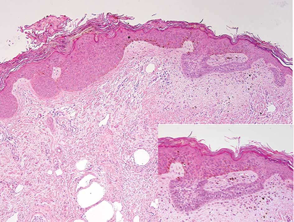
Histopathological study of the biopsy specimen
revealed hyperkeratosis and proliferation of basaloid and squamoid
cells in the acanthotic epidermis. These basaloid and squamoid
cells were without atypia, and mitotic figures were rarely noted
(Fig. 1). These were typical
histopathological findings for SK. There were some buds composed of
follicular germinative cell-like immature basaloid cells directly
attached to the undersurface of the SK (Fig. 1). These immature basaloid cells had
large elongated nuclei and scant cytoplasm, and showed peripheral
palisading (Fig. 1, inset). Mitotic
figures and apoptotic bodies were scattered. There were areas of
retraction of the stroma around the tumor buds. These
histopathological findings corresponded to the superficial type of
BCC. Therefore, a diagnosis of the superficial type of BCC arising
within SK was ultimately made based on the biopsy specimen.
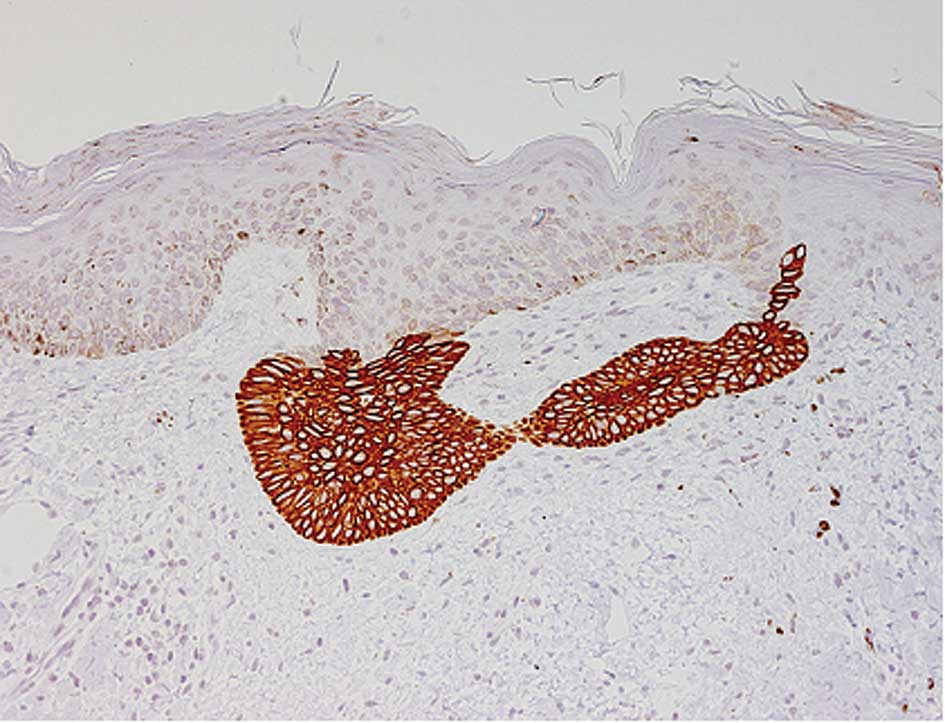
Immunohistochemical studies revealed that CK17, CK19
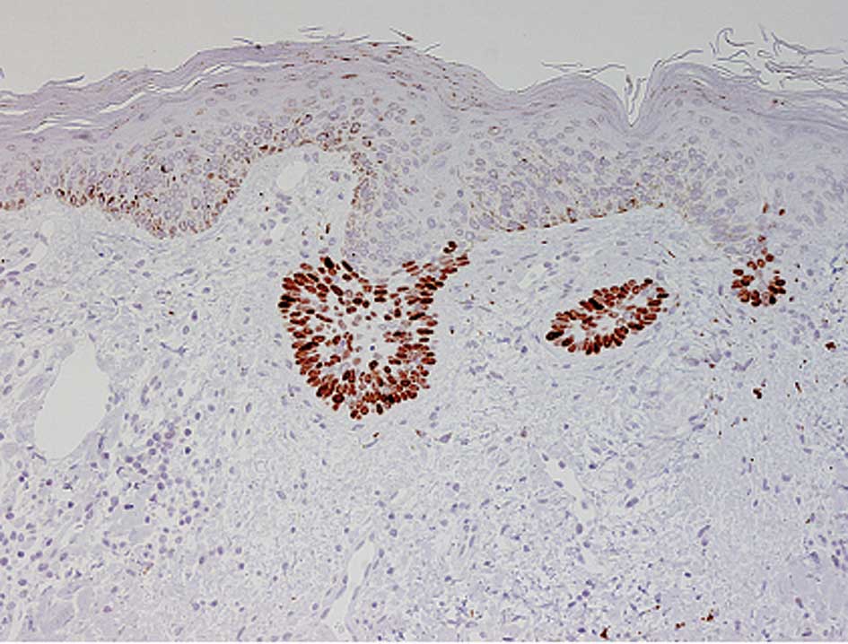
and SOX9 were expressed in the BCC, but not in the SK (Fig. 2). Furthermore, the overexpression of
p53 protein was observed in the BCC, but not in the SK (Fig. 3).
The histopathological and immunohistochemical
findings of the surgically resected specimen were identical to
those of the biopsy specimen.
Discussion
The development of a malignant neoplasm in
association with SK was previously identified. BCC, squamous cell
carcinoma and malignant melanoma have been documented to occur
within SK, and BCC is the most common malignant neoplasm occurring
within SK (1,2). The predominant histological subtype of
BCC associated with SK is the superficial type, as observed in the
present case (2).
This report is the first to describe the
immunohistochemical characteristics of BCC arising within SK. We
found that CK17, CK19 and SOX9 were expressed in the BCC, but not
in the SK. Findings of previous reports revealed that CK17 and CK19
were expressed in the outer root sheath of hair follicles as well
as in most cases of BCC (3), but
not in SK (4). SOX9 belongs to the
SOX gene family, members of which are known to be involved in a
large number of developmental processes. Recent studies showed that
SOX 9 was expressed in the outer root sheath of hair follicles,
sweat and sebaceous glands, as well as BCC (5,6);
however, its expression was rarely observed in SK (6). Thus, the immunohistochemical findings
of the present case are typical for SK and BCC.
BCC is considered to originate from follicular
germinative cells, and various immunohistochemical studies have
indicated that the outer root sheath of hair follicles may be a
possible origin of BCC (3). In
addition, SK is also thought to be a benign skin appendage neoplasm
showing follicular differentiation, especially follicular
infundibula. Therefore, Cascajo et al speculated that there
was a pathogenic relationship between SK and BCC, with respect to a
common follicular origin (2).
However, the immunohistochemical characteristics of BCC and SK in
the present case were different, suggesting that BCC does not arise
directly from SK, but instead, that SK is the nidus of the
carcinoma, resulting in the abutment of SK with BCC.
Furthermore, the results of the present case suggest
that immunohistochemical surveillance of the expression of CK17,
CK19, SOX 9, and p53 protein is useful in differentiating minute
BCC, especially the superficial type, from the non-neoplastic hair
buds, as it is not always easy to distinguish between them.
References
|
1
|
Lim C: Seborrheic keratoses with
associated lesions: a retrospective analysis of 85 lesions.
Australas J Dermatol. 47:109–113. 2006. View Article : Google Scholar : PubMed/NCBI
|
|
2
|
Cascajo CD, Reichel M and Sanchez JL:
Malignant neoplasms associated with seborrheic keratosis. An
analysis of 54 cases. Am J Dermatopathol. 18:278–282. 1996.
View Article : Google Scholar : PubMed/NCBI
|
|
3
|
Ishida M, Kushima R and Okabe H:
Immunohistochemical demonstration of D2–40 in basal cell carcinomas
of the skin. J Cutan Pathol. 35:926–930. 2008.
|
|
4
|
Liu HN, Chang YT and Chen CC: Differential
of hidroacanthoma simplex from clonal seborrheic keratosis – an
immunohistochemical study. Am J Dermatopathol. 26:188–193.
2004.
|
|
5
|
Krahl D and Sellheyer K: Sox9, more than a
marker of the outer root sheath: spatiotemporal expression pattern
during human cutaneous embyogenesis. J Cutan Pathol. 37:350–356.
2010. View Article : Google Scholar : PubMed/NCBI
|
|
6
|
Vidal VPI, Ortonne N and Schedl A: SOX9
expression is a general marker of basal cell carcinoma and
adnexal-related neoplasms. J Cutan Pathol. 35:373–379. 2008.
View Article : Google Scholar : PubMed/NCBI
|