Introduction
Head and neck squamous cell carcinoma (HNSCC) is a
frequently occurring cancer worldwide. HNSCC exhibits heterogeneous
characteristics: some HNSCC exhibit a well-differentiated, low
invasive, bland phenotype, whereas others exhibit a highly invasive
phenotype, often with lymph nodal and/or distant metastasis, and
the sensitivity to chemotherapy or radiation therapy varies
according to the individual case. Moreover, as background risk
factors, the involvement of environmental exposure of the patient,
including tobacco smoking and alcohol consumption, make it more
complex to understand the biological behavior of HNSCC.
HNSCC occurs as a result of cumulative genetic or
epigenetic alterations in cancer-related genes, oncogenes and tumor
suppressor genes (TSGs). Despite recent advances in molecular
pathology, little is known regarding the genes or pathways that
play key roles in HNSCC (1). To
establish a more concise understanding of the biological
characteristics of HNSCC, our group has been working on a
genome-wide analysis with the aim of identifying the genes that act
as the principle drivers of carcinogenesis, invasion and
metastasis. The focus has been on the abnormal function of the
major tumor suppressor molecules. Previously, we reported on the
decreased function in the ING family (2–6), and
the possible tumor suppressor role of Dickkopf (Dkk)-3, which is
the tumor suppressor against oncogenic Wnt/β-catenin signaling in
HNSCC (7,8).
Wnt/β-catenin signaling is one of the most crucial
pathways in carcinogenesis in HNSCC. Recent cancer stem cell
research revealed that the side population, which includes cancer
stem cells in HNSCC, demonstrates abnormal Wnt/β-catenin signaling,
and that such cells possess highly invasive potential (9). The Dkk family functions as a negative
regulator of Wnt signaling, and thus acts as a tumor suppressor
(10). As previously reported, the
down-regulation of Dkk-3 in mRNA expression is observed as a common
event in several types of malignancies, including glioma (11), hepatocellular carcinoma (12), breast cancer (13), melanoma (14), prostate (15) and gastrointestinal cancer (16). The decrease in mRNA expression
induction may also decrease Dkk-3 protein expression, which may
lead to the loss of tumor suppressor functions, resulting in
carcinogenesis.
However, of note, the mRNA down-regulation of Dkk-3
may vary depending on the tissue of origin. For instance, Dkk3 mRNA
expression is not decreased in certain esophageal squamous cell
carcinoma cell lines (16).
Furthermore, Dkk-3 protein expression is conserved in esophageal
squamous cell carcinoma tissue samples (17), indicating the existence of crucial
and specific roles of Dkk-3 in squamous epithelium origin, while
the protein expression status of Dkk-3 in HNSCC has yet to be
reported.
In the present study, we investigated the protein
expression of Dkk-3 in a HNSCC tissue sample and HNSCC cell lines,
as well as the correlation between the clinicopathological
characteristics and patient prognosis by statistical analysis. We
report notable findings regarding Dkk-3 expression and discuss its
possible role in HNSCC carcinogenesis and progression.
Materials and methods
Tissue samples and patients
In total, 90 cases of HNSCC tissue samples and 10
cell lines were used for this study. The tissue sample was obtained
from the Okayama University Tumor bank (Professor Kenji Shimizu)
and Okayama University Hospital Surgical Pathology Unit (Okayama,
Japan) between 1994 and 2008. The details of the patient data,
including gender, age, history of smoking, alcohol consumption, TNM
staging, incidence of lymph nodal metastasis and the condition with
or without chemotherapy or radiation therapy, are shown in Table I.
 | Table IClinicopathological characteristics of
patients. |
Table I
Clinicopathological characteristics of
patients.
| Parameter | Number | % |
|---|
| Age (years) mean ±
SD | 67.28±11.28 | |
| Gender |
| Male | 63 | 70.0 |
| Female | 27 | 30.0 |
| Tumor site |
| Tongue | 28 | 31.1 |
| Gingiva | 23 | 25.6 |
| Buccal mucosa | 10 | 11.1 |
| Mouth floor | 8 | 8.9 |
| Larynx | 8 | 8.9 |
| Hypopharynx | 7 | 7.8 |
| Mesopharynx | 4 | 4.4 |
| Oropharynx | 1 | 1.1 |
| Lower lip | 1 | 1.1 |
| T stage |
| T1 | 16 | 17.8 |
| T2 | 33 | 36.7 |
| T3 | 17 | 18.9 |
| T4 | 24 | 26.7 |
| N stage |
| N0 | 49 | 54.4 |
| N1 | 17 | 18.9 |
| N2 | 21 | 23.3 |
| N3 | 3 | 3.3 |
| TNM stage |
| I | 14 | 15.6 |
| II | 22 | 24.4 |
| III | 17 | 18.9 |
| IV | 37 | 41.1 |
|
Differentiation |
| Well | 53 | 58.9 |
| Moderate | 26 | 28.9 |
| Poor | 11 | 12.2 |
The patients included 63 males and 27 females.
Follow-up duration periods were between 1 and 105 months. During
follow-up, 13 patients with local cancer recurrence, 25 with lymph
nodal/distant metastasis and 2 with both recurrence and metastasis,
were observed. Of the 90 patients, 25 patients succumbed to cancer.
The tumors were surgically excised from gingiva (N=23), buccal
mucosa (N=10), the mouth floor (N=8), tongue (N=28), lower lip
(N=1), larynx (N=8), hypopharynx (N=7), mesopharynx (N=4) and
oropharynx (N=1) after informed consent was obtained from each
patient. The tissue samples were fixed with 10% neutral-buffered
formalin and processed for paraffin-embedded sections. Serial
sections (4 μm) were used for hematoxylin and eosin (H&E)
sections and immunohistochemical analysis.
Cell lines
The cell lines HSC-2, HSC-3, HSC-4 and Ca9-22 were
provided by RIKEN BRC through the National Bio- Resource Project of
the The Ministry of Education, Culture, Sports, Science and
Technology (MEXT), Japan. The cell lines UT-SCC-12A, UT-SCC-12B,
UT-SCC-16A, UT-SCC-16B, UT-SCC-60A and UT-SCC-60B, which were
paired and originally established cell lines from the primary and
metastatic cancers of the same patients, were kindly provided by Dr
Reidar Grenman, University of Turku, Finland.
The cell lines were maintained in Dulbecco’s
modified Eagle’s medium (Gibco, Tokyo, Japan) with antibiotics and
an antimycotic agent, including 100 U/ml penicillin, 100 μg/ml
streptomycin and 25 mg/ml amphotericin B (Gibco), in a
CO2 incubator with an atmosphere of 95% air plus 5%
CO2 at 37˚C. When the cells became confluent, total
protein was extracted for Western blotting as reported in a
previous study (18).
Western blotting
Western blotting was performed according to the
usual procedure. Cell extracts were boiled for 5 min in sodium
dodecyl sulfate (SDS) gel-loading buffer (0.1 M Tris-HCl, pH 6.8,
20% glycerol, 2.5% SDS, 0.05% bromophenol blue and 5%
β-mercaptoethanol). Equal amounts of each protein sample were then
loaded and separated on 12.5% SDS-polyacrylamide gels and blotted
onto polyvinylidene difluoride (PVDF) membranes. Following the
blockade of non-specific binding by soaking the PVDF membranes in
5% skim milk, proteins were detected using anti-Dkk-3 (R&D
Systems, Minneapolis, MN, USA) and anti-β-actin (Abcam, Cambridge,
MA, USA). Proteins were visualized using the ECL Plus Western
blotting detection system (Amersham, Arlington Heights, IL, USA)
according to the manufacturer’s instructions.
Immunohistochemistry
Immunohistochemistry was carried out according to
the manufacturer’s instructions using goat polyclonal antibody
against human Dkk-3 (R&D Systems). Briefly, cut tissue sections
were deparaffinized in xylene for 20 min, and then dehydrated in
graded alcohol solutions. The endogenous peroxidase activity was
blocked by immersing the sections in 3% H2O2
in methanol for 30 min. For antigen retrieval, the sections were
heated in 0.01 mol/l citrate buffer (pH 6.0) for 15 min. The
sections were then treated with 10% normal rabbit serum for 30 min,
followed by incubation with primary Dkk-3 antibody at 4˚C
overnight. Identification of an immunoreactive site was achieved by
subsequent incubation with a biotinylated secondary antibody for 30
min, followed by detection using the avidin-biotin complex (ABC)
method (Vectastain ABC kit; Funakoshi, Tokyo, Japan). Sections were
then reacted with 0.02% 3,3′-diaminobenzidine (Wako, Osaka, Japan)
with 0.006% H2O2 in 0.05 ml/l Tris-HCl
(pH7.6). The sections were counterstained by Mayer’s hematoxylin
and mounted. The specimens were checked by two pathologists, and
scored as positive if there was any positive reaction, and
otherwise scored as negative.
Statistical analysis
Pearson’s Chi-square test and Fisher’s exact test
were used to evaluate the correlation between the Dkk-3 protein
expression and clinicopathological characteristics. The
Kaplan-Meier analysis was performed to determine the survival
analysis. For comparison of survival between the Dkk-3-positive and
-negative groups, the log-rank test was used. The duration of
disease-free survival (DFS) was determined from the day following
surgery to the initial recurrence or metastasis evaluated by
clinical examination. Overall survival (OS) in months was
calculated from the day following surgery to the last follow-up
appointment. To determine the detailed involvement of Dkk-3
expression in DFS, metastasis-free survival (MFS) and
recurrence-free survival (RFS) were also examined. MFS was
determined from the day following surgery to the initial
metastasis-evaluated clinical examination. RFS was determined from
the day following surgery to the initial recurrence of the
surgically excised cancer. For multivariate analysis, a Cox
proportional hazard model was used. Computations were performed
using PASW Statistics 18 (SPSS Inc. Chicago, IL, USA). P<0.05
was considered statistically significant.
Results
Expression of Dkk-3 in HNSCC
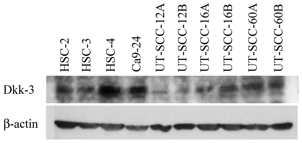
Results of the Western blot analysis revealed that
the HNSCC-derived cell lines expressed Dkk-3 protein to a varying
degree. The HSC-4 and Ca9-22 cells exhibited intense Dkk-3
expression, whereas the UT-SCC-12A and UT-SCC-12B cells were
comparatively weak. UT-SCC- 12A/12B, UT-SCC-16A/16B and
UT-SCC-60A/60B were paired cell lines, which were derived from the
primary cancer/metastatic cancer of the same patients. Of note, the
paired cell lines expressed Dkk-3 regardless of their
primary/metastatic origin (Fig.
1).
Dkk-3 expression was also detected in the tissue
samples. Using immunohistochemistry, the majority of the HNSCC
tissue samples exhibited a positive reaction for Dkk-3. Of the 90
cases, 76 cases (84.4%) were positive, whereas only 14 cases
(14.6%) were negative. A positive reaction was detected in the
invasive cancer nests and the dysplastic epithelium continuing to
the cancer. Dkk-3 was present in the cytoplasm and plasma membrane
of the tumor cells as well as in blood vessels around the tumor
nest and in the lymphatic vessels, but no nuclear expression was
observed. The expression of Dkk-3 in the metastatic lymph node was
also examined in certain cases. Intense Dkk-3 expression was also
observed in tumor cells in the metastatic lymph nodes (Fig. 2).
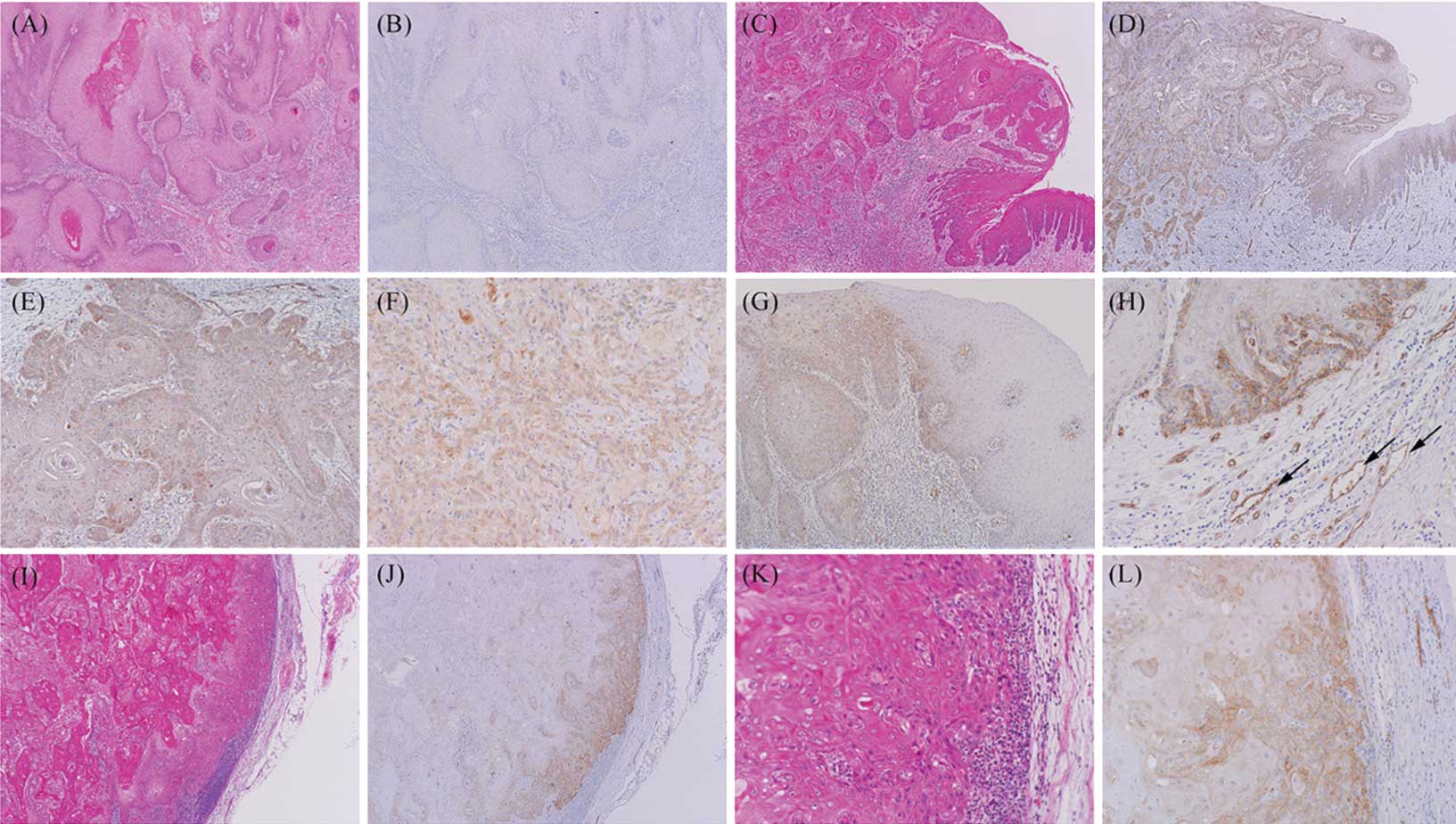 | Figure 2Dkk-3 protein expression was evaluated
as negative (A and B) and positive (C and D). Dkk-3 protein
expression was observed in the cytoplasm and plasma membrane of the
cancer cells, regardless of histological differentiation (E and F,
well-differentiated SCC and poorly differentiated SCC,
respectively). (G) Normal and dysplastic epithelium, adjacent to
the invasive cancer region, also expressed Dkk-3 to varying
degrees. (H) Dkk-3 expression was also observed in adjacent
microvessels around the cancer nests, including both blood vessels
and lymphatic vessels (arrows). (I-K) Dkk-3 expression was observed
in metastatic cancer cells of the lymph nodes. Original
magnification: A-D, G, J and I, ×40; E, F, H, K and L, ×100,
respectively. SCC, squamous cell carcinoma. |
Dkk-3 expression and clinicopathological
characteristics
The relationship between Dkk-3 expression and
various clinicopathological characteristics was examined. No
significant association was found between Dkk-3 and any of the
parameters investigated (Table
II). However, a tendency of correlation was shown for smoking
habits (p=0.170), TNM stage (p=0.154) and existence of the
chemotherapy (p=0.097). In relation to smoking, Dkk-3-positive
expression was observed in 37 cases out of 44 smoking patients
(84.1%), whereas the non-smoking patient positivity was 68.8%
(11/16 cases). Eight patients out of 14 cases (57.1%) were from
early TNM stage (I–II), whereas 48 (63.2%) of the Dkk-3-positive
cases (N=76) were from late TNM stage (III–IV). As for
chemotherapy, the majority of the Dkk-3-positive patients (54/75
cases, 88.5%) did not undergo chemotherapy. Of note, the Dkk-3 (−)
group included no cases of metastasis and only 2 cases (2/13,
15.4%) of local recurrence, whereas 25 patients with lymph
nodal/distant metastasis and 2 patients with local recurrence and
metastasis belong to the Dkk-3 (+) group. The number of patients
who succumbed to the disease in the follow-up period in the Dkk-3
(+) and Dkk-3 (−) groups were 23/25 (92.0%) and 2/25 (8.0%),
respectively.
| Table IIRelationship between Dkk-3 protein
expression and clinicopathological characteristics. |
Table II
Relationship between Dkk-3 protein
expression and clinicopathological characteristics.
| Parameters | Dkk-3
expression | |
|---|
|
| |
|---|
| Dkk-3 (+)
(N=76) | Dkk-3 (−)
(N=14) | p-value |
|---|
| Gender |
| Male | 52 (68.4%) | 11 (78.6%) | 0.338 |
| Female | 24 (31.6%) | 3 (21.4%) | |
| Age |
| <65 | 24 (31.6%) | 4 (28.6%) | 1.000 |
| ≥65 | 52 (68.4%) | 10 (71.4%) | |
| Smokinga |
| Yes | 37 (77.1%) | 7 (58.3%) | 0.170 |
| No | 11 (22.9%) | 5 (41.7%) | |
| Alcohol
consumptiona |
| Yes | 31 (64.6%) | 7 (58.3%) | 0.466 |
| No | 17 (35.4%) | 5 (41.7%) | |
| TNM stage |
| I–II | 28 (36.8%) | 8 (57.1%) | 0.154 |
| III–IV | 48 (63.2%) | 6 (42.9%) | |
| T stage |
| T1–T2 | 40 (52.6%) | 9 (64.3%) | 0.421 |
| T3–T4 | 36 (47.4%) | 5 (35.7%) | |
| N stage |
| N (−) | 40 (52.6%) | 9 (64.3%) | 0.421 |
| N (+) | 36 (47.4%) | 5 (35.7%) | |
|
Differentiation |
| Well | 43 (56.6%) | 10 (71.4%) | 0.299 |
| Moderate-poor | 33 (43.6%) | 4 (28.6%) | |
| Radiation
therapya |
| Yes | 22 (29.7%) | 5 (35.7%) | 0.438 |
| No | 52 (70.3%) | 9 (64.3%) | |
|
Chemotherapya |
| Yes | 21 (28.0%) | 7 (50.0%) | 0.097 |
| No | 54 (72.0) | 7 (50.0%) | |
| Previous cancer
historya |
| Yes | 18 (25.0%) | 5 (35.7%) | 0.300 |
| No | 54 (75.0%) | 9 (64.3%) | |
Dkk-3 expression and survival
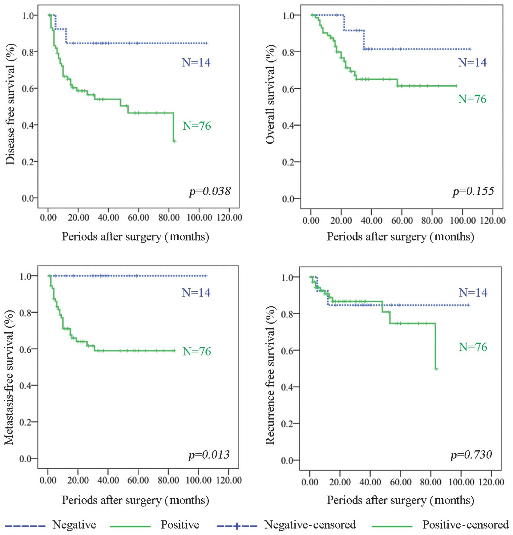
Survival analysis by the Kaplan-Meier curves
indicated that patients without Dkk-3 expression have a
significantly longer DFS than patients with Dkk-3 expression
(p=0.038). No significant association was found between OS and
Dkk-3 expression; however, similarly to DFS, patients without Dkk-3
expression presented a tendency for longer OS than those with Dkk-3
expression (p=0.155). As for MFS and RFS, the Dkk-3 (−) group
showed significantly longer MFS (p=0.013), whereas no significant
difference was found in RFS (p=0.730) (Fig. 3). The Cox proportional hazard model
for survival analysis revealed that the Dkk-3 expression status was
the independent prognostic marker for DFS (p=0.004), OS (p=0.001)
and MFS (p=0.015) (Table
III).
| Table IIICox proportional hazard model for
survival analysis. |
Table III
Cox proportional hazard model for
survival analysis.
| Disease-free
survival | Overall
survival |
|---|
|
|
|
|---|
| Variables | p-value | HR | 95% CI | p-value | HR | 95% CI |
|---|
| | |
| | |
|
|---|
| | | Lower | Upper | | | Lower | Upper |
|---|
| Gender | 0.440 | 1.881 | 0.378 | 9.344 | 0.332 | 2.565 | 0.383 | 17.199 |
| Age | 0.285 | 0.578 | 0.212 | 1.579 | 0.340 | 0.594 | 0.204 | 1.733 |
| Smoking | 0.696 | 1.399 | 0.259 | 7.550 | 0.767 | 1.320 | 0.210 | 8.308 |
| Alcohol | 0.125 | 0.407 | 0.129 | 1.282 | 0.564 | 0.684 | 0.188 | 2.484 |
| TNM stage | 0.923 | 0.000 | 0.000 | 2.95E+101 | 0.929 | 0.000 | 0.000 | 2.1E+122 |
| T stage | 0.934 | 30233.381 | 0.000 | 1.6E+111 | 0.942 | 56904.112 | 0.000 | 8.1E+132 |
| N stage | 0.198 | 3.234 | 0.540 | 19.354 | 0.095 | 6.755 | 0.716 | 63.733 |
|
Differentiation | 0.243 | 2.188 | 0.588 | 8.149 | 0.086 | 3.967 | 0.822 | 19.134 |
| Radiation | 0.002 | 0.120 | 0.031 | 0.46 | 0.034 | 0.225 | 0.057 | 0.897 |
| Chemotherapy | 0.232 | 0.484 | 0.147 | 1.592 | 0.184 | 0.407 | 0.108 | 1.535 |
| Cancer history | 0.962 | 0.964 | 0.215 | 4.325 | 0.581 | 1.793 | 0.226 | 14.26 |
| Dkk-3
expression | 0.004 | 29.631 | 2.965 | 296.135 | 0.010 | 11.282 | 1.785 | 71.291 |
|
| Metastasis-free
survival | Recurrence-free
survival |
|
|
|
| Variables | p-value | HR | 95% CI | p-value | HR | 95% CI |
| | |
| | |
|
| | | Lower | Upper | | | Lower | Upper |
|
| Gender | 0.242 | 2.780 | 0.501 | 15.421 | 0.942 | 0.000 | 0.000 | 2.5E131 |
| Age | 0.428 | 0.650 | 0.224 | 1.885 | 0.267 | 0.012 | 0.000 | 29.177 |
| Smoking | 0.820 | 1.225 | 0.212 | 7.061 | 0.904 | 2.348E8 | 0.000 | 1.0E145 |
| Alcohol | 0.120 | 0.402 | 0.128 | 1.266 | 0.997 | 1.855 | 0.000 | 1.1E132 |
| TNM stage | 0.935 | 0.000 | 0.000 | 5367E112 | 0.815 | 0.000 | 0.000 | 2.6E161 |
| T stage | 0.942 | 22231.109 | 0.000 | 9.71E121 | 0.898 | 1.142E11 | 0.000 | 9.8E179 |
| N stage | 0.260 | 2.805 | 0.467 | 16.867 | 0.853 | 461597.142 | 0.000 | 4.13E65 |
|
Differentiation | 0.378 | 1.845 | 0.473 | 7.195 | 0.739 | 6.062E9 | 0.000 | 2.73E67 |
| Radiation | 0.002 | 0.112 | 0.028 | 0.446 | 0.945 | 36.285 | 0.000 | 8.26E45 |
| Chemotherapy | 0.706 | 0.777 | 0.210 | 2.884 | 0.746 | 0.000 | 0.000 | 6.37E48 |
| Cancer history | 0.868 | 0.881 | 0.200 | 3.894 | 0.968 | 4601.361 | 0.000 | 9.8E180 |
| Dkk-3
expression | 0.015 | 17.774 | 1.755 | 179.986 | 0.362 | 5000.076 | 0.000 | 4.43E11 |
Discussion
HNSCC is an unmanageable cancer, which exhibits
variation both biologically and clinically. Certain cases of HNSCC
show a highly invasive phenotype, which gives rise to metastasis in
lymph nodes and/or distant organs and exhibits chemoresistance,
while others do not. To improve our understanding of the biological
mechanisms of HNSCC, which may lead to the establishment of a new
cancer therapy strategy, the search for specific and powerful genes
or pathways in HNSCC has been ongoing.
We focused on Wnt/β-catenin signaling and its
negative regulator molecules as one of the most promising targets.
Wnt/β-catenin signaling is tightly regulated by antagonists, such
as the Dkk family, Wnt inhibitory factor (WIF) and secreted
frizzled related protein (sFRP). When Wnt ligands bind to their
receptor complex, frizzled and LRP5/6, β-catenin translocation into
the nucleus occurs, resulting in the transcription of c-myc, c-jun,
fra-1 and cyclin D1 (19). In
cancer conditions, the aberrant expression of Wnt or
down-regulation of Wnt antagonists has been repeatedly reported in
several types of malignancy (20–22).
Further investigation has revealed that down-regulation of Dkk-3 in
cancers was due to epigenetic silencing by DNA methylation
(23–28). However, paradoxically, Dkk-3 has
been found to be overexpressed in hepatoblastomas and
hepatocellular carcinomas (29),
suggesting that the function of Dkk-3 may differ depending on the
tissue of origin. Moreover, to the best of our knowledge, Dkk-3
expression in HNSCC has not yet been reported. Based on this fact,
we firstly investigated Dkk-3 protein expression in a large number
of HNSCC tissue samples and cell lines, and assessed the
correlation between Dkk-3 protein expression and clinical
aspects.
Notably, our data varied from those of previous
studies of other malignancies, suggesting that Dkk-3 may not act as
a tumor suppressor in HNSCC. Results of the Western blot analysis
revealed that all the cell lines studied (10/10) exhibited a
positive reaction to a varying degree. Dkk-3 expression is present
in primary and metastatic cancer. These data suggest that Dkk-3 DNA
methylation is not likely to be involved in HNSCC carcinogenesis,
whereas this event is common in other organs. Reflecting the
protein expression in cell lines, 84.4% (76/90) of HNSCC tissue
samples demonstrated Dkk-3 expression. Supporting our data, certain
reports (16,17) have noted Dkk-3 expression in
esophageal squamous cell carcinoma. As for Dkk-3 expression in
cancer cell lines, it is reported that 8 out of the 13 cell lines
expressed Dkk-3 mRNA, and that its protein expression was also
conserved in 16.9% of the esophageal squamous cell carcinoma tissue
samples (16). Recently, Dkk-3 has
been reported to be expressed and up-regulated in esophageal
squamous cell carcinoma (17).
Taken together, specific Dkk-3 expression is thought to be a common
event in squamous epithelium in the head and neck region, which
would be a significant and noteworthy finding in understanding the
biological characteristics of HNSCC.
In addition, we have examined the association
between Dkk-3 expression and clinicopathological characteristics
together with survival analyses to gain a better understanding of
the specific Dkk-3 expression in HNSCC. The patients were divided
into two groups based on the Dkk-3 expression profile, Dkk-3 (+)
and Dkk-3 (−). The Chi-square test results revealed no significant
correlation between Dkk-3 expression and the clinicopathological
parameters (Table II). A
comparison was made of the DFS and OS between the Dkk-3 (+) and
Dkk-3 (−) groups. The Dkk-3 (−) group tended to show longer OS
(p=0.155) and significantly longer DFS (p=0.038). MFS of the Dkk-3
(−) group was significantly longer than that of the Dkk-3 (+) group
(p=0.013), whereas RFS showed no significant difference between the
two groups. Thus, this longer MFS may contribute to longer DFS in
the Dkk-3 (−) group.
The patient group includes numerous confounding
factors, including varying tumor stage, lymph nodal status,
chemotherapy and/or radiation therapy. Therefore, a Cox
proportional hazard model analysis has been performed in order to
exclude the bias to clarify whether Dkk-3 is an independent
prognostic factor. The results showed that no Dkk-3 expression was
an independent prognostic biomarker for DFS, OS and MFS (p=0.004,
p=0.010 and p=0.015, respectively), together with existence of
radiation therapy (DFS, p=0.002; OS, p=0.034; and MFS,
p=0.002).
Taking into consideration the fact that Dkk-3 is a
known tumor suppressor and that its expression is generally reduced
in cancer tissues, our findings are thought to be relatively
paradoxical. Moreover, our results demonstrate that the Dkk-3 (−)
group did not experience lymph nodal/distant metastasis, and that
Dkk-3 (−) patients have a significantly lower risk of presenting
metastasis. This finding suggests that Dkk-3 may be involved in the
metastatic process and may be used as a prognostic marker in HNSCC.
In this context, Dkk-3 appears to act as an oncogene, playing a
role in cancer metastasis in HNSCC.
The possibility of oncogenic properties of Dkk-3 has
yet to be investigated. However, another finding in this study may
aid the explanation of the current results. In the
immunohistochemical analyses, Dkk-3 expression was also observed in
the small blood/lymphatic vessels around the tumor nests. Recently,
the vascular expression of Dkk-3 around cancer cells has become a
topic of debate. Dkk-3 is reportedly up-regulated in the tumor
endothelium of colorectal cancer. This augmentation of Dkk-3
expression is correlated with an increase in the number of
microvessels, suggesting that Dkk-3 is a marker for
neo-angiogenesis in colorectal cancer (30). Moreover, Untergasser et al
demonstrated that the number of blood vessels expressing Dkk-3 is
increased in glioma, high-grade non-Hodgkin’s lymphoma, melanoma
and colorectal cancer in comparison with the blood vessels located
in non-tumor tissues. Overexpression or inhibition of Dkk-3 in
endothelial colony-forming cells does not affect their
proliferation or migration, but tube formation in matrigel may
increase following Dkk-3 overexpression and decrease following
Dkk-3 down-regulation (31). Thus,
it is possible that Dkk-3 may be a differentiation factor involved
in the remodeling of the tumor vasculature. Neo-angiogenesis is
critical for tumor growth (32–34)
and improved prognosis for patients without Dkk-3 in HNSCC may be
correlated to the effect of Dkk-3 on tumor vasculature. Such
hypotheses may also be in agreement with the significant
relationship found between Dkk-3 and metastasis.
In conclusion, we have shown that Dkk-3 expression
is conserved in HNSCC, and that this expression may be used as a
prognostic factor for metastasis risk in HNSCC. In the present
study, we confirmed a noteworthy phenomenon. However, the detailed
functions of Dkk-3 and the mechanism pertaining to why Dkk-3 is
specifically conserved in HNSCC remains unclear. Further
investigation including functional analyses may clarify these
points.
Acknowledgements
This work was partially supported by a grant-in-aid
for scientific researches from the Ministry of Education, Culture,
Sports, Science and Technology (Japan), 22791766 (to N.K.),
22•00130 (to M.L.), 20791515 (to H.T.) and 21592326 (to H.N.).
References
|
1
|
Ha PK, Chang SS, Glazer CA, Califano JA
and Sidransky D: Molecular techniques and genetic alterations in
head and neck cancer. Oral Oncol. 45:335–339. 2009. View Article : Google Scholar : PubMed/NCBI
|
|
2
|
Gunduz M, Ouchida M, Fukushima K, Hanafusa
H, Etani T, Nishioka S, Nishizaki K and Shimizu K: Genomic
structure of the human ING1 gene and tumor-specific mutations
detected in head and neck squamous cell carcinomas. Cancer Res.
60:3143–3146. 2000.PubMed/NCBI
|
|
3
|
Gunduz M, Ouchida M, Fukushima K, Ito S,
Jitsumori Y, Nakashima T, Nagai N, Nishizaki K and Shimizu K:
Allelic loss and reduced expression of the ING3, a candidate tumor
suppressor gene at 7q31, in human head and neck cancers. Oncogene.
21:4462–4470. 2002. View Article : Google Scholar : PubMed/NCBI
|
|
4
|
Gunduz M, Nagatsuka H, Demircan K, Gunduz
E, Cengiz B, Ouchida M, Tsujigiwa H, Yamachika E, Fukushima K,
Beder L, et al: Frequent deletion and down-regulation of ING4, a
candidate tumor suppressor gene at 12p13, in head and neck squamous
cell carcinomas. Gene. 356:109–117. 2005. View Article : Google Scholar : PubMed/NCBI
|
|
5
|
Cengiz B, Gunduz E, Gunduz M, Beder LB,
Tamamura R, Bagci C, Yamanaka N, Shimizu K and Nagatsuka H: Tumor
specific mutation and downregulation of ING5 detected in oral
squamous cell carcinoma. Int J Cancer. 127:2088–2094. 2010.
View Article : Google Scholar : PubMed/NCBI
|
|
6
|
Borkosky SS, Gunduz M, Nagatsuka H, Beder
LB, Gunduz E, Ali MA, Rodriguez AP, Cilek MZ, Tominaga S, Yamanaka
N, Shimizu K and Nagai N: Frequent deletion of ING2 locus at 4q35.1
associates with advanced tumor stage in head and neck squamous cell
carcinoma. J Cancer Res Clin Oncol. 135:703–713. 2009. View Article : Google Scholar : PubMed/NCBI
|
|
7
|
Katase N, Gunduz M, Beder L, Gunduz E,
Lefeuvre M, Hatipoglu OF, Borkosky SS, Tamamura R, Tominaga S,
Yamanaka N, et al: Deletion at Dickkopf (dkk)-3 locus (11p15.2) is
related with lower lymph node metastasis and better prognosis in
head and neck squamous cell carcinomas. Oncol Res. 17:273–282.
2008. View Article : Google Scholar : PubMed/NCBI
|
|
8
|
Katase N, Gunduz M, Beder LB, Gunduz E, Al
Sheikh Ali M, Tamamura R, Yaykasli KO, Yamanaka N, Shimizu K and
Nagatsuka H: Frequent allelic loss of Dkk-1 locus (10q11.2) is
related with low distant metastasis and better prognosis in head
and neck squamous cell carcinomas. Cancer Invest. 28:103–110. 2010.
View Article : Google Scholar : PubMed/NCBI
|
|
9
|
Song J, Chang I, Chen Z, Kang M and Wang
CY: Characterization of side population in HNSCC: highly invasive,
chemoresistant and abnormal Wnt signaling. PLoS One. 5:e114562010.
View Article : Google Scholar : PubMed/NCBI
|
|
10
|
Lee EJ, Jo M, Rho SB, Park K, Yoo YN, Park
J, Chae M, Zhang W and Lee JH: Dkk3, downregulated in cervical
cancer, functions as a negative regulator of beta-catenin. Int J
Cancer. 124:287–297. 2009. View Article : Google Scholar : PubMed/NCBI
|
|
11
|
Mizobuchi Y, Matsuzaki K, Kuwayama K,
Kitazato K, Mure H, Kageji T and Nagahiro S: REIC/Dkk-3 induces
cell death in human malignant glioma. Neuro Oncol. 10:244–253.
2008. View Article : Google Scholar : PubMed/NCBI
|
|
12
|
Yang B, Du Z, Gao YT, Lou C, Zhang SG, Bai
T, Wang YJ and Song WQ: Methylation of Dickkopf-3 as a prognostic
factor in cirrhosis-related hepatocellular carcinoma. World J
Gastroenterol. 16:755–763. 2010. View Article : Google Scholar : PubMed/NCBI
|
|
13
|
Veeck J, Wild PJ, Fuchs T, Schüffler PJ,
Hartmann A, Knüchel R and Dahl E: Prognostic relevance of
Wnt-inhibitory factor-1 (WIF1) and Dickkopf-3 (DKK3) promoter
methylation in human breast cancer. BMC Cancer. 9:2172009.
View Article : Google Scholar : PubMed/NCBI
|
|
14
|
Kuphal S, Lodermeyer S, Bataille F,
Schuierer M, Hoang BH and Bosserhoff AK: Expression of Dickkopf
genes is strongly reduced in malignant melanoma. Oncogene.
25:5027–5036. 2006. View Article : Google Scholar : PubMed/NCBI
|
|
15
|
Zenzmaier C, Untergasser G, Hermann M,
Dirnhofer S, Sampson N and Berger P: Dysregulation of Dkk-3
expression in benign and malignant prostatic tissue. Prostate.
68:540–547. 2008. View Article : Google Scholar : PubMed/NCBI
|
|
16
|
Maehata T, Taniguchi H, Yamamoto H, Nosho
K, Adachi Y, Miyamoto N, Miyamoto C, Akutsu N, Yamaoka S and Itoh
F: Transcriptional silencing of Dickkopf gene family by CpG island
hypermethylation in human gastrointestinal cancer. World J
Gastroenterol. 14:2702–2714. 2008. View Article : Google Scholar : PubMed/NCBI
|
|
17
|
Zhang Y, Dong WG, Yang ZR, Lei XF and Luo
HS: Expression of Dickkopf-3 in esophageal squamous cell carcinoma.
Zhonghua Nei Ke Za Zhi. 49:325–327. 2010.PubMed/NCBI
|
|
18
|
Yamachika E, Tsujigiwa H, Shirasu N, Ueno
T, Sakata Y, Fukunaga J, Mizukawa N, Yamada M and Sugahara T:
Immobilized recombinant human bone morphogenetic protein-2 enhances
the phosphorylation of receptor-activated Smads. J Biomed Mater Res
A. 88:599–607. 2009. View Article : Google Scholar : PubMed/NCBI
|
|
19
|
Kikuchi A, Kishida S and Yamamoto H:
Regulation of Wnt signaling by protein-protein interaction and
post-translational modifications. Exp Mol Med. 38:1–10. 2006.
View Article : Google Scholar : PubMed/NCBI
|
|
20
|
Kurayoshi M, Oue N, Yamamoto H, Kishida M,
Inoue A, Asahara T, Yasui W and Kikuchi A: Expression of Wnt-5a is
correlated with aggressiveness of gastric cancer by stimulating
cell migration and invasion. Cancer Res. 66:10439–10448. 2006.
View Article : Google Scholar : PubMed/NCBI
|
|
21
|
Sato H, Suzuki H, Toyota M, Nojima M,
Maruyama R, Sasaki S, Takagi H, Sogabe Y, Sasaki Y, Idogawa M, et
al: Frequent epigenetic inactivation of DICKKOPF family genes in
human gastrointestinal tumors. Carcinogenesis. 28:2459–2466. 2007.
View Article : Google Scholar : PubMed/NCBI
|
|
22
|
Suzuki H, Toyota M, Carraway H, Gabrielson
E, Ohmura T, Fujikane T, Nishikawa N, Sogabe Y, Nojima M, Sonoda T,
et al: Frequent epigenetic inactivation of Wnt antagonist genes in
breast cancer. Br J Cancer. 98:1147–1156. 2008. View Article : Google Scholar : PubMed/NCBI
|
|
23
|
Götze S, Wolter M, Reifenberger G, Müller
O and Sievers S: Frequent promoter hypermethylation of Wnt pathway
inhibitor genes in malignant astrocytic gliomas. Int J Cancer.
126:2584–2593. 2010.PubMed/NCBI
|
|
24
|
Ding Z, Qian YB, Zhu LX and Xiong QR:
Promoter methylation and mRNA expression of Dkk-3 and WIF-1 in
hepatocellular carcinoma. World J Gastroenterol. 15:2595–2601.
2009. View Article : Google Scholar : PubMed/NCBI
|
|
25
|
Fujikane T, Nishikawa N, Toyota M, Suzuki
H, Nojima M, Maruyama R, Ashida M, Ohe-Toyota M, Kai M, Nishidate
T, et al: Genomic screening for genes upregulated by demethylation
revealed novel targets of epigenetic silencing in breast cancer.
Breast Cancer Res Treat. 122:699–710. 2010. View Article : Google Scholar : PubMed/NCBI
|
|
26
|
Veeck J, Bektas N, Hartmann A, Kristiansen
G, Heindrichs U, Knüchel R and Dahl E: Wnt signaling in human
breast cancer: expression of the putative Wnt inhibitor Dickkopf-3
(DKK3) is frequently suppressed by promoter hypermethylation in
mammary tumours. Breast Cancer Res. 10:R822008. View Article : Google Scholar : PubMed/NCBI
|
|
27
|
Licchesi JD, Westra WH, Hooker CM, Machida
EO, Baylin SB and Herman JG: Epigenetic alteration of Wnt pathway
antagonists in progressive glandular neoplasia of the lung.
Carcinogenesis. 29:895–904. 2008. View Article : Google Scholar : PubMed/NCBI
|
|
28
|
Hirata H, Hinoda Y, Nakajima K, Kikuno N,
Yamamura S, Kawakami K, Suehiro Y, Tabatabai ZL, Ishii N and Dahiya
R: Wnt antagonist gene polymorphisms and renal cancer. Cancer.
115:4488–4503. 2009. View Article : Google Scholar : PubMed/NCBI
|
|
29
|
Pei Y, Kano J, Iijima T, Morishita Y,
Inadome Y and Noguchi M: Overexpression of Dickkopf 3 in
hepatoblastomas and hepatocellular carcinomas. Virchows Arch.
454:639–646. 2009. View Article : Google Scholar : PubMed/NCBI
|
|
30
|
Zitt M, Untergasser G, Amberger A, Moser
P, Stadlmann S, Zitt M, Müller HM, Mühlmann G, Perathoner A,
Margreiter R, Gunsilius E and Ofner D: Dickkopf-3 as a new
potential marker for neoangiogenesis in colorectal cancer:
expression in cancer tissue and adjacent non-cancerous tissue. Dis
Markers. 24:101–109. 2008. View Article : Google Scholar : PubMed/NCBI
|
|
31
|
Untergasser G, Steurer M, Zimmermann M,
Hermann M, Kern J, Amberger A, Gastl G and Gunsilius E: The
Dickkopf-homolog 3 is expressed in tumor endothelial cells and
supports capillary formation. Int J Cancer. 122:1539–1547. 2008.
View Article : Google Scholar : PubMed/NCBI
|
|
32
|
Folkman J, Bach M, Rowe JW, Davidoff F,
Lambert P, Hirsch C, Goldberg A, Hiatt HH, Glass J and Henshaw E:
Tumor angiogenesis - therapeutic implications. N Engl J Med.
285:1182–1186. 1971. View Article : Google Scholar : PubMed/NCBI
|
|
33
|
Bergers G and Benjamin LE: Tumorigenesis
and the angiogenic switch. Nat Rev Cancer. 3:401–410. 2003.
View Article : Google Scholar
|
|
34
|
Carmeliet P and Jain RK: Angiogenesis in
cancer and other diseases. Nature. 407:249–257. 2000. View Article : Google Scholar : PubMed/NCBI
|