Introduction
Advances in chemotherapy for metastatic colorectal
cancer have lead to improvements in overall survival (1,2). As
the worldwide guidelines for colorectal cancer treatment have
recommended, FOLFOX, which consists of a combination of
5-fluorouracil (5-FU)/leucovorin (LV) and oxaliplatin, is one of
the first-line standard chemotherapies for metastatic colorectal
cancer, as well as FOLFIRI, consisting of 5-FU/LV and irinotecan. A
number of reports showed that the response rate to FOLFOX is
approximately 20–50% (1,3–5). These
increases in the response rate have markedly improved the overall
survival in metastatic colorectal cancer patients. However, the
remaining patients do not benefit from the FOLFOX treatment. The
patients who receive chemotherapy usually suffer from adverse
events including myelosuppression, nausea, diarrhea and peripheral
paresthesia (1–3). Numerous patients undergoing FOLFOX
treatment have complained of peripheral neuropathy induced by
oxaliplatin. If the efficacy of FOLFOX treatment were favorable,
the benefits of treatment are likely to be greater than the
disadvantages of the adverse events caused by the anti-cancer
drugs. However, if we were able to recognize unfavorable outcomes
in advance when patients with metastatic colorectal cancer receive
FOLFOX treatment, we would avoid ineffective treatment and go
directly to more efficacioius chemotherapeutic drugs. To perform
chemotherapy more effectively, predictive markers of
chemosensitivity are required.
Candidates for predictive markers of the efficacy of
chemotherapy on various types of cancer have been investigated.
Regarding colorectal cancer treatment, genes related to the drug
metabolism pathway of 5-FU were previously studied intensively
(6–10). Thymidylate synthase (TS), thymidine
phosphorylase (TP), dihydropyrimidine dehydrogenase (DPD) and
orotate phosphoribosyltransferase (OPRT) are key enzymes involved
in DNA and RNA synthesis (11). Of
these, TS expression appears to be a more useful indicator for the
prediction of chemosensitivity and prognosis. Previous reports
indicated that colorectal cancer patients with low levels of TS
expression had better prognosis than those with high levels of TS
expression (6,8–10). By
contrast, patients with a high level of TS expression who underwent
curative resection of colorectal cancer had more effective
resection when the patients received 5-FU-based treatment as
adjuvant chemotherapy (12,13). TP is identical to platelet-derived
endothelial cell growth factor (PD-ECGF) and is crucial in
angiogenesis in various types of cancer (14). The clinical significance of a high
TP expression is associated with resistance to 5-FU-based
chemotherapy and poor prognosis in colorectal cancer (7,14).
Excision repair cross-complementing 1 (ERCC1) is involved in the
repair of damaged DNA (15). ERCC1
expression is reportedly correlated with resistance to
platinum-based drugs, including oxaliplatin (9,10).
When we predict the efficacy of chemotherapy for
metastatic lesions, two methods may be used: one prediction method
is to estimate using the expression profiles of primary lesions and
the other method involves investigating metastatic lesions
directly. There appears to be no consensus on which method is
better in the identificaiton of predictive markers of
chemosensitivity. Although a previous report (16) demonstrated that TS mRNA expression
differed depending on the organs in which metastatic lesions
developed, there is a possibility that even if metastatic lesions
developed in the same organ, the mRNA expression profiles may be
different due to the development pattern of metastasis: synchronous
or metachronous metastasis. Previous reports (17–19)
have shown the relationship of mRNA expression between the primary
lesions and corresponding liver metastatic lesions in colorectal
cancer. However, obtaining specimens from metastatic tumors appears
to be difficult in clinical practice. Ideally, the efficacy of
chemotherapy for metastatic lesions is predicted by examining gene
expression in the resected primary lesions.
In the present study, we investigated the
correlation of TS, TP, DPD, OPRT and ERCC1 mRNA expression in a
primary lesion, and the comparison between the expression of
primary lesions and those of corresponding synchronous or
metachronous liver metastatic lesions using laser capture
microdissection and real-time PCR methods. Moreover, we analyzed
the relationship between the response of FOLFOX treatment and the
levels of TS, TP, DPD, OPRT and ERCC1 mRNA expression.
Materials and methods
Clinical samples of patients
Formalin-fixed paraffin- embedded (FFPE) specimens
obtained from 70 patients (24 females and 46 males) with primary
colorectal cancer who underwent surgery at our institute between
2005 and 2009 were used to investigate the correlation of TS, TP,
DPD, OPRT and ERCC1 mRNA expression in primary colorectal cancer.
The median age of the patients was 65 years (range, 32–85). The
carcinomas at the time of primary tumor resection were staged
according to the UICC classification. These cases included stage I,
2 cases; stage II, 17 cases; stage III, 9 cases; and stage IV, 42
cases.
Of the 70 patients, 30 patients with liver
metastasis, which included 15 synchronous and 15 metachronous liver
metastases, underwent biopsy or potentially curative resection of
liver metastatic tumors. Of 15 patients with metachronous liver
metastasis, 10 patients received adjuvant chemotherapy with oral
5-FU drugs for 6 months following the resection of primary lesions.
Previous adjuvant chemotherapy was completed at least 6 months
prior to inclusion. FFPE specimens of liver metastatic tumors in
these 30 patients were examined for the expression profiles of TS,
TP, DPD, OPRT and ERCC1 mRNA. We analyzed the alteration of these
mRNA levels and the correlation of the expression of these enzymes
in primary lesions and liver metastatic lesions.
Of the 70 patients, 45 patients with unresectable
liver metastasis (39 synchronous and 6 metachronous metastasis) who
received mFOLFOX6 (5) as the
first-line chemotherapy were used to investigate the relationship
between the mRNA level of each enzyme and the efficacy of mFOLFOX6
treatment. The response to mFOLFOX6 treatment was evaluated during
4 to 6 courses of the treatment according to the Response
Evaluation Criteria in Solid Tumors (RECIST version 1.1). We
defined complete response (CR), partial response (PR) and stable
disease (SD) as a responder group, and progressive disease (PD) as
a non-responder group. The response was observed as follows: CR in
one patient, PR in 20 patients, SD in 13 patients, and PD in 11
patients.
This study was performed in accordance with the
ethics guidelines for clinical research with the approval of our
institutional ethics committee. Informed consent was obtained from
the individuals included in the study.
Laser capture microdissection and RNA
extraction
A representative FFPE tumor specimen was selected by
a pathologist following examination of the hematoxylin and
eosin-stained slides. Sections (10-μm) were stained with neutral
fast red to enable visualization of histology for laser capture
microdissection (P.A.L.M. Microlaser Technologies AG, Munich,
Germany), which was performed to ensure that only tumor cells were
studied. RNA was isolated from FFPE specimens using a novel,
proprietary procedure (Response Genetics, Los Angeles, CA, USA).
Following RNA isolation, cDNA was obtained from each sample
according to a previously described procedure (20).
PCR quantification of mRNA
expression
Target cDNA sequences were amplified by quantitative
PCR using a fluorescence-based real-time detection method (ABI
PRISM 7900, Applied Biosystems, Foster City, CA, USA). The 25 μl
PCR reaction mixture contained 600 nmol/l of each primer, 200
nmol/l each of dATP, dCTP and dGTP, 400 μmol/l dUTP, 5.5 mmol/l
MgCl2, and 1X TaqMan buffer containing a reference dye
(all reagents were supplied by Applied Biosystems). The PCR
conditions were 50°C for 10 sec and 95°C for 10 min, followed by 42
cycles at 95°C for 15 sec and 60°C for 1 min. The information on
the primer sequence and probes used for TS, TP, DPD, OPRT and ERCC1
were described in previous reports (17,18).
The level of expression for each mRNA is the ratio against the
amounts of β-actin used as an internal control.
Statistical analysis
The Pearson product-moment correlation coefficient
was used to determine the correlations between the expression of
each of the studied mRNA molecules and other molecules in primary
colorectal cancer, and to analyze the correlation of expression of
each of the studied mRNA molecules between primary colorectal
cancer and corresponding liver metastatic cancer. To compare the
mRNA level of each molecule between the responder group and the
non-responder group, the Wilcoxon signed-rank test was used. The
paired t-test was used to investigate the alteration of the mRNA
level of each molecule in the primary tumor with that in the liver
metastatic tumor. The level of mRNA expression was expressed as the
mean ± standard deviation. A statistically significant difference
was noted at P<0.05. Statistical analysis was performed with
Prism 5 software (GraphPad Software, Inc., La Jolla, CA, USA).
Results
Correlation of TS, TP, DPD, OPRT and
ERCC1 mRNA expression in 70 primary lesions of colorectal cancer
patients
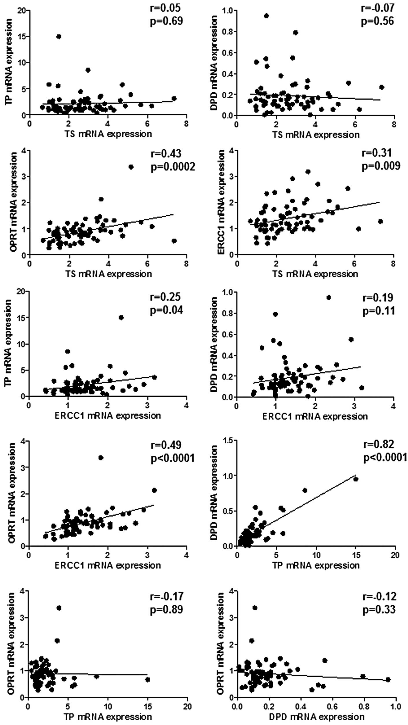
TS mRNA expression positively correlated with OPRT
and ERCC1 mRNA expression but the tendency was weak (r=0.43,
p=0.0002; r=0.31, p=0.009, respectively) (Fig. 1). There was a marked and positive
correlation between TP and DPD mRNA expression (r=0.82;
p<0.0001) and a weak correlation between TP and ERCC1 mRNA
expression (r=0.25; p=0.04) (Fig.
1). ERCC1 mRNA expression was correlated weakly with OPRT mRNA
expression (r=0.49; p<0.0001) (Fig.
1). No other significant correlations were observed.
Alteration of TS, TP, DPD, OPRT and ERCC1
mRNA expression in primary lesions and liver metastatic
lesions
A comparison between the levels of TS, TP, DPD, OPRT
and ERCC1 mRNA expression in primary lesions (n=30) and that in
synchronous (n=15) or metachronous (n=15) liver metastatic lesions
was performed. Results showed that OPRT and ERCC1 mRNA expression
was significantly decreased in synchronous liver metastatic lesions
as compared to primary lesions (p=0.036 and p=0.044, respectively)
(Table I). No other significant
differences were found in primary and liver metastatic lesions.
 | Table IThe level of mRNA expression in
primary colorectal cancer and liver metastatic cancer. |
Table I
The level of mRNA expression in
primary colorectal cancer and liver metastatic cancer.
| Synchronous liver
metastasis (n=15) | Metachronous liver
metastasis (n=15) |
|---|
|
|
|
|---|
| Primary lesions | Liver metastatic
lesions | P-value | Primary lesions | Liver metastatic
lesions | P-value |
|---|
| TS | 2.92±1.6 | 2.47±1.32 | 0.14 | 3.03±1.6 | 3.4±2.2 | 0.23 |
| TP | 3.41±3.57 | 3.61±4.72 | 0.79 | 1.61±0.8 | 2.05±1.41 | 0.12 |
| DPD | 0.23±0.24 | 0.3±0.29 | 0.16 | 0.19±0.12 | 0.22±0.11 | 0.15 |
| OPRT | 1.14±0.78 | 0.81±0.45 | 0.036 | 0.92±0.28 | 0.86±0.52 | 0.34 |
| ERCC1 | 1.72±0.7 | 1.45±0.48 | 0.044 | 1.5±0.62 | 1.31±0.65 | 0.19 |
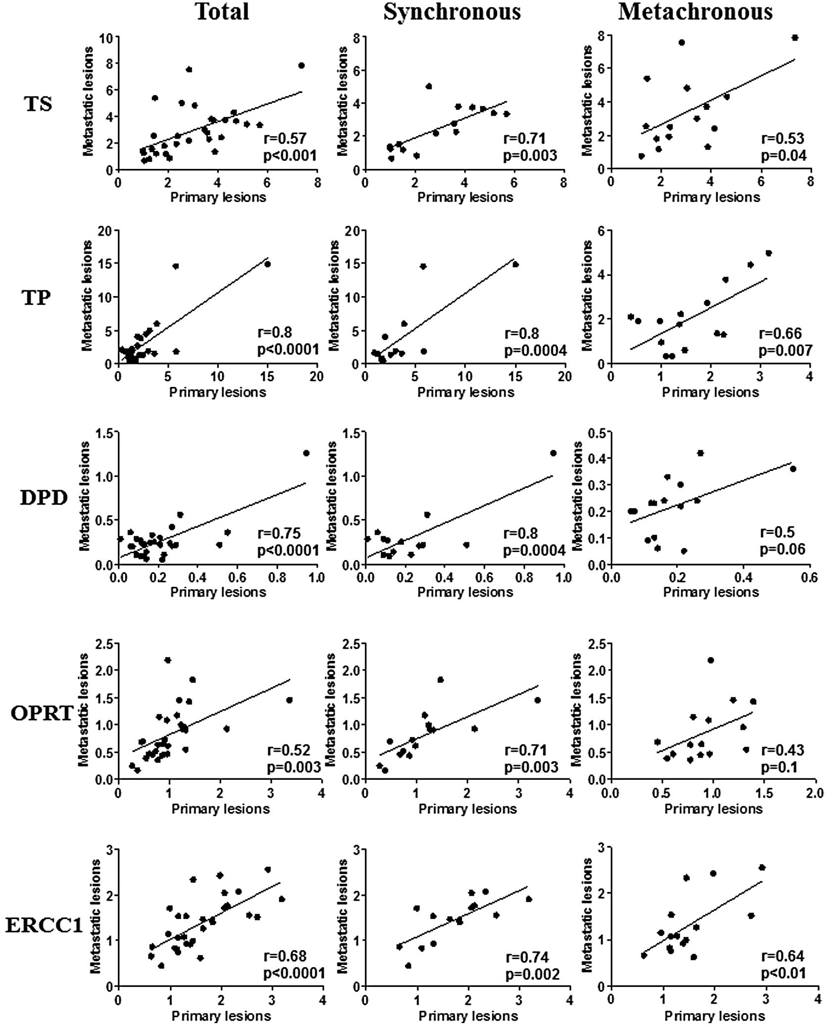
Correlation of TS, TP, DPD, OPRT and
ERCC1 mRNA expression in primary lesions and corresponding
synchronous and metachronous liver metastatic lesions
The expression of all of the genes in primary
colorectal cancer lesions positively correlated with those in
corresponding liver metastatic lesions (Fig. 2). Marked correlations were found
between TP and DPD mRNA expression in primary lesions and that in
liver metastatic lesions (r=0.8; p<0.0001, r=0.75; p<0.0001,
respectively) (Fig. 2). Weak
correlations between primary lesions and liver metastatic lesions
were found in TS, DPD and ERCC1 mRNA expression (r=0.57;
p<0.001, r=0.52; p=0.003, r=0.68; p<0.0001, respectively)
(Fig. 2). When the metastatic
pattern was divided into synchronous metastasis and metachronous
metastasis, there were marked correlations between primary lesions
and synchronous liver metastatic lesions in TS, TP, DPD, OPRT and
ERCC1 mRNA expression (Fig. 2).
Regarding metachronous liver metastatic lesions, there were weak
correlations between primary lesions and liver metastatic lesions
in TS, TP and ERCC1 mRNA expression, while no correlations were
observed in TP and DPD mRNA expression (Fig. 2).
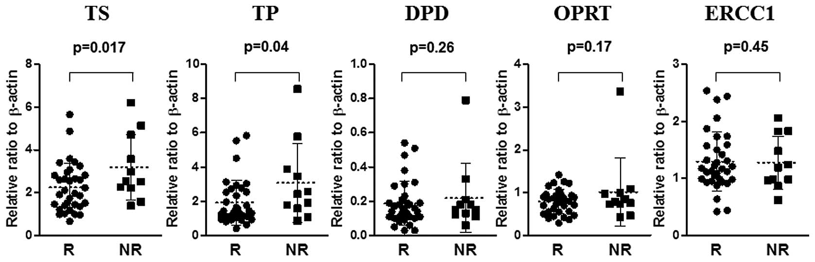
The relationship between the response of
FOLFOX and the level of TS, TP, DPD, OPRT and ERCC1 mRNA expression
in primary colorectal cancers
The level of TS and TP mRNA in the responder group
(n=34) was significantly lower than that in the non-responder group
(n=11) (p=0.017 and p=0.04, respectively) (Fig. 2). No significant differences were
found between the level of DPD, OPRT and ERCC1 mRNA in the
responder group and in the non-responder group (Fig. 3).
Discussion
Previously, 5-FU-based chemotherapy was performed
for metastatic colorectal cancer patients and has also been
administered to patients who underwent curative surgery for
colorectal cancer as adjuvant chemotherapy (21). However, predictive markers for
chemosensitivity to 5-FU and prognostic factors have been
investigated to select eligible patients with colorectal cancer.
Among genes that are correlated with the 5-FU metabolic pathway,
TS, TP, DPD and OPRT expression have been intensively assessed by
real-time PCR, enzyme-linked immunosorbent assay (ELISA), and
immunohistochemical methods. Thus, a number of investigators have
reported the relationship between these expression profiles and
clinical significance in predicting the efficacy of 5-FU treatment
for metastatic colorectal cancer (6–10) and
curatively resected colorectal cancer patients (12,13).
Recent standard chemotherapy for metastatic colorectal cancer has
constituted multiple combinations of anti-cancer drugs with 5-FU.
FOLFOX treatment has been recommended for metastatic colorectal
cancer patients as the first- or second-line chemotherapy (1–4) and
for stage III colon cancer patients as adjuvant chemotherapy
(22). Oxaliplatin is classified as
one of the platinum-based chemotherapeutic agents, along with
cisplatin. However, its function is different from that of
cisplatin as it has a 1,2-diaminocyclohexane carrier ligand in its
chemical structure (23). Its
cytotoxic effects are induced by the direct binding to interstrand
DNA, interfering with DNA transcription and replication. ERCC1 is
essentially involved in the recognition and removal of the
platinum-DNA adducts (15). Thus,
high levels of ERCC1 expression in cancer tissues attenuate the
chemosensitivity of oxaliplatin (24,25).
In the present study, we examined the mRNA
expression of the 5-FU/oxaliplatin metabolism-related genes,
including TS and ERCC1, in colorectal cancer patients with liver
metastasis receiving FOLFOX treatment as first-line chemotherapy.
Although there are only a few reports that have focused on the
expression levels of those mRNAs to predict the efficacy of FOLFOX
treatment, our results show that the levels of TS and TP mRNA
expression in the responder group was significantly lower than that
in the non-responder group, and was similar to previous reports
(9,10). Shirota et al demonstrated
that low levels of TS and ERCC1 mRNA expression in liver metastatic
tumors or recurrent colorectal tumor masses had favorable responses
and prolonged survival compared with high levels of expression in
colorectal cancer patients receiving a combination of 5-FU and
oxaliplatin chemotherapy as the second-or third-line treatment
using the laser capture microdissection method (9). Using immunohistochemistry, findings of
another report have shown that a negative TS expression had a
better response for 5-FU/oxaliplatin than a positive TS expression,
while ERCC1 expression was not associated with the response to
5-FU/oxaliplatin chemotherapy as the first-line chemotherapy
(10). Our study may be the first
report to demonstrate the correlation between TS and TP mRNA
expression of primary tumors and the efficacy of the first-line
FOLFOX treatment in colorectal cancer with liver metastasis using
the laser capture microdissection method. Prior to the era of
FOLFOX treatment for metastatic colorectal cancer, patients with
colorectal cancer receiving 5-FU treatment were surveyed to
identify the predictive markers for chemotherapy outcome. Since
then, TS and TP expression have been considered to be useful
indicators to predict the efficacy of chemotherapy and prognosis in
metastatic colorectal cancer patients (6–8). Our
results suggest that even if 5-FU treatment changes to FOLFOX
treatment, the evaluation of TS and TP mRNA expression in primary
colorectal cancer tissues is likely to be useful in the prediction
of the efficacy of FOLFOX treatment in colorectal cancer patients
with liver metastasis, although it may be difficult to evaluate the
efficacy of oxaliplatin by ERCC1 expression alone.
Regarding the comparison between the mRNA level of
TS, TP, DPD, OPRT and ERCC1 expression in primary colorectal cancer
lesions and that in synchronous or metachronous liver metastatic
lesions, OPRT and ERCC1 mRNA levels were significantly decreased in
synchronous liver metastatic lesions and no alteration of the TS
and TP mRNA levels was observed. Although these investigations have
previously been reported (16–19,26,27),
the results remain controversial. This discrepancy may be caused by
the difference in methodology and study population. As for TS mRNA
expression, almost all reports have demonstrated supportive results
that there was not a significant alteration between primary tumor
and liver metastatic tumor (17,19,26,27),
while it has been reported that the TS mRNA levels in metachronous
liver metastases were higher than those in primary tumors (18). This evidence led us to the
conclusion that examining the TS mRNA level in primary colorectal
cancer tissue as a predictive indicator of chemosensitivity is
sufficient and it is not necessary to know the TS mRNA level in
liver metastatic cancer tissue.
We further demonstrated the significant correlation
of TS, TP, DPD, OPRT and ERCC1 mRNA levels between primary
colorectal cancer lesions and liver metastatic lesions. The
correlation between synchronous liver metastatic lesions and
primary lesions was stronger than that of metachronous liver
metastatic lesions with regard to the mRNA level of the genes that
we examined. This difference means the acquisition of tumor
heterozygosity when the metachronous liver metastasis developed,
and it may be caused by the effect of adjuvant chemotherapy with
oral administration of 5-FU in the stage III colorectal cancer
patients following curative resection of colorectal cancer.
Previous studies (17–19) that analyzed the correlation of mRNA
expression between primary colorectal cancer lesions and liver
metastatic lesions have demonstrated similar results to our current
results. In particular, the significant correlation of TS mRNA
expression was consistent with our results. These experiments were
performed by almost identical methods using FFPE samples. Another
study (26) has shown that no
correlations of TS, TP, DPD and OPRT mRNA expression were found in
primary colorectal tumors and corresponding metastatic liver tumors
using frozen tissues. In this case, the different results may be
explained by the different materials used for RNA extraction. When
FFPE samples are used through the laser capture microdissection
method, the amount of mRNA expression reflects almost all cancer
cells. However, it is likely that the frozen tissues include
interstitial component cells with the exception of cancer cells.
For this reason, the difference between component cells of primary
colorectal cancer tissues and liver metastatic cancer tissues could
affect these results. Therefore, when a comparison of mRNA
expression among different organs is performed, the RNA extraction
method through laser capture microdissection appears to be better
since the mRNA expression of target genes reflects the product of
cancer cells without interstitial component cells.
When the mRNA level of all molecules was compared
among primary colorectal cancer tissues, there were significant
correlations among TS, OPRT and ERCC1, and a strong correlation was
observed between TP and DPD. The correlation between TS and OPRT
mRNA levels has already been documented in a previous report
(26). These enzymes may work in a
coordinated manner as a critical step in the de novo pathway
of DNA synthesis. Thus, the alteration of OPRT mRNA expression may
be associated with that of TS mRNA expression. Regarding the
correlation between TS and OPRT expression, the combination of OPRT
with a high expression and TS with a low expression is considered
to be effective for chemotherapy (28). Moreover, a weak correlation was
found between TS and ERCC1 mRNA expression. These results suggest
that the measurement of only TS mRNA expression is adequate to
predict the efficacy of chemotherapy without the measurement of
OPRT and ERCC1 mRNA expression. The strong correlation between TP
and DPD has been described in numerous previous reports (17,19,26,29–32).
This consistent result indicated that this correlation is adequate
to examine either TP or DPD mRNA expression. In terms of clinical
significance, the lower expression of TP and DPD had a better
response to 5-FU treatment for colorectal cancer patients (8).
Progress in systemic chemotherapy, including
molecular-targeted therapy for metastatic colorectal cancer,
enables conversion therapy. As such, unresectable metastatic tumors
become resectable (33). A strong
correlation has been reported between response rates and the liver
resection rate in patients with isolated liver metastases (34). The addition of bevacizumab (35) to the FOLFOX and FOLFIRI regimens, or
the addition of cetuximab (36) in
cases with wild-type KRAS expression, has been suggested to be
effective. Oncologists tend to spend their efforts on how to use
monoclonal antibodies to increase the response rate for metastatic
colorectal cancer. However, when we consider the root of
chemotherapy, it remains important to predict the efficacy of
FOLFOX, which is a basic regimen of multi-drug combination therapy.
Furthermore, the present results showing that the mRNA expression
profiles in primary colorectal cancer lesions correlated strongly
with those in synchronous liver metastatic lesions would be useful
to predict the efficacy of FOLFOX for metastatic lesions. As a
result, an increase in the probability of actual conversion therapy
is likely to occur.
Taken together, our current results indicate that
the measurement of the TS and TP mRNA levels may estimate the
ERCC1, OPRT and DPD mRNA levels in primary colorectal cancer and
may be useful in the prediction of the efficacy of FOLFOX treatment
in liver metastatic lesions, particularly synchronous liver
metastatic lesions, reflecting the mRNA expression profiles of all
molecules in primary colorectal cancer tumor. This evidence may
contribute to the clinical application for future strategies,
including conversion therapy.
References
|
1
|
de Gramont A, Figer A, Seymour M, Homerin
M, Hmissi A, Cassidy J, Boni C, Cortes-Funes H, Cervantes A, Freyer
G, Papamichael D, Le Bail N, Louvet C, Hendler D, de Braud F,
Wilson C, Morvan F and Bonetti A: Leucovorin and fluorouracil with
or without oxaliplatin as first-line treatment in advanced
colorectal cancer. J Clin Oncol. 18:2938–2947. 2000.
|
|
2
|
Tournigand C, André T, Achille E, Lledo G,
Flesh M, Mery-Mignard D, Quinaux E, Couteau C, Buyse M, Ganem G,
Landi B, Colin P, Louvet C and de Gramont A: FOLFIRI followed by
FOLFOX6 or the reverse sequence in advanced colorectal cancer: a
randomized GERCOR study. J Clin Oncol. 22:229–237. 2004. View Article : Google Scholar : PubMed/NCBI
|
|
3
|
Goldberg RM, Sargent DJ, Morton RF, Fuchs
CS, Ramanathan RK, Williamson SK, Findlay BP, Pitot HC and Alberts
SR: A randomized controlled trial of fluorouracil plus leucovorin,
irinotecan, and oxaliplatin combinations in patients with
previously untreated metastatic colorectal cancer. J Clin Oncol.
22:23–30. 2004. View Article : Google Scholar
|
|
4
|
Reddy GK, Gibson AD and Price N: Evolution
of FOLFOX regimens in the treatment of advanced colorectal cancer.
Clin Colorectal Cancer. 4:296–299. 2005. View Article : Google Scholar : PubMed/NCBI
|
|
5
|
Shimizu T, Satoh T, Tamura K, Ozaki T,
Okamoto I, Fukuoka M and Nakagawa K:
Oxaliplatin/fluorouracil/leucovorin (FOLFOX4 and modified FOLFOX6)
in patients with refractory or advanced colorectal cancer:
post-approval Japanese population experience. Int J Clin Oncol.
12:218–223. 2007. View Article : Google Scholar
|
|
6
|
Leichman CG, Lenz HJ, Leichman L,
Danenberg K, Baranda J, Groshen S, Boswell W, Metzger R, Tan M and
Danenberg PV: Quantitation of intratumoral thymidylate synthase
expression predicts for disseminated colorectal cancer response and
resistance to protracted-infusion fluorouracil and weekly
leucovorin. J Clin Oncol. 15:3223–3229. 1997.
|
|
7
|
Metzger R, Danenberg K, Leichman CG,
Salonga D, Schwartz EL, Wadler S, Lenz HJ, Groshen S, Leichman L
and Danenberg PV: High basal level gene expression of thymidine
phosphorylase (platelet-derived endothelial cell growth factor) in
colorectal tumors is associated with nonresponse to 5-fluorouracil.
Clin Cancer Res. 4:2371–2376. 1998.
|
|
8
|
Salonga D, Danenberg KD, Johnson M,
Metzger R, Groshen S, Tsao-Wei DD, Lenz HJ, Leichman CG, Leichman
L, Diasio RB and Danenberg PV: Colorectal tumors responding to
5-fluorouracil have low gene expression levels of dihydropyrimidine
dehydrogenase, thymidylate synthase, and thymidine phosphorylase.
Clin Cancer Res. 6:1322–1327. 2000.
|
|
9
|
Shirota Y, Stoehlmacher J, Brabender J,
Xiong YP, Uetake H, Danenberg KD, Groshen S, Tsao-Wei DD, Danenberg
PV and Lenz HJ: ERCC1 and thymidylate synthase mRNA levels predict
survival for colorectal cancer patients receiving combination
oxaliplatin and fluorouracil chemotherapy. J Clin Oncol.
19:4298–4304. 2001.
|
|
10
|
Kim SH, Kwon HC, Oh SY, Lee DM, Lee S, Lee
JH, Roh MS, Kim DC, Park KJ, Choi HJ and Kim HJ: Prognostic value
of ERCC1, thymidylate synthase, and glutathione S-transferase pi
for 5-FU/oxaliplatin chemotherapy in advanced colorectal cancer. Am
J Clin Oncol. 32:38–43. 2009. View Article : Google Scholar : PubMed/NCBI
|
|
11
|
Koopman M, Venderbosch S, Nagtegaal ID,
van Krieken JH and Punt CJ: A review on the use of molecular
markers of cytotoxic therapy for colorectal cancer, what have we
learned? Eur J Cancer. 45:1935–1949. 2009. View Article : Google Scholar : PubMed/NCBI
|
|
12
|
Edler D, Glimelius B, Hallström M,
Jakobsen A, Johnston PG, Magnusson I, Ragnhammar P and Blomgren H:
Thymidylate synthase expression in colorectal cancer: a prognostic
and predictive marker of benefit from adjuvant fluorouracil-based
chemotherapy. J Clin Oncol. 20:1721–1728. 2002. View Article : Google Scholar : PubMed/NCBI
|
|
13
|
Kornmann M, Schwabe W, Sander S, Kron M,
Sträter J, Polat S, Kettner E, Weiser HF, Baumann W, Schramm H,
Häusler P, Ott K, Behnke D, Staib L, Beger HG and Link KH:
Thymidylate synthase and dihydropyrimidine dehydrogenase mRNA
expression levels: predictors for survival in colorectal cancer
patients receiving adjuvant 5-fluorouracil. Clin Cancer Res.
9:4116–4124. 2003.
|
|
14
|
Takebayashi Y, Akiyama S, Akiba S, Yamada
K, Miyadera K, Sumizawa T, Yamada Y, Murata F and Aikou T:
Clinicopathologic and prognostic significance of an angiogenic
factor, thymidine phosphorylase, in human colorectal carcinoma. J
Natl Cancer Inst. 88:1110–1117. 1996. View Article : Google Scholar
|
|
15
|
Altaha R, Liang X, Yu JJ and Reed E:
Excision repair cross complementing-group 1: Gene expression and
platinum resistance. Int J Mol Med. 14:959–970. 2004.PubMed/NCBI
|
|
16
|
Yamada H, Ichikawa W, Uetake H, Shirota Y,
Nihei Z, Sugihara K and Hirayama R: Thymidylate synthase gene
expression in primary colorectal cancer and metastatic sites. Clin
Colorectal Cancer. 1:169–173. 2008. View Article : Google Scholar
|
|
17
|
Kuramochi H, Hayashi K, Uchida K, Miyakura
S, Shimizu D, Vallbohmer D, Park S, Danenberg KD, Takasaki K and
Danenberg PV: 5-fluorouracil-related gene expression levels in
primary colorectal cancer and corresponding liver metastasis. Int J
Cancer. 119:522–526. 2006. View Article : Google Scholar : PubMed/NCBI
|
|
18
|
Kobayashi H, Sugihara K, Uetake H, Higuchi
T, Yasuno M, Enomoto M, Iida S, Azuma M, Mori R, Omori A, Lenz HJ,
Danenberg KD and Danenberg PV: Messenger RNA expression of TS and
ERCC1 in colorectal cancer and matched liver metastasis. Int J
Oncol. 33:1257–1262. 2008.PubMed/NCBI
|
|
19
|
Sameshima S, Tomozawa S, Kojima M, Koketsu
S, Motegi K, Horikoshi H, Okada T, Kon Y and Sawada T:
5-Fluorouracil-related gene expression in primary sites and hepatic
metastases of colorectal carcinomas. Anticancer Res. 28:1477–1481.
2008.PubMed/NCBI
|
|
20
|
Lord RV, Salonga D, Danenberg KD, Peters
JH, DeMeester TR, Park JM, Johansson J, Skinner KA, Chandrasoma P,
DeMeester SR, Bremner CG, Tsai PI and Danenberg PV: Telomerase
reverse transcriptase expression is increased early in the
Barrett’s metaplasia, dysplasia, adenocarcinoma sequence. J
Gastrointest Surg. 4:135–142. 2000.PubMed/NCBI
|
|
21
|
Midgley R and Kerr D: Colorectal cancer.
Lancet. 353:391–399. 1999. View Article : Google Scholar
|
|
22
|
André T, Boni C, Mounedji-Boudiaf L,
Navarro M, Tabernero J, Hickish T, Topham C, Zaninelli M, Clingan
P, Bridgewater J, Tabah-Fisch I and de Gramont A; Multicenter
International Study of Oxaliplatin/5-Fluorouracil/Leucovorin in the
Adjuvant Treatment of Colon Cancer (MOSAIC) Investigators.
Oxaliplatin, fluorouracil, and leucovorin as adjuvant treatment for
colon cancer. N Engl J Med. 350:2343–2351. 2004.
|
|
23
|
Raymond E, Faivre S, Woynarowski JM and
Chaney SG: Oxaliplatin: mechanism of action and antineoplastic
activity. Semin Oncol. 25:4–12. 1998.PubMed/NCBI
|
|
24
|
Gossage L and Madhusudan S: Current status
of excision repair cross complementing-group 1 (ERCC1) in cancer.
Cancer Treat Rev. 33:565–577. 2007. View Article : Google Scholar : PubMed/NCBI
|
|
25
|
Arnould S, Hennebelle I, Canal P, Bugat R
and Guichard S: Cellular determinants of oxaliplatin sensitivity in
colon cancer cell lines. Eur J Cancer. 39:112–119. 2003. View Article : Google Scholar : PubMed/NCBI
|
|
26
|
Inokuchi M, Uetake H, Shirota Y, Yamada H,
Tajima M and Sugihara K: Gene expression of 5-fluorouracil
metabolic enzymes in primary colorectal cancer and corresponding
liver metastasis. Cancer Chemother Pharmacol. 53:391–396. 2004.
View Article : Google Scholar : PubMed/NCBI
|
|
27
|
Okumura K, Shiomi H, Mekata E, Kaizuka M,
Endo Y, Kurumi Y and Tani T: Correlation between chemosensitivity
and mRNA expression level of 5-fluorouracil-related metabolic
enzymes during liver metastasis of colorectal cancer. Oncol Rep.
15:875–882. 2006.
|
|
28
|
Fujii R, Seshimo A and Kameoka S:
Relationships between the expression of thymidylate synthase,
dihydropyrimidine dehydrogenase, and orotate
phosphoribosyltransferase and cell proliferative activity and
5-fluorouracil sensitivity in colorectal carcinoma. Int J Clin
Oncol. 8:72–78. 2003. View Article : Google Scholar
|
|
29
|
Kinoshita M, Kodera Y, Hibi K, Nakayama G,
Inoue T, Ohashi N, Ito Y, Koike M, Fujiwara M and Nakao A: Gene
expression profile of 5-fluorouracil metabolic enzymes in primary
colorectal cancer: potential as predictive parameters for response
to fluorouracil-based chemotherapy. Anticancer Res. 27:851–856.
2007.
|
|
30
|
Mori K, Hasegawa M, Nishida M, Toma H,
Fukuda M, Kubota T, Nagasue N, Yamana H, Hirakawa YS, Chung K,
Ikeda T, Takasaki K, Oka M, Kameyama M, Toi M, Fujii H, Kitamura M,
Murai M, Sasaki H, Ozono S, Makuuchi H, Shimada Y, Onishi Y, Aoyagi
S, Mizutani K, Ogawa M, Nakao A, Kinoshita H, Tono T, Imamoto H,
Nakashima Y and Manabe T: Expression levels of thymidine
phosphorylase and dihydropyrimidine dehydrogenase in various human
tumor tissues. Int J Oncol. 17:33–38. 2000.PubMed/NCBI
|
|
31
|
Uchida K, Danenberg PV, Danenberg KD and
Grem JL: Thymidylate synthase, dihydropyrimidine dehydrogenase,
ERCC1, and thymidine phosphorylase gene expression in primary and
metastatic gastrointestinal adenocarcinoma tissue in patients
treated on a phase I trial of oxaliplatin and capecitabine. BMC
Cancer. 8:3862008. View Article : Google Scholar
|
|
32
|
Collie-Duguid ES, Johnston SJ, Boyce L,
Smith N, Cowieson A, Cassidy J, Murray GI and McLeod HL: Thymidine
phosphorylase and dihydropyrimidine dehydrogenase protein
expression in colorectal cancer. Int J Cancer. 94:297–301. 2001.
View Article : Google Scholar : PubMed/NCBI
|
|
33
|
Adam R, Delvart V, Pascal G, Valeanu A,
Castaing D, Azoulay D, Giacchetti S, Paule B, Kunstlinger F,
Ghémard O, Levi F and Bismuth H: Rescue surgery for unresectable
colorectal liver metastases downstaged by chemotherapy: a model to
predict long-term survival. Ann Surg. 240:644–657. 2004.PubMed/NCBI
|
|
34
|
Folprecht G, Grothey A, Alberts S, Raab HR
and Köhne CH: Neoadjuvant treatment of unresectable colorectal
liver metastases: correlation between tumour response and resection
rates. Ann Oncol. 16:1311–1319. 2005. View Article : Google Scholar : PubMed/NCBI
|
|
35
|
Kabbinavar FF, Hambleton J, Mass RD,
Hurwitz HI, Bergsland E and Sarkar S: Combined analysis of
efficacy: the addition of bevacizumab to fluorouracil/leucovorin
improves survival for patients with metastatic colorectal cancer. J
Clin Oncol. 23:3706–3712. 2005. View Article : Google Scholar : PubMed/NCBI
|
|
36
|
Saltz LB, Meropol NJ, Loehrer PJ Sr,
Needle MN, Kopit J and Mayer RJ: Phase II trial of cetuximab in
patients with refractory colorectal cancer that expresses the
epidermal growth factor receptor. J Clin Oncol. 22:1201–1208. 2004.
View Article : Google Scholar : PubMed/NCBI
|