Introduction
Serum proteins often serve as indicators of disease
and are a rich source of cancer biomarkers. Certain biomarkers used
for the diagnosis, progression and prognosis of cancer have been
identified by either surface-enhanced laser desorption/ionization
(SELDI-TOF-MS) (1–7) or matrix-assisted laser
desorption/ionization time-of-flight mass spectrometry
(MALDI-TOF-MS). SELDI is a useful and powerful platform which may
be used to analyze raw mixture samples directly and identify
biomarkers in a large number of samples. It provides rapid
screening ability and superior visibility of results, avoiding the
complication of individual differences (8).
Gastric carcinoma is one of the most common cancer
malignancies in Asia. Currently, the most effective treatment for
gastric carcinoma is surgical resection; however, prognosis often
remains poor due to the advanced stage of cancer progression at
diagnosis. Therefore, the identification of novel biomarkers for
gastric carcinoma, particularly in the early stages, is urgently
required.
Potential biomarkers for gastric carcinoma diagnosis
were previously reported, including serum amyloid A (SAA) (7), which is also elevated in various types
of cancer (9). However, in previous
studies, a limited number of samples was used and there was an
absence of information on the clinicopathological staging in the
patients. In our previous study, sera from a number of gastric
carcinoma patients and healthy individuals were screened by
ProteinChip array and a classification tree with four biomarkers
was established (10). The
sensitivity and specificity in blind tests are better than any of
the clinical serological tests for gastric carcinoma. However,
among the biomarkers discovered by SELDI in various cancers, only a
few have been identified, due to technical limitations. In the
present study, a new strategy was developed to directly identify
the new peptide biomarkers on chip instead of purified biomarkers
by MALDI-TOF or other methods.
Materials and methods
Samples
Samples were obtained from patients in the Taizhou
Municipal Hospital, China. The sera of the patients were obtained
prior to implementation of any therapeutic measures. The diagnoses
were confirmed by postsurgical pathology. The total gastric cancer
group consisted of 70 patients at different clinical stages (as
defined by the International Union against Cancer, UICC, 1997):
stage I (n=l0), stage II (n=l9), stage III (n=11) and stage IV
(n=30). The median age of the patients was 57 years (range, 32–89).
There were 44 males and 28 females. The median ages of patients in
the stage I, II, III and IV groups were 53 years (range, 37–77), 64
years (range, 32–77), 58 years (range, 36–71) and 55 years (range,
32–89), respectively. The samples were collected in 10-ml tubes in
the early morning before breakfast and then the sera were
immediately separated and stored at −80°C until use. The serum
samples showed normal clotting time. The study and use of clinical
samples was approved by the local ethics committee of medical
research, and oral consent was obtained from all subjects.
Analysis of ProteinChip array
After thawing and 2 min of centrifugation (10,000
rpm), 5 μl serum sample without fractionated treatment was added
into 10 μl 0.5% U9 (9 mol/l urea, 0.2% CHAPS (3[(3-cholamidopropyl)
dimethylammonio]-l-propanesulfonate), 0.1% DTT (DL-dithiothreitol)
in a 96-well plate and incubated for 30 min at 4°C with 600 rpm
vigorous agitation. The ProteinChip array cassette was placed into
a 96-well bioprocessor, and 200 μl NaAc (50 mmol/l, pH 4.0) was
added into each well and incubated for 5 min at 4°C with 600 rpm
vigorous agitation. The liquid was removed and the procedure was
repeated once. NaAC (185 μl) was added into each well in the
96-well plate (600 rpm, 2 min) and 100 μl samples from the
different patient groups were added separately into different wells
of the ProteinChip array cassette (600 rpm, l h). After the content
from each well was removed, the wells were washed with 200 μl NaAC
(pH 4.0, 600 rpm, 5 min). The procedure was repeated two more
times. Each spot was washed with 200 μl high-performance liquid
chromatography (HPLC), which was removed immediately. The procedure
was repeated once. After air drying, 5 μl elute was diluted with 5
μl SPA (saturated solution of sinapinic acid in 50% acetonitrile
with 0.5% trifluoroacetic acid).
The resulting mixture (2 μl) was aspirated and
spotted onto the gold-coated ProteinChip arrays. After air-drying
for approximately 5 min at room temperature, the chips were placed
in the protein biological system II-C (PBS II-C) ProteinChip Reader
(Ciphergen Biosystems, Inc., Fremont, CA, USA), and data were
collected by an average of 150 laser shots, with a detector
sensitivity of 8 and a laser intensity of 190. Mass calibration was
performed using an all-in-one peptide reference standard comprising
vasopressin (1,084.2 Da), somatostatin (1,637.9 Da), bovine insulin
β chain (3,495.9 Da), human insulin recombinant (5,807.6 Da) and
hirudin (7,033.6 Da) (Ciphergen Biosystems, Inc.). The default
background subtraction was applied, and the peak intensities were
normalized using the total ion current from a mass charge of
1,000–50,000 Da. A biomarker detection software package (Ciphergen
Biomarker Wizards, Ciphergen Biosystems, Inc.) was used to
autodetect protein peaks. Protein peaks were selected based on a
first pass of a signal to noise ratio of 3 and a minimum peak
threshold of 20% of all spectra. This process was completed with a
second pass of peak selection at 0.2% of the mass window, and the
estimated peaks were added. These selected protein peaks were
averaged as clusters and were exported to a commercially available
software package (Biomarker Patterns, Ciphergen Biosystems, Inc.)
for further classification analysis.
The profiling spectra of serum samples were
normalized using total ion current normalization by Ciphergen's
ProteinChip software (version 3.1). Peak labeling was performed
using Biomarker Wizard software 3.1 (Ciphergen Biosystems, Inc.). A
two-sample t-test was used to compare mean normalized intensities
between the case and control groups. P<0.05 was considered
statistically significant.
High-performance liquid chromatography
(HPLC) analysis
The HPLC separation system consisted of a Shimadzu
LC-10ATVP infusion pump, a Shimadzu DGU-14A degasser, a Shimadzu
SPD-10A ultraviolet detector, a Rheodyne 7725i admission valve, a
Phenomenex Nucleosil C18 chromatographic column (250×4.6 mm ID) and
a WDL-95 chromatographic workstation. Linear and gradient elution
was carried out with 5% acetonitrile water solution containing 0.1%
trifluoroacetic acid to 60% acetonitrile water solution containing
0.1% trifluoroacetic acid at a rate of 1.0 ml/min within 0–30 min.
Separation and detection were carried out under chromatographic
conditions that were measured at UV 220 nm. The effluent of the
chromatographic peak was collected in centrifuge tubes for
subsequent mass spectrometry analysis.
Matrix-assisted laser
desorption/ionization time-of-flight mass spectrometry
MALDI-TOF mass spectra were obtained with a
MALDI-TOF mass spectrometer with delayed extraction [Voyager Elite;
Applied Biosystems, Inc. (ABI), Framingham, MA, USA]. UV light from
a nitrogen laser with a 337-nm emission wavelength was used for
irradiation. A typical spectrum was collected at an extraction
voltage of 20 kV. Delayed (175 ns) extraction mode was used for all
acquisitions. The laser power was adjusted to a level just above
the threshold for signal appearance to minimize head group loss.
Each phospholipid mass spectrum was collected by averaging data
from 250 laser shots. For sample plate spotting, 3 μl IMAC-eluted
phospholipids were mixed with 3 μl matrix, deposited onto the MALDI
plate, and allowed to air-dry. The MALDI matrix used in the studies
was a recently developed solid ionic crystal matrix consisting of
20 mg paranitroaniline (PNA) and butyric acid at a 1:2 molar ratio
dissolved in ethanol.
The spectra were collected using a two-point
calibration of protonated lyso-PC at a mass/charge ratio (m/z) of
496.34 and protonated DMPC at m/z 678.51. Phospholipids in the
biological samples were assigned according to their molecular
weights, as derived from either the monoisotopic protonated
molecules, monoisotopic sodium adducts, and/or postsource decay
(PSD) productions in MALDI-TOF mass spectra.
Database search
Proteins with theoretical matching enzymatic
peptides were searched against the ExPASy database (http://us.expasy.org/tools/tagident.html) software
according to the m/z data of the peptides and proper setting of
search parameters. The protein of interest was determined according
to the search results and the relative molecular weight of the
protein.
Results
Protein peaks with molecular weights of
11.1–11.9 kDa were significantly increased in the patients with
gastric carcinoma
The CM10 ProteinChip was used to carry out the
measurements on the serum samples from 70 gastric carcinoma
patients and 30 healthy adults. The protein spectra were normalized
according to the total ionic strength. Biomarker Wizard software
was used to analyze the protein spectra in addition to automatic
peak marking, and the signal to noise ratio was set to 3. The
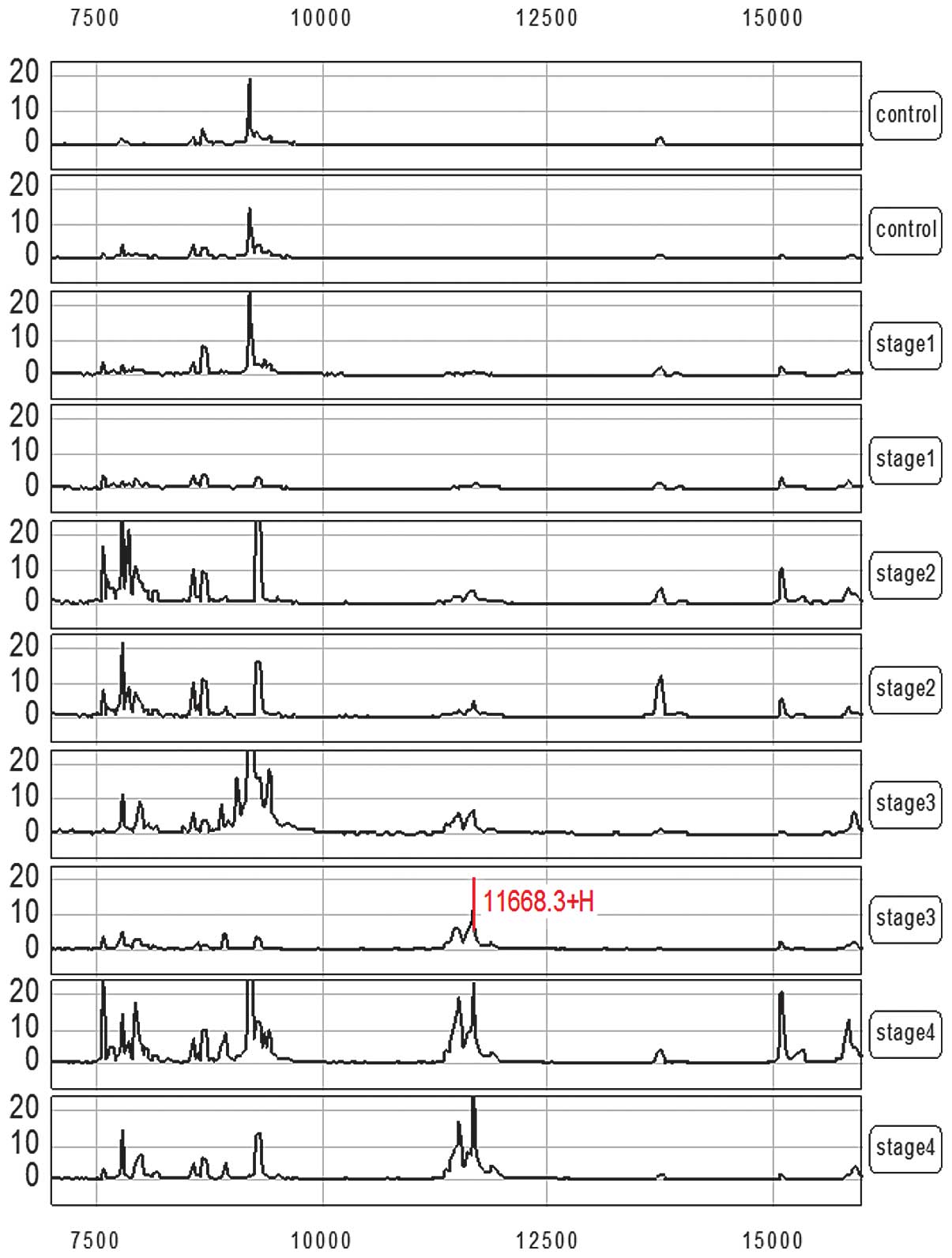
result showed that the average strength of a group of protein peaks
with molecular weights of 11.1–11.9 kDa in the gastric carcinoma
patients was 18.21±21.05 (mean ± SD), which was significantly
higher than that of the normal control at 0.65±0.55 (mean ± SD)
(P<0.01; Fig. 1). Corresponding
analyses were carried out according to the expression level (peak
intensity) of the group of protein peaks with molecular weights of
11.1–11.9 kDa in combination with the clinicopathological staging
of the patients. This group of protein peaks was positively
correlated with the course of gastric carcinoma (Figs. 1 and 2), and the peak intensity gradually
increased with the aggravation of the patient's condition.
Protein peaks with molecular weights of
11.1–11.9 kDa on the chip were identified as SAA1
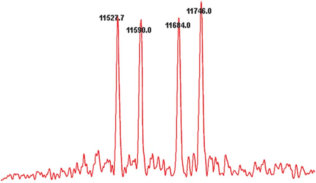
The serum of gastric carcinoma was separated and
detected using HPLC, and the effluent of the chromatographic peak
was collected in centrifuge tubes. Approximately 1 μl solution was
dropped on the sample target after the effluent of the
chromatographic peak was mixed with the matrix solution. The
solution was introduced into the laser desorption/ionization
time-of-flight mass spectrometer for analysis once the solvent had
evaporated and the sample had crystallized. Signals from 10–50
individual scans were added together to obtain the mass spectra
(Fig. 3). Proteins with theoretical
matching enzymatic peptides were searched against the ExPASy
database (http://us.expasy.org/tools/tagident.html) software
according to the m/z data of the peptides and proper setting of
search parameters, and the group of protein peaks were found to
belong to SAA1.
Discussion
The identification of new biomarkers of human
cancers has become more successful due to the development of new
high-throughput techniques in the field of proteomics. SELDI-TOF-MS
is a promising novel proteomic tool for the detection of
biomarkers. It demonstrates superior characteristics including
reliable high-throughput ability, speed, few sample requirements,
and the ability to analyze complex biological mixtures directly.
The efficacy of SELDI in the identification of biomarkers has been
proven in cancers of the lung (11), pancreas (12), breast (13), prostate (14) and ovary (15), as well as in other diseases
(16,17). However, the disadvantage of SELDI is
that it cannot directly identify the biomarkers on chips; therefore
it is not widely used in the study of molecular mechanisms. In the
present study, one biomarker that may have potential value for
diagnosing and monitoring the progression of gastric carcinoma was
identified using a new strategy, and its efficacy was found to be
consistent with that observed in the study by Cho et al
(18). This technique may be
applicable in a number of circumstances and facilitate on-chip
identification.
SAA is an acute-phase reactant, whose level in blood
is elevated in response to trauma, infection, inflammation and
neoplasia (18–20). However, SAA is considered to be an
acute-phase protein existing as various isoforms at a molecular
mass of 11.1–11.9 kDa. SAA was found to be significantly higher in
patients with metastatic disease than in those with limited disease
(21), and may therefore serve as a
useful biomarker to monitor relapse, metastasis and prognosis in
certain types of cancer (12).
Recently, the correlation between SAA and gastric carcinoma was
reported by Chan et al (7).
Results of that study revealed that the SAA level in patients with
gastric carcinoma was higher than the SAA level in patients with
adenocarcinoma and other diseases, although it is not correlated
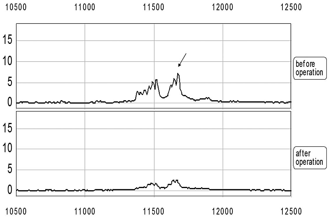
with tumor size or clinical stage. However, our results suggest
that SAA is a more significant biomarker of disease and prognosis
in certain types of cancer since it is one of the few biomarkers
that corresponds significantly with the progression of gastric
carcinoma and may be effectively decreased following medical
treatments.
Results of this study showed the average strength of
a group of protein peaks with molecular weights of 11.1–11.9 kDa in
the patients of gastric carcinoma to be 18.21±21.05 (mean ± SD),
which was significantly higher than the strength of the normal
control, at 0.65±0.55 (mean ± SD) (P<0.01). Corresponding
analyses were carried out according to the expression level (peak
intensity) of the group of protein peaks with molecular weights of
11.1–11.9 kDa in combination with the clinicopathological staging
of the patients. This group of protein peaks was found to be
positively correlated with the course of gastric carcinoma.
Moreover, the peak intensity gradually increased with the
aggravation of the patient's condition.
In conclusion, based on the studies of other authors
and our previous study, we suggest that SAA may serve as an
effective serum biomarker for the detection of gastric carcinoma
and its recurrence.
Acknowledgements
This study was funded by Zhejiang Medicine, Health
and Science grant 2010KYB127, and Zhejiang Gongyixing Applied
Technology grant 2011C33045. The authors also thank the Department
of Laboratory Medicine, Taizhou Municipal Hospital for its
support.
References
|
1
|
Le L, Chi K, Tyldesley S, et al:
Identification of serum amyloid A as a biomarker to distinguish
prostate cancer patients with bone lesions. Clin Chem. 51:695–707.
2005. View Article : Google Scholar : PubMed/NCBI
|
|
2
|
Yu Y, Chen S, Wang LS, et al: Prediction
of pancreatic cancer by serum biomarkers using surface-enhanced
laser desorption/ionization-based decision tree classification.
Oncology. 68:79–86. 2005. View Article : Google Scholar
|
|
3
|
Traub F, Feist H, Kreipe HH and Pich A:
SELDI-MS-based expression profiling of ductal invasive and lobular
invasive human breast carcinomas. Pathol Res Pract. 201:763–770.
2005. View Article : Google Scholar : PubMed/NCBI
|
|
4
|
Cho WC and Ngan RK: Biomarker discovery in
nasopharyngeal carcinoma using proteinchip profiling. J Hong Kong
Coll Radiol. 11:63–68. 2008.
|
|
5
|
Cho WC: Proteomics technologies and
challenges. Genomics Proteomics Bioinformatics. 5:77–85. 2007.
View Article : Google Scholar
|
|
6
|
Sasazuki S, Inoue M, Sawada N, et al:
Plasma levels of C-reactive protein and serum amyloid A and gastric
cancer in a nested case-control study: Japan Public Health
Center-based prospective study. Carcinogenesis. 31:712–718. 2010.
View Article : Google Scholar
|
|
7
|
Chan DC, Chen CJ, Chu HC, et al:
Evaluation of serum amyloid A as a biomarker for gastric cancer.
Ann Surg Oncol. 14:84–93. 2007. View Article : Google Scholar : PubMed/NCBI
|
|
8
|
Cho WC: Research progress in SELDI-TOF-MS
and its clinical applications. Sheng Wu Gong Cheng Xue Bao.
22:871–876. 2006. View Article : Google Scholar : PubMed/NCBI
|
|
9
|
Woong-Shick A, Sung-Pil P, Su-Mi B, et al:
Identification of hemoglobin-alpha and -beta subunits as potential
serum biomarkers for the diagnosis and prognosis of ovarian cancer.
Cancer Sci. 96:197–201. 2005. View Article : Google Scholar : PubMed/NCBI
|
|
10
|
Liang Y, Fang M, Li J, et al: Serum
proteomic patterns for gastric lesions as revealed by SELDI mass
spectrometry. Exp Mol Pathol. 81:176–180. 2006. View Article : Google Scholar : PubMed/NCBI
|
|
11
|
Valle RP, Chavany C, Zhukov TA and
Jendoubi M: New approaches for biomarker discovery in lung cancer.
Expert Rev Mol Diagn. 3:55–67. 2003. View Article : Google Scholar : PubMed/NCBI
|
|
12
|
Yokoi K, Shih LC, Kobayashi R, et al:
Serum amyloid A as a tumor marker in sera of nude mice with
orthotopic human pancreatic cancer and in plasma of patients with
pancreatic cancer. Int J Oncol. 27:1361–1369. 2005.PubMed/NCBI
|
|
13
|
Laronga C, Becker S, Watson P, et al:
SELDI-TOF serum profiling for prognostic and diagnostic
classification of breast cancers. Dis Markers. 19:229–238. 2003.
View Article : Google Scholar : PubMed/NCBI
|
|
14
|
Adam BL, Qu Y, Davis JW, et al: Serum
protein fingerprinting coupled with a pattern-matching algorithm
distinguishes prostate cancer from benign prostate hyperplasia and
healthy men. Cancer Res. 62:3609–3614. 2002.
|
|
15
|
Petricoin EF, Ardekani AM, Hitt BA, et al:
Use of proteomic patterns in serum to identify ovarian cancer.
Lancet. 359:572–577. 2002. View Article : Google Scholar : PubMed/NCBI
|
|
16
|
Cui JF, Liu YK, Zhou HJ, et al: Screening
serum hepatocellular carcinoma-associated proteins by SELDI-based
protein spectrum analysis. World J Gastroenterol. 14:1257–1262.
2008. View Article : Google Scholar
|
|
17
|
Ge Z, Zhu YL, Zhong X, Yu JK and Zheng S:
Discovering differential protein expression caused by CagA-induced
ERK pathway activation in AGS cells using the SELDI-ProteinChip
platform. World J Gastroenterol. 14:554–562. 2008. View Article : Google Scholar : PubMed/NCBI
|
|
18
|
Cho WC, Yip TT, Ngan RK, et al:
ProteinChip array profiling for identification of disease- and
chemotherapy-associated biomarkers of nasopharyngeal carcinoma.
Clin Chem. 53:241–250. 2007. View Article : Google Scholar : PubMed/NCBI
|
|
19
|
Malle E and De Beer FC: Human serum
amyloid A (SAA) protein: a prominent acute-phase reactant for
clinical practice. Eur J Clin Invest. 26:427–435. 1996. View Article : Google Scholar : PubMed/NCBI
|
|
20
|
Kokubun M, Imafuku Y, Okada M, Ohguchi Y,
Ashikawa T, Yamada T and Yoshida H: Serum amyloid A (SAA)
concentration varies among rheumatoid arthritis patients estimated
by SAA/CRP ratio. Clin Chim Acta. 360:92–102. 2005. View Article : Google Scholar : PubMed/NCBI
|
|
21
|
Weinstein PS, Skinner M, Sipe JD, Lokich
JJ, Zamcheck N and Cohen AS: Acute-phase proteins or tumour
markers: the role of SAA, SAP, CRP and CEA as indicators of
metastasis in a broad spectrum of neoplastic diseases. Scand J
Immunol. 19:193–198. 1984. View Article : Google Scholar : PubMed/NCBI
|