Introduction
Malignant tumors, which have become a prominent
health care issue, are the most serious current threat to human
life and health of all major diseases. According to the World
Health Organization, 7.6 million individuals worldwide succumbed to
malignant tumors (approximately 13% of all mortalities) in 2008.
Mortalities from cancer worldwide are likely to continue to
increase to over 11 million in 2030 (1). Thus, malignant tumors have become a
serious threat and the number one cause of mortality. However,
cancer mortality may be reduced if early detection and treament
occurs, and the real challenge is to develop novel markers that are
suitable for early diagnosis and prophylactic screening.
Auto-antibodies are present in the blood of patients
who are affected by malignant tumors (2,3).
Numerous antibodies against autologous cell proteins may be
detected in the sera of patients with various types of cancer,
which may have been present in the patient in the early stages of
cancer. These antibodies are directed against a group of autologous
cellular antigens, generally known as tumor-associated antigens
(TAAs) (4–6). Auto-antibodies circulate for a longer
time than other polypeptides, since they are stable in the serum
and are often produced in large amounts. Therefore, serum profiling
of circulating auto-antibodies is considered an attractive method
in the diagnosis of cancer at early stages. Several markers, such
as Her2, BPAG1 and GRP78, have been detected and proposed for
immunotherapeutic approaches (7–9), such
as high-titer HER-2/neu protein-specific antibody, which was
detected in patients with early stage breast cancer (7). The levels of anti-BPAG1
auto-antibodies were significantly higher in the sera of melanoma
patients than in those of the healthy volunteers, and anti-BPAG1
auto-antibodies were detected in melanoma patients at the early and
advanced stages of disease (8).
However, these particular markers are only suitable for patients
with specific cancers, but not for all types of cancer.
Astrocyte elevated gene-1 (AEG-1, also known as MTDH
and Lyric), a novel gene that was cloned in 2002 (10), is a transmembrane protein and an
important genetic determinant involved in the regulation of
multiple events in tumorigenesis. Expression analysis has revealed
that AEG-1 is overexpressed in a number of types of cancer
including breast, liver, esophageal and prostate cancer, as well as
in melanoma and malignant glioma in comparison to their normal
counterparts (11–16). AEG-1 has emerged as a potentially
crucial mediator in several aspects of tumor progression in recent
years (17,18). Gain-of-function and loss-of-function
studies demonstrate a significant role of AEG-1 in the process of
tumorigenesis. Overexpression of AEG-1 is crucial in the
maintenance of the malignant phenotype in human malignancies
(13,17,19–22).
AEG-1 promoted tumorigenesis by modulating multiple signal
transduction pathways and altering global gene expression changes.
The mechanism of AEG-1 in the development and prognosis of tumors
is not fully elucidated.
Therefore, this study was designed to evaluate the
presence of anti-AEG-1 auto-antibody immunity in a broader spectrum
of tumors, such as hepatic carcinoma, and breast, rectal, lung and
gastric cancer.
Materials and methods
Patient information and serum
specimens
Following informed consent, serum samples were
obtained at the time of initial diagnosis from 98 patients with
breast cancer, 96 with hepatic carcinoma, 88 with rectal cancer,
113 with lung cancer, and 88 with gastric cancer (260 females and
223 males; median age 53 years; range 21–81), at the Tangdu
Hospital of the Fourth Military Medical University between 2009 and
2011. None of the cancer patients in this study had received
preoperative radiation therapy or chemotherapy. For the use of
these clinical materials for research purposes, prior consent from
patients and approval from the Institute Research Medical Ethics
Committee of Tangdu Hospital were obtained. Sera were stored in
aliquots at −80°C until use.
Normal blood donors
Control serum samples were obtained from normal
donors who contributed blood at the Tangdu Hospital of Fourth
Military Medical University. Samples from 115 females and 115 males
were stored at −80°C. The age range of the control group was
between 20 and 70 years old.
Cloning of the AEG-1 cDNA and
construction of the prokaryotic expression vector
To construct the prokaryotic expression vector, a
543 bp AEG-1 gene fragment (from 1140 to 1682 bp, part of the
extracellular domain) that encodes the lung-homing domain (amino
acid 381–443) (23) was amplified
using the PCR method and the human lung cancer cell line A549
(purchased from ATCC, Manassas, VA, USA) cDNA as a template. The
primers which introduced NcoI and SacI restriction
enzyme sites were as follows: sense:
5′-GCCCATGGTAGTTTCTTCAGGATTG-3′ (1140–1154); and antisense:
5′-CCGGAGCTCGTTATCTTCACCTTGC-3′ (1682–1696). The PCR products were
excised from agarose gel and isolated using the gel extraction kit
(Invitrogen, Branchburg, NJ, USA). The purified AEG-1 fragment was
inserted into a pGSTag expression vector. The recombinant vector
was designated as pGSTag-AEG-1-lung-homing.
AEG-1-lung-homing domain peptide
expression and purification
BL21 (DE3) pLysS codon + E.coli were
transformed with a pGSTag-AEG-1-lung-homing recombinant vector, and
the transformants were selected on LB plates containing ampicillin.
Colonies were grown overnight at 37°C in LB broth supplemented with
100 mg/ml ampicillin. The overnight culture was diluted 25-fold
with fresh LB medium and cultured at 37°C until an optical density
(OD) at 600 nm of 0.6 was achieved. The protein expression was
induced by the addition of 1 mM IPTG (Sigma) and was incubated at
37°C for 4 h. Bacteria were collected by centrifugation and lysed
by sonication. The suspensions were collected by centrifugation at
12,000 rpm for 20 min at 4°C. To purify GST-AEG-1-lung-homing
fusion protein, 50% slurry of glutathione sepharose 4B beads was
added and incubated for 30 min at room temperature with gentle
agitation (GE Healthcare Bio-Sciences AB, Sweden), and then
centrifuged at 500 g for 5 min to pellet the beads. To remove the
unbound proteins after washing, 0.5 ml glutathione elution buffer
per ml bed volume was added, incubated at room temperature for 10
min, and centrifuged at 500 g for 5 min to collect the supernatant.
Fusion protein purity was checked by 12% SDS-PAGE and further
confirmed by western blot analysis using anti-AEG-1 antibody.
Protein concentrations were determined by densitometric analysis
using bovine serum albumin (BSA) as the standard (Bio-Rad,
Hercules, CA, USA).
Enzyme-linked immunosorbent assay (ELISA)
for the detection of anti-AEG-1 auto-antibody in the sera of
different cancer patients
Anti-AEG-1 auto-antibody in sera from cancer
patients was detected by analyzing the experimental sera response
to purified GST-AEG-1-lung-homing peptide by ELISA as described
previously (24). In these
analyses, plates were incubated with 150 μl/well purified
GST-AEG-1-lung-homing peptide at a concentration of 4 μg/ml
overnight at 4°C. Plates incubated with 150 μl/well purified GST
protein were used as a negative control. Following incubation, the
wells were blocked with 1% BSA 200 μl/well for 2 h at 37°C. The
plates were washed with PBST (0.5% Tween), and 50 μl human sera
from various cancer patients (at dilutions of 1:1, 1:5, 1:10, 1:25,
1:50, 1:100 and 1:1,000) were added per well and incubated for 2 h
at 37°C. After washing, peroxidase-conjugated goat anti-human IgG
(HRP) was added to the wells at a 1:5,000 dilution in PBST and
incubated for 1 h at 37°C. Following the final wash, OPD developing
reagent was added. The reaction was terminated with 2 M sulfuric
acid, and the OD was read at 490 nm.
Statistical analysis
Each experiment was repeated 3 times, and all data
were indicated as the means ± standard deviation (± s). Student's
t-test was utilized to evaluate the difference between treated and
control groups (p<0.05).
Results
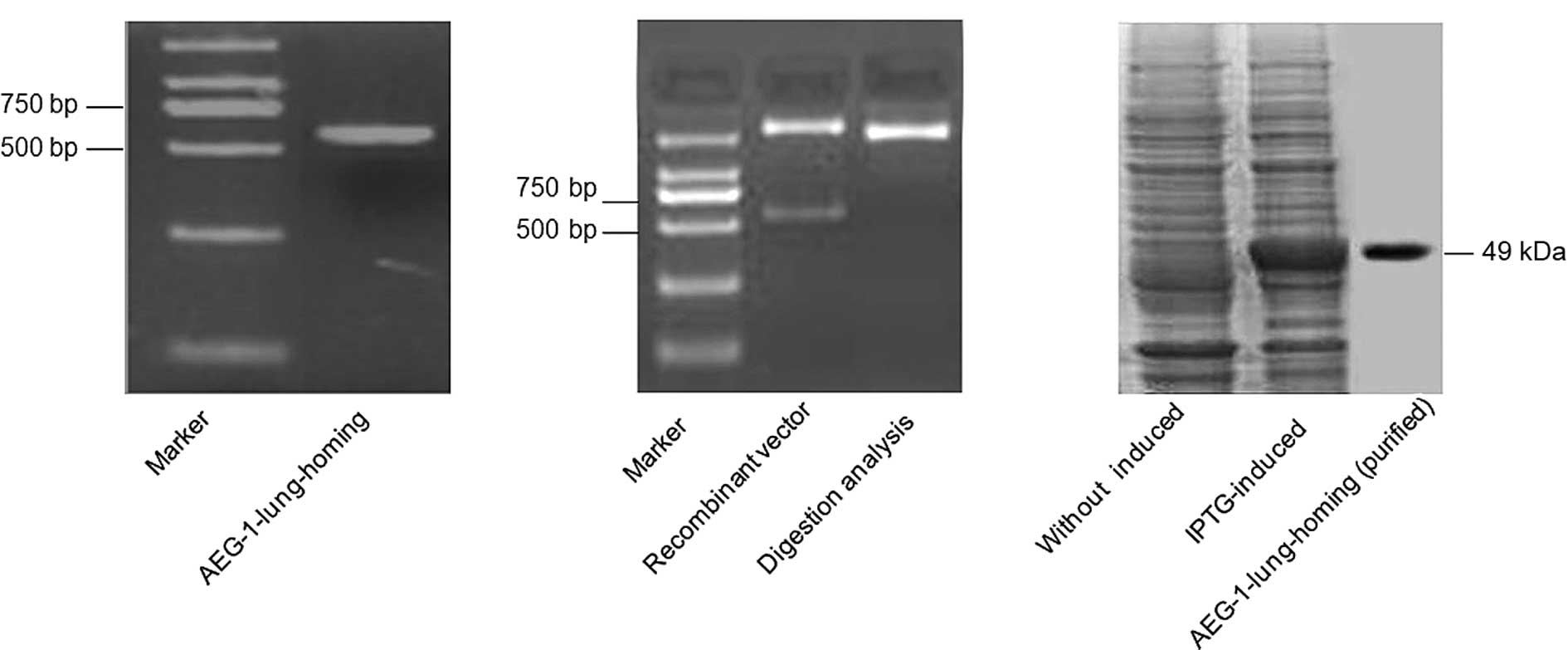
Design of expression vectors, and
expression and purification of fusion proteins
To express the lung-homing domain of extracellular
AEG-1, the prokaryotic expression vector of
pGSTag-AEG-1-lung-homing was constructed. The GST-AEG-1-lung-homing
fusion peptide was expressed from a bacterial source, and it was
purified by glutathione sepharose 4B beads (Fig. 1). Analysis of expression products by
western blot analysis with anti-AEG-1 antibody revealed that the
recombinant proteins exhibited the expected size of 49 kDa and were
highly enriched under native conditions with reproducibly high
levels of expression (7.5 mg/l culture).
ELISA detection of anti-AEG-1
auto-antibodies in sera from various cancer patients
Patients with different types of cancer had
antibodies directed against the AEG-1 oncogenic protein. Sera were
obtained from various cancer patients, including 98 with breast
cancer, 96 with hepatic carcinoma, 88 with rectal cancer, 113 with
lung cancer and 88 with gastric cancer at the time of initial
diagnosis and the samples were analyzed for the presence of
antibodies directed against the AEG-1 oncoprotein. Antibodies at
titers ≥1:50 were detected in 238 of 483 (49%) cancer patients.
Antibodies detected in patients with different types of cancer were
as follows: 44 of 98 (45%) in breast cancer patients, 48 of 96
(50%) in hepatic carcinoma patients, 51 of 113 (45%) in lung cancer
patients, 43 of 88 (49%) in rectal cancer patients, and 52 of 88
(59%) in gastric cancer patients, compared with 0 of 230 (0%)
normal individuals (Table I,
p<0.01). These results demonstrated that AEG-1-Abs were present
in a broad range of malignant tumors.
 | Table IPositive rates of serum anti-AEG-1
auto-antibody in different types of cancer patients and normal
individuals. |
Table I
Positive rates of serum anti-AEG-1
auto-antibody in different types of cancer patients and normal
individuals.
| Sample | Total sample
number | AEG-1 auto-antibody
positive number | N (%) | P-valuea |
|---|
| Breast cancer | 98 | 44 | 45 | <0.01 |
| Hepatic
carcinoma | 96 | 48 | 50 | <0.01 |
| Retal cancer | 88 | 43 | 49 | <0.01 |
| Lung cancer | 113 | 51 | 45 | <0.01 |
| Gastric cancer | 88 | 52 | 59 | <0.01 |
| Total cancer | 483 | 238 | 49 | <0.01 |
| Normal | 230 | 0 | 0 | |
Association between anti-AEG-1
auto-antibody levels and the clinicopathological characteristics of
cancer patients
AEG-1-Abs at titer ≥1:50 were detected in 238 of 483
(49%) cancer patients (260 females and 223 males). The positive
rate of AEG-1-Abs in female patients was the same as that in male
patients. No significant difference was observed between the
patients with or without metastasis (p>0.05). However, AEG-1-Abs
at titer ≥1:50 were detected in 168 of 287 (59%) of patients <60
years old, but it was only detected in 70 of 196 (36%) of patients
who were >60 years old (Table
II, P<0.01). These results demonstrated that the positive
rate of AEG-1-Abs decreases with age.
| Table IIAssociation between the
clinicopathological characteristics of cancer patients and the
presence of anti-AEG-1 auto-antibodies. |
Table II
Association between the
clinicopathological characteristics of cancer patients and the
presence of anti-AEG-1 auto-antibodies.
| Sample | Total sample
number | Anti-AEG-1
auto-antibody positive number | N (%) | P-value |
|---|
| Gender | | | | 0.599 |
| Male | 223 | 107 | 48 | |
| Female | 260 | 131 | 50 | |
| Metastasis | | | | 0.872 |
| Yes | 98 | 49 | 50 | |
| No | 385 | 189 | 49 | |
| Age (years) | | | | <0.01 |
| <60 | 287 | 168 | 59 | |
| ≥60 | 196 | 70 | 36 | |
Anti-AEG-1 auto-antibody levels in
different clinical TNM stages of patients
The positive rate of antibodies to AEG-1 was
different in the various TNM stages of patients. In stage I and II
cancer patients, antibodies were detected in 40 of 127 (31%) in all
cancer patients at titers ≥1:50 or greater, 9 of 30 (30%) in breast
cancer patients, 8 of 31 (26%) in hepatic carcinoma patients, 6 of
20 (30%) in lung cancer patients, 8 of 23 (35%) in rectal cancer
patients, and 9 of 23 (39%) in gastric cancer patients. However, in
stage III and IV patients, there were 198 of 356 (56%) in all
cancer patients, and 35 of 68 (51%) in breast cancer patients, 40
of 65 (62%) in hepatic carcinoma patients, 37 of 68 (54%) in lung
cancer patients, 43 of 90 (48%) in rectal cancer patients, and 43
of 65 (64%) in gastric cancer patients (Table III, P<0.01). Our results
revealed that the levels of AEG-1-Abs had a significant correlation
with the stage of patients, and may be used as a novel marker of
the progression of cancer patients.
| Table IIIAntibodies to AEG-1 detected at the
time of diagnosis in different stage patients. |
Table III
Antibodies to AEG-1 detected at the
time of diagnosis in different stage patients.
| Sample | AEG-1-Abs positive
case n (%) in stage I and II | AEG-1-Abs positive
case n (%) in stage III and IV | P-value |
|---|
| Breast cancer | 9 (30) | 35 (51) | 0.049 |
| Hepatic
carcinoma | 8 (26) | 40 (62) | 0.001 |
| Rectal cancer | 6 (30) | 37 (54) | 0.055 |
| Lung cancer | 8 (35) | 43 (48) | 0.264 |
| Gastric cancer | 9 (39) | 43 (64) | 0.023 |
| Total cancer | 40 (31) | 198 (56) | <0.01 |
Discussion
Investigators have previously demonstrated that the
sera of cancer patients contained antibodies that were capable of
reacting with TAAs (15,16). The types of TAAs that induce these
auto-antibody responses are varied and include the tumor-suppressor
p53 (25,26) and oncogene products, such as
HER-2/neu proteins (20). Human
AEG-1 mRNA encodes a single-pass transmembrane oncoprotein with a
molecular mass of 64 kDa (2,9).
Studies on AEG-1 from our laboratory and others (11–16)
have shown that AEG-1 was overexpressed in almost all types of
malignant tumors, and expression analysis of cancer samples and
normal samples has established that AEG-1 acts a potential TAA.
Thus, we presumed that this oncoprotein may induce the
auto-antibody response in the serum of cancer patients, and these
AEG-1-Abs may be potential markers for the diagnosis and prognostic
evaluation of cancer patients.
In this study, the GST-AEG-1-lung-homing fusion
protein (amino acid 271–451 NP_848927) which contained the
lung-homing domain of AEG-1 was initally expressed and purified.
Using the ELISA assay, peptides were used as a capture agent to
detect AEG-1 auto-antibody responses in sera from 483 patients
suffering from different types of cancer, and sera from 230 normal
blood donors were used as controls. This is the first report
regarding the detection of AEG-1-Abs in cancer patients. Our
results demonstrate that antibody reactivity correlates with the
presence of cancer. Antibodies at titers ≥1:50 or greater were
detected in 238 of 483 (49%) cancer patients, as well as in hepatic
carcinoma (50%), lung (45%), breast (45%), gastric (59%) and rectal
(49%) cancer patients. Furthermore, the positive rates of AEG-1-Abs
decreased with age (Table I,
P<0.01).
The identification of TAAs that elicit an immune
response is important for clinical applications both for the early
diagnosis of tumors and for the immunotherapy strategy design. In
this study, AEG-1-Abs was also detected in some patients with
early-stage cancer. In stage I and II cancer patients, AEG-1-Abs
were detected in 40 of 127 (31%) cases at titers ≥1:50 including 9
of 30 (30%) breast cancer patients, 8 of 31 (26%) hepatic carcinoma
patients, 6 of 20 (30%) lung cancer patients, 8 of 23 (35%) rectal
cancer patients, and 9 of 23 (39%) gastric cancer patients. These
results demonstrated that the potential utility of auto-antibody
against AEG-1 could be used as an early diagnostic biomarker for a
broad spectrum of cancer patients.
The identification of tumor antigens is also
important for clinical therapy, such as cancer vaccines. Many
tumor-associated antigens are excellent targets for immunotherapy
and vaccine design. Although there are certain limitations to
vaccine therapy, including the short half-life of infused
antibodies, the generation of immunity to the antibody construct
and the inability of antibodies to penetrate solid tumors, our
results provided evidence that AEG-1-Abs may be detected in the
sera of a broad range of cancer patients, and suggested that it is
possible to induce substantial immunity against AEG-1 by
immunization boosting with AEG-1 vaccines, which are paving the way
for further investigation of AEG-1-based immunotherapy
approaches.
To the best of our knowledge, this report is the
first to show that AEG-1-Abs, as a novel marker of malignant
tumors, may be detected in the sera of different cancer patients.
However, more studies are required to develop this marker into a
more sophisticated method that can be used clinically.
Acknowledgements
We thank the Department of Clinical Laboratory of
the Tangdu Hospital of Fourth Military Medical University for
samples used in this study. This study was supported by a grant
from the National Natural Science Foundation of China (no.
81001195).
References
|
1
|
Ferlay J, Shin HR, Bray F, Forman D,
Mathers C and Parkin DM: GLOBOCAN 2008 v1.2, Cancer Incidence and
Mortality. Worldwide: IARC CancerBase No. 10. Lyon, France.
http://globocan.iarc.fr.
|
|
2
|
Caron M, Choquet-Kastylevsky G and
Joubert-Caron R: Cancer immunomics using autoantibody signatures
for biomarker discovery. Mol Cell Proteomics. 6:1115–1122. 2007.
View Article : Google Scholar : PubMed/NCBI
|
|
3
|
Desmetz C, Cortijo C, Mange A and Solassol
J: Humoral response to cancer as a tool for biomarker discovery.
Proteomics. 72:982–988. 2009. View Article : Google Scholar : PubMed/NCBI
|
|
4
|
Zhang JY, Megliorino R, Peng XX, Tan EM,
Chen Y and Chan EKL: Antibody detection using tumor-associated
antigen mini-array in immunodiagnosing human hepatocellular
carcinoma. Hepatology. 46:107–114. 2007. View Article : Google Scholar
|
|
5
|
Tan EM and Zhang JY: Autoantibodies to
tumor-associated antigens: reporters from the immune system.
Immunol Rev. 222:328–340. 2008. View Article : Google Scholar : PubMed/NCBI
|
|
6
|
Lu HL, Goodell V and Disis ML: Humoral
immunity directed against tumor-associated antigens as potential
biomarkers for the early diagnosis of cancer. J Proteome Res.
7:1388–1394. 2008. View Article : Google Scholar : PubMed/NCBI
|
|
7
|
Disis ML, Pupa SM, Gralow JR, Dittadi R,
Menard S and Cheever MA: High-titer HER-2/neu protein-specific
antibody can be detected in patients with early-stage breast
cancer. J Clin Oncol. 15:3363–3367. 1997.PubMed/NCBI
|
|
8
|
Shimbo T, Tanemura T, Yamazaki T, Tamai K,
Katayama I and Kaneda Y: Serum anti-BPAG1 auto-antibody is a novel
marker for human melanoma. PLoS One. 5:e10566–e10573. 2010.
View Article : Google Scholar : PubMed/NCBI
|
|
9
|
Defresne F, Bouzin C, Guilbaud C, et al:
Differential influence of anticancer treatments and angiogenesis on
the seric titer of autoantibody used as tumor and metastasis
biomarker. Neoplasia. 12:562–570. 2010.PubMed/NCBI
|
|
10
|
Su ZZ, Kang DC, Chen Y, et al:
Identification and cloning of human astrocyte genes displaying
elevated expression after infection with HIV-1 or exposure to HIV-1
envelope glycoprotein by rapid subtraction hybridization. Oncogene.
21:3592–3602. 2002. View Article : Google Scholar
|
|
11
|
Kang DC, Su ZZ, Sarkar D, et al: Cloning
and characterization of HIV-1-inducible astrocyte elevated gene-1,
AEG-1. Gene. 353:8–15. 2005. View Article : Google Scholar : PubMed/NCBI
|
|
12
|
Yoo BK, Emdad L, Su ZZ, et al: Astrocyte
elevated gene-1 regulates hepatocellular carcinoma development and
progression. J Clin Invest. 119:465–477. 2009. View Article : Google Scholar : PubMed/NCBI
|
|
13
|
Hu GH, Chong RA, Yang QF, et al: MTDH
activation by 8q22 genomic gain promotes chemoresistance and
metastasis of poor-prognosis breast cancer. Cancer Cell. 15:9–20.
2009. View Article : Google Scholar : PubMed/NCBI
|
|
14
|
Kikuno N, Shiina H, Urakami S, et al:
Knockdown of astrocyte-elevated gene-1 inhibits prostate cancer
progression through upregulation of FOXO3a activity. Oncogene.
26:7647–7655. 2007. View Article : Google Scholar : PubMed/NCBI
|
|
15
|
Yu CP, Chen K, Zheng HQ, et al:
Overexpression of astrocyte elevated gene-1 (AEG-1) is associated
with esophageal squamous cell carcinoma (ESCC) progression and
pathogenesis. Carcinogenesis. 30:894–901. 2009. View Article : Google Scholar : PubMed/NCBI
|
|
16
|
Lee SG, Jeon HY, Su ZZ, et al: Astrocyte
elevated gene-1 contributes to the pathogenesis of neuroblastoma.
Oncogene. 28:2476–2484. 2009. View Article : Google Scholar : PubMed/NCBI
|
|
17
|
Emdad L, Sarkar D, Su ZZ, et al: Astrocyte
elevated gene-1: recent insights into a novel gene involved in
tumor progression, metastasis and neurodegeneration. Pharmacol
Therapeut. 114:155–170. 2007. View Article : Google Scholar : PubMed/NCBI
|
|
18
|
Lawrence N and Chin KL: The metastasis
problem gets stickier. Cancer Cell. 15:1–2. 2009. View Article : Google Scholar
|
|
19
|
Sarkar D, Park ES, Emdad L, Lee SG, Su ZZ
and Fisher PB: Molecular basis of nuclear factor-κB activation by
astrocyte elevated gene-1. Cancer Res. 68:1478–1484. 2008.
|
|
20
|
Emdad L, Sarkar D, Su ZZ, et al:
Activation of the nuclear factor κB pathway by astrocyte elevated
gene-1: implications for tumor progression and metastasis. Cancer
Res. 66:1509–1516. 2006.
|
|
21
|
Emdad L, Lee SG, Su ZZ, et al: Astrocyte
elevated gene-1 (AEG-1) functions as an oncogene and regulates
angiogenesis. Proc Natl Acad Sci USA. 15:21300–21305. 2009.
View Article : Google Scholar : PubMed/NCBI
|
|
22
|
Khuda II, Koide N, Noman ASM, et al:
Astrocyte elevated gene-1 (AEG-1) is induced by lipopolysaccharide
as toll-like receptor 4 (TLR4) ligand and regulates TLR4 signaling.
Immunol. 128:e700–e706. 2009. View Article : Google Scholar : PubMed/NCBI
|
|
23
|
Brown DM and Ruoslahti E: Metadherin, a
cell surface protein in breast tumors that mediates lung
metastasis. Cancer Cell. 5:365–374. 2004. View Article : Google Scholar : PubMed/NCBI
|
|
24
|
Dong J, Zeng BH, Xu LH, et al: Anti-CDC25B
auto antibody predicts poor prognosis in patients with advanced
esophageal squamous cell carcinoma. J Transl Med. 8:81–88. 2010.
View Article : Google Scholar : PubMed/NCBI
|
|
25
|
Soussi T: p53 antibodies in the sera of
patients with various types of cancer. Cancer Res. 60:1777–1788.
2000.PubMed/NCBI
|
|
26
|
Crawford LV, Pim DC and Bulbrook RD:
Detection of antibodies against the cellular protein p53 in sera
from patients with breast cancer. Int J Cancer. 30:403–408. 1982.
View Article : Google Scholar : PubMed/NCBI
|