Introduction
Desmoplastic small round cell tumor (DSRCT) is a
recently recognized clinicopathological entity that is most common
in adolescent males and usually affects the abdominal cavity
(1). Since first being described in
1989 by Gerald and Rosai (2), DSRCT
has been reported in various types of organs, including lung
(3), ovary (4,5),
pleura (6–8), tunica vaginalis (9–11),
soft tissues and bone (12,13), parotid gland (14), central nervous system (15,16)
and kidney (17,18). DSRCT is a rare but aggressive
malignancy with a poor prognosis and its etiopathogenesis remains
unknown. The tumor has a characteristic histology, with extensive
stromal tissue surrounding islands of small and undifferentiated
cells revealing the desmoplastic appearance (19). DSRCT exhibits a unique
immunohistochemical profile, characterized by the coexpression of
epithelial (keratin and epithelial membrane antigen), mesenchymal
(vimentin) and myogenic (desmin) markers (1). The reciprocal chromosomal
translocation t(11;22)(p13;q12) is specific for DSRCT and this
translocation results in the fusion of the Ewing sarcoma gene
(EWS) on chromosome 22 with the Wilms' tumor suppressor gene
(WT1) on chromosome 11 (20). Undifferentiated tumors often lack
the appropriate histopathological features to provide an accurate
diagnosis. In the present study, we investigated the expression of
molecular markers and EWS-WT1 gene fusion in
paraffin-embedded material from four cases of DSRCT.
Patients and methods
Patients
A total of four male patients from Sun Yat-Sen
University Cancer Center (Guangzhou, China) were involved in this
study. The patients included three outpatients and a hospital
patient. from The patients had provided informed consent for use of
tumor specimens. The study was approved by the Institute Research
Medical Ethics Committee of Sun Yat-Sen University.
Staining
A review of the clinical data and pathological
sections of all cases was performed, representative sections were
then selected and immunohistochemically stained. All antibody
assays were performed using a standard two-step technique. The
pretreatment methods, primary antibodies and their working
dilutions used in the present study are listed in Table I. Fluorescence in situ
hybridization (FISH) was used to detect the EWS-WT1 fusion.
Sections from representative samples were cut into positively
charged slices at 4 μm thickness. The FISH assays began with
deparaffinization of the sections followed by target retrieval
(boiled in citrate buffer for 15 min) and pepsin (4 mg/ml)
digestion at 37°C for 15 min. For the FISH break apart strategy,
the commercial EWSR1 dual color break apart set was used (Vysis
Inc., Downers Grove, IL, USA). This combines a 500-kbp Spectrum
Orange-labeled probe on the centromeric side of the 7-kbp EWSR1
breakpoint region between exons 7 and 10 of the EWS gene with an
1,100-kbp Spectrum Green-labeled probe localizing on the telomeric
side of this breakpoint region. For the FISH fusion approach, a
paired set of laboratory-prepared rhodamine-labeled EWS and
fluoroisothiocyanate (FITC)-labeled WT-1 were used. The FISH probes
were diluted 1:50 in DenHyb buffer (Insitus Laboratories,
Albuquerque, NM, USA), hybridization mix (10 μl/slide) was applied
to the sections, followed by simultaneous denaturing of the probe
and target at 90°C for 13 min. Overnight hybridization at 37°C
occurred in a humidified chamber, followed by post-hybridization
washes in 50% formamide 1X and 2X SSC.
4′,6-diamidino-2-phenylindole (DAPI; 0.5 μl/ml; Insitus
Laboratories) was used as a nuclear counterstain. Green and red
fluorescent signals were counted in cellular tumor regions with
appropriate filters. Sections with sufficient hybridization
efficiency (i.e., the majority of nuclei presented a signal) were
considered informative, and at least 100 intact and the result was
evaluated by two reviewers.
 | Table IPretreatment and working dilutions of
primary antibodies used in immunohistochemistry. |
Table I
Pretreatment and working dilutions of
primary antibodies used in immunohistochemistry.
| Antigen detected | Pretreatment | Working dilution | Manufacturer (Cat.
No.) |
|---|
| CK | HP EDTA | 1:250 | Dako (M-3515) |
| Vimentin | - | 1:400 | Dako (M-0725) |
| Desmin | HP CB | 1:150 | Santa Cruz
(sc-70961) |
| CAM5.2 | HP EDTA | Ready for use | ZETA (Z2018) |
| WT-1 | HP EDTA | 1:80 | Dako (M-3561) |
| EMA | HP CB | 1:200 | Dako (M-0613) |
| NSE HP | EDTA | 1:150 | Dako (M-0873) |
| CD56 | HP EDTA | 1:200 | Dako (M-7304) |
| CD99 | HP EDTA | 1:100 | Dako (IM-3601) |
| CK5/6 | HP EDTA | 1:250 | Dako (M-7237) |
| Myogenin | HP EDTA | 1:100 | Dako (M-3559) |
| MyoD1 | HP EDTA | 1:100 | Dako (M-3512) |
| Calretinin | HP EDTA | 1:200 | Dako (M-7245) |
| CD117 | HP EDTA | 1:150 | Dako (A-4502) |
| CD34 | HP EDTA | 1:200 | Dako (M-7165) |
| HMB45 | HP CB | 1:80 | Abcam (ab-787) |
| CEA | HP CB | 1:300 | Dako (M-7072) |
Results
Clinical data
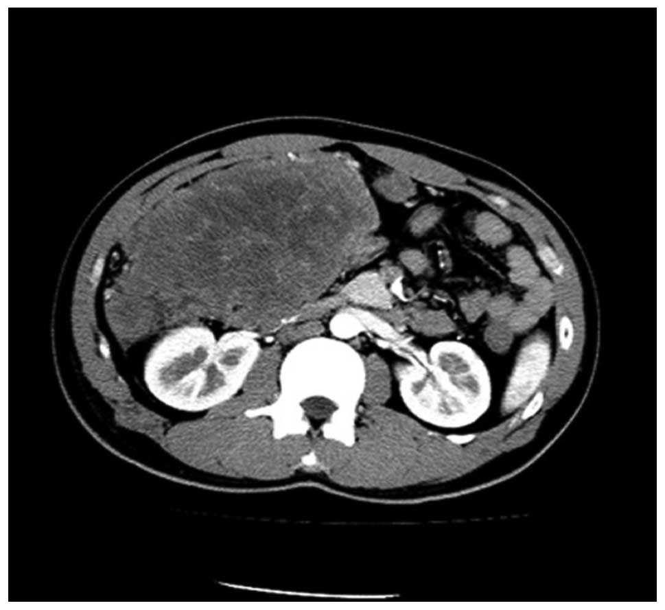
The eldest patient was 52 and the youngest was 26
years old. B-mode ultrasonography and imageology data showed three
tumors situated in the abdominal cavity and one in the pelvic
cavity (Fig. 1). Clinical symptoms
included abdominal distention, stomach ache and a lump was felt. A
few patients had nausea and constipation and one suffered
intestinal obstruction. One tumor involved the liver and another
involved the intestine. All clinical conditions are listed in
Table II.
| Table IIClinical characteristics and follow-up
of the four DSRCT patients. |
Table II
Clinical characteristics and follow-up
of the four DSRCT patients.
| Case | Gender | Age (years) | Location of
tumor | Size (cm × cm) | Follow-up |
|---|
| 1 | Male | 28 | Abdominal cavity | 11.5×8 | Deceased |
| 2 | Male | 26 | Abdominal cavity | 12×9 | Deceased |
| 3 | Male | 28 | Pelvic cavity | 13.5×12 | Deceased |
| 4 | Male | 52 | Abdominal cavity | 15×10 | Relapsed |
Macroscopic pathological features
The tumors were 8–15 cm in diameter (mean tumor
diameter, 13 cm), solid and gray-white, with an appearance of
nodosity or sublobes, and hemorrhage or necrosis was observed in
three patients.
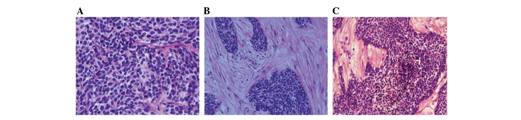
Microscopic pathological features
The neoplastic cells were arranged compactly and
were small, round or orbicular-ovate or short-fusiform with round
or orbicular-ovate hyperchromatic nuclei containing inconspicuous
nucleoli (Fig. 2A). The
karyokinesis was clearly observed and the cytoplasm was diminished.
The cells were gathered into nests or clusters with a large amount
of hyperplastic and pyknotic fibrous connective tissue among them
(Fig. 2B). Necrosis and hemorrhage
were observed in certain neoplastic cell nests (Fig. 2C).
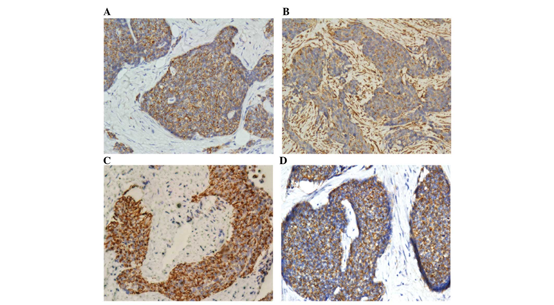
Immunohistochemical features
As shown in Table
III, the tumor cells of all four DSRCTs expressed CK, vimentin,
desmin and CAM5.2 (Fig. 3).
Cytoplasm immunoreactivity for desmin presented as punctiform
located alongside the nuclei. Certain DSRCTs also expressed WT-1,
EMA, NSE, CD56, CD99 and CK5/6. Staining was negative for myogenin,
MyoD1, calretinin, CD117, CD34, HMB45 and CEA (Table III).
| Table IIIExpression of antibodies used in
immunohistochemical analysis of the four DSRCT patients. |
Table III
Expression of antibodies used in
immunohistochemical analysis of the four DSRCT patients.
| Antigen detected | Case 1 | Case 2 | Case 3 | Case 4 |
|---|
| CK | + | + | + | + |
| Vimentin | + | + | + | + |
| Desmin | + | + | + | + |
| CAM5.2 | + | + | + | + |
| WT-1 | + | − | − | + |
| EMA | − | + | + | + |
| NSE | − | + | − | + |
| CD56 | − | + | − | + |
| CD99 | + | + | + | − |
| CK5/6 | + | − | + | − |
| Myogenin | − | − | − | − |
| MyoD1 | − | − | − | − |
| Calretinin | − | − | − | − |
| CD117 | − | − | − | − |
| CD34 | − | − | − | − |
| HMB45 | − | − | − | − |
| CEA | − | − | − | − |
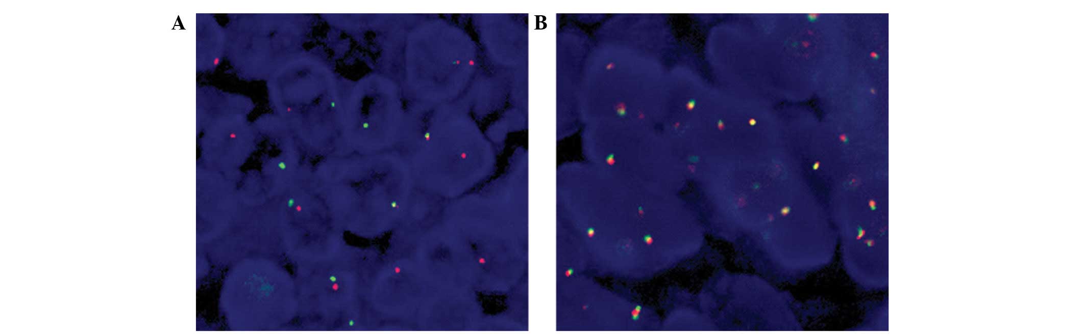
FISH
EWS-WT1 fusion transcripts were detected in
three of four cases using paraffin-embeded tissue. The EWS-R1
break-apart cocktail revealed separation of signals which is
indicative of the translocation involving the EWS locus (Fig. 4).
Discussion
DSRCT occurs predominantly in adolescents and
children with an average age of 21 years, ranging from 3 to 52
years old. The male:female ratio is approximately 5:1. A previous
study revealed that 95% of the cases occurred within the abdominal
and pelvic cavities, and less than 5% of cases occur in tissues,
including the pleura, adjacent to the testis, brain, liver, lung,
mediastinum, paranasal sinuses, ovaries and pancreas (21). Patients with DSRCTs typically
present with a large abdominal mass that is widely disseminated at
the time of diagnosis. There is often extensive regional spread to
the lymph nodes and peritoneal seeding. Metastases may be present
in the liver, lung and bone.
Imaging often reveals a large nodular
intra-abdominal and/or pelvic mass, usually located on the omentum
or mesentery, with no clear parenchymal organs of primary lesions.
The tumor is often a large gray mass, with multiple nodes or lobes,
solid and often growing along the plasma membrane. The sections
revealed hemorrhage, cystic degeneration or necrosis.
Microscopically, DSRCT has a characteristic
appearance of sharply demarcated islands and cords of mitotically
active small cells embedded in a dense desmoplastic stroma. The
stroma may be associated with hyalinization degeneration and
spindle-shaped cells arranged inside as a parallel or fascicular
structure which has a fibroblast-like or myofibroblast morphology
(22).
Immunohistochemical staining revealed a distinct
polyphenotypic pattern consisting of the coexpression of epithelial
(cytokeratin, epithelial membrane antigen, CAM5.2), mesenchymal
(vimentin, desmin) and neural (neuron-specific enolase) markers. A
number of cases have also been reported to express CgA, Syn, CD99,
WT-1 and other markers (5). Desmin
is of diagnostic value due to the characteristic expression and
punctate staining of the nuclei of the tumor cells.
Cytogenetic studies have demonstrated a
characteristic reciprocal chromosomal translocation,
t(11;22)(p13;q12). The translocation of t(11;22)(p13;q12) is
specific for DSRCT, regardless of its anatomical location (9,23,24).
Chimeric EWS-WT1 RNA messages encoded in DSRCTs exhibit
significant molecular diversity. EWS combinatorial variability,
internal deletion, exon skipping of the coding region sequence and
random nucleotide insertions contribute to the heterogeneity of
these tumor-specific gene products (25). The chimeric transcript corresponding
to the fusion gene product may be detected by the reverse
transcription-polymerase chain reaction (RT-PCR) (23), which is highly sensitivitive and
specific, and not only suitable for fresh or paraffin-embedded
tissue, but may also be applied to fine needle aspiration.
With regard to differential diagnosis, DSRCT must be
distinguished from other small round cell tumors, including Ewing's
sarcoma/primitive neuroectodermal tumors (PNET), neuroblastoma,
rhabdomyosarcoma, malignant mesothelioma, small cell carcinoma,
lymphoma, synovial sarcoma and gastrointestinal stromal tumor.
Ewing's sarcoma/PNET is typically positive for CD99 and vimentin,
but negative for cytokeratins and myogenic markers. The
characteristic reciprocal chromosomal translocation in Ewing's
sarcoma/PNET, t(11;22)(q24;q12), is different from DSRCT, which has
a translocation of t(11;22)(p13;q12). Neuroblastoma occurs mainly
in children of less than 5 years old and the stroma is rich in
nerve fiber network, often showing ganglion cell differentiation.
Tumor cells are negative for CK, desmin and WT1. Rhabdomyosarcoma
has often been observed in striated muscle cell differentiation and
tumor cells also express MSA, MyoD1 and other myogenic markers,
with the exception of desmin. Small cell carcinoma demonstrates
immunoreactivity with epithelial markers, including TTF-1 and
neuroendocrine markers, but is negative for myogenic markers.
Lymphoma often demonstrates a diffuse growth pattern and
immunostains show reactivity of lymphoma cells to lymphoid markers
but negativity to epithelial and myogenic markers. In contrast to
malignant mesothelioma, DSRCT cells are negative for CK5/6,
calretinin and other mesothelial markers, and mesothelioma tumor
cells are negative for desmin. Approximately 90% of synovial
sarcoma is detected characteristically by SYT-SSX1/2 chromosomal
translocation. Similarly, gastrointestinal stromal tumors are
detected by the characteristic c-kit gene abnormality.
DSRCT is a highly invasive tumor with a poor
prognosis. Rapid progression includes early seeding, as well as
hematogenous and lymphatic metastasis. The tumor often metastasizes
to the liver, lungs and lymph nodes. Only 29% of patients survive
up to three years and 18% have a five-year survival rate. No ethnic
predisposition or other known risk factors have been identified
specifically for the disease (26).
To date, early surgical treatment of patients remains the only
efficient way to obtain a radical cure. However, most patients are
in an advanced stage at the time of treatment and are rarely able
to obtain radical resection (27).
Several studies suggest a total resection of the mass followed by
postoperative multiagent chemotherapy and radiation therapy as the
best therapeutic option (26). The
ideal therapeutic strategy for treating DSRCT remains unclear due
to the rarity of the tumor.
In conclusion, DSRCT is a rare, but highly
aggressive malignancy and the prognosis in these patients is
extremely poor. Further characterization of the structural and
functional attributes of the EWS-WT1 gene fusion may lead to
an understanding of its role in tumor development and yield insight
into the therapy of DSRCT. A wider availability of specific
molecular markers of the tumor would provide auxiliary methods for
primary diagnosis and treatment. Prospective genetic therapies
focused on developing targeted immunotherapy, moreover, a greater
awareness of this disease among pathologists, oncologists, surgeons
and biologists, may greatly aid the design of a more effective
therapy.
References
|
1
|
Chang F: Desmoplastic small round cell
tumors: cytologic, histologic, and immunohistochemical features.
Arch Pathol Lab Med. 130:728–732. 2006.PubMed/NCBI
|
|
2
|
Gerald WL and Rosai J: Case 2.
Desmoplastic small round cell tumor with divergent differentiation.
Pediatr Pathol. 9:177–183. 1989. View Article : Google Scholar : PubMed/NCBI
|
|
3
|
Syed S, Haque AK, Hawkins HK, Sorensen PH
and Cowan DF: Desmoplastic small round cell tumor of the lung. Arch
Pathol Lab Med. 126:1226–1228. 2002.PubMed/NCBI
|
|
4
|
Young RH, Eichhorn JH, Dickersin GR and
Scully RE: Ovarian involvement by the intra-abdominal desmoplastic
small round cell tumor with divergent differentiation: a report of
three cases. Hum Pathol. 23:454–464. 1992. View Article : Google Scholar : PubMed/NCBI
|
|
5
|
Slomovitz BM, Girotra M, Aledo A, Saqi A,
Soslow RA, Spigland NA and Caputo TA: Desmoplastic small round cell
tumor with primary ovarian involvement: case report and review.
Gynecol Oncol. 79:124–128. 2000. View Article : Google Scholar : PubMed/NCBI
|
|
6
|
Bian Y, Jordan AG, Rupp M, Cohn H,
McLaughlin CJ and Miettinen M: Effusion cytology of desmoplastic
small round cell tumor of the pleura. A case report. Acta Cytol.
37:77–82. 1993.PubMed/NCBI
|
|
7
|
Parkash V, Gerald WL, Parma A, Miettinen M
and Rosai J: Desmoplastic small round cell tumor of the pleura. Am
J Surg Pathol. 19:659–665. 1995. View Article : Google Scholar : PubMed/NCBI
|
|
8
|
Choi JK, van Hoeven K, Brooks JJ and Gupta
PK: Desmoplastic small round cell tumor presenting in pleural fluid
and accompanied by desmin-positive mesothelial cells. Acta Cytol.
39:377–378. 1995.PubMed/NCBI
|
|
9
|
Rodriguez E, Sreekantaiah C, Gerald W,
Reuter VE, Motzer RJ and Chaganti RS: A recurring translocation,
t(11;22)(p13;q11.2), characterizes intra-abdominal desmoplastic
small round-cell tumors. Cancer Genet Cytogenet. 69:17–21. 1993.
View Article : Google Scholar : PubMed/NCBI
|
|
10
|
Roganovich J, Bisogno G, Cecchetto G,
D'Amore ES and Carli M: Paratesticular desmoplastic small round
cell tumor: case report and review of the literature. J Surg Oncol.
71:269–272. 1999. View Article : Google Scholar : PubMed/NCBI
|
|
11
|
Kawano N, Inayama Y, Nagashima Y, Miyagi
Y, Uemura H, Saitoh K, Kubota Y, Hosaka M, Tanaka Y and Nakatani Y:
Desmoplastic small round-cell tumor of the paratesticular region:
report of an adult case with demonstration of EWS and WT1 gene
fusion using paraffin-embedded tissue. Mod Pathol. 12:729–734.
1999.
|
|
12
|
Antonescu CR, Gerald WL, Magid MS and
Ladanyi M: Molecular variants of the EWS-WT1 gene fusion in
desmoplastic small round cell tumor. Diagn Mol Pathol. 7:24–28.
1998. View Article : Google Scholar : PubMed/NCBI
|
|
13
|
Adsay V, Cheng J, Athanasian E, Gerald W
and Rosai J: Primary desmoplastic small cell tumor of soft tissues
and bone of the hand. Am J Surg Pathol. 23:1408–1413. 1999.
View Article : Google Scholar : PubMed/NCBI
|
|
14
|
Wolf AN, Ladanyi M, Paull G, Blaugrund JE
and Westra WH: The expanding clinical spectrum of desmoplastic
small round-cell tumor: a report of two cases with molecular
confirmation. Hum Pathol. 30:430–435. 1999. View Article : Google Scholar : PubMed/NCBI
|
|
15
|
Tison V, Cerasoli S, Morigi F, Ladanyi M,
Gerald WL and Rosai J: Intracranial desmoplastic small-cell tumor.
Report of a case. Am J Surg Pathol. 20:112–117. 1996. View Article : Google Scholar : PubMed/NCBI
|
|
16
|
Neder L, Scheithauer BW, Turel KE, Arnesen
MA, Ketterling RP, Jin L, Moynihan TJ, Giannini C and Meyer FB:
Desmoplastic small round cell tumor of the central nervous system:
report of two cases and review of the literature. Virchows Arch.
454:431–439. 2009. View Article : Google Scholar : PubMed/NCBI
|
|
17
|
Su MC, Jeng YM and Chu YC: Desmoplastic
small round cell tumor of the kidney. Am J Surg Pathol.
28:1379–1383. 2004. View Article : Google Scholar : PubMed/NCBI
|
|
18
|
Egloff AM, Lee EY, Dillon JE and Callahan
MJ: Desmoplastic small round cell tumor of the kidney in a
pediatric patient: sonographic and multiphase CT findings. AJR Am J
Roentgenol. 185:1347–1349. 2005. View Article : Google Scholar : PubMed/NCBI
|
|
19
|
Leuschner I, Radig K and Harms D:
Desmoplastic small round cell tumor. Semin Diagn Pathol.
13:204–212. 1996.PubMed/NCBI
|
|
20
|
Gerald WL, Rosai J and Ladanyi M:
Characterization of the genomic breakpoint and chimeric transcripts
in the EWS-WT1 gene fusion of desmoplastic small round cell tumor.
Proc Natl Acad Sci USA. 92:1028–1032. 1995. View Article : Google Scholar : PubMed/NCBI
|
|
21
|
Gerald WL, Miller HK, Battifora H,
Miettinen M, Silva EG and Rosai J: Intra-abdominal desmoplastic
small round-cell tumor. Report of 19 cases of a distinctive type of
high-grade polyphenotypic malignancy affecting young individuals.
Am J Surg Pathol. 15:499–513. 1991. View Article : Google Scholar
|
|
22
|
Ferlicot S, Coué O, Gilbert E, Beuzeboc P,
Servois V, Klijanienko J, Delattre O and Vielh P: Intraabdominal
desmoplastic small round cell tumor: report of a case with fine
needle aspiration, cytologic diagnosis and molecular confirmation.
Acta Cytol. 45:617–621. 2001. View Article : Google Scholar
|
|
23
|
Ladanyi M and Gerald W: Fusion of the EWS
and WT1 genes in the desmoplastic small round cell tumor. Cancer
Res. 54:2837–2840. 1994.PubMed/NCBI
|
|
24
|
Sawyer JR, Tryka AF and Lewis JM: A novel
reciprocal chromosome translocation t(11;22)(p13;q12) in an
intraabdominal desmoplastic small round-cell tumor. Am J Surg
Pathol. 16:411–416. 1992. View Article : Google Scholar : PubMed/NCBI
|
|
25
|
Liu J, Nau MM, Yeh JC, Allegra CJ, Chu E
and Wright JJ: Molecular heterogeneity and function of EWS-WT1
fusion transcripts in desmoplastic small round cell tumors. Clin
Cancer Res Sep. 6:3522–3529. 2000.PubMed/NCBI
|
|
26
|
Koniari K, Mahera H, Nikolaou M, Chatzis
O, Glezakou O, Magiasis V and Kiratzis G: Intraabdominal
desmoplastic small round cell tumor: Report of a case and
literature review. Int J Surg Case Rep. 2:293–296. 2011. View Article : Google Scholar : PubMed/NCBI
|
|
27
|
Kushner BH, LaQuaglia MP, Wollner N,
Meyers PA, Lindsley KL, Ghavimi F, Merchant TE, Boulad F, Cheung
NK, Bonilla MA, et al: Desmoplastic small round-cell tumor:
prolonged progression-free survival with aggressive multimodality
therapy. J Clin Oncol. 14:1526–1531. 1996.PubMed/NCBI
|