Introduction
Dermatofibrosarcoma protuberans (DFSP) is a
relatively rare distinct dermal and subcutaneous neoplasm, which is
generally regarded as a low-grade sarcoma (1). Although DFSP is characterized
clinically as locally aggressive with a high rate of local
recurrence, distant metastases are rare. Several histopathological
variants of DFSP, including pigmented DFSP and fibrosarcomatous
DFSP, have been reported. Pigmented DFSP, also known as Bednar
tumor, is characterized by the presence of melanin-containing
dendritic cells within the tumor. The incidence of this variant is
quite low, accounting for less than 5% of all DFSP cases (2). Fibrosarcomatous DFSP (FS-DFSP) is a
further variant, characterized by areas histopathologically
indistinguishable from fibrosarcoma, and with a clinically
increased incidence of metastasis (3). Fibrosarcomatous pigmented DFSP is
extremely rare; only five cases have been reported in the English
language literature (3–7). In the present study, we report a
further case of this extremely rare lesion and discuss the
clinicopathological features.
Patients and methods
Case report
A 51-year-old Japanese man presented with a
slow-growing nodular cutaneous mass in his left upper arm, which
was first detected two months earlier. The tumor was reddish to
purplish in color and measured 4×4 cm in size. Assessment of the
biopsy specimen indicated DFSP; thus, total resection of the tumor
with 3-cm margins was performed. This study was approved by the
Ethics Committee of Shiga University of Medical Science. Written
informed consent was obtained from the patient prior to the
study.
The postoperative course was uneventful, and no
recurrence or metastasis was observed during 2 months of medical
follow-up.
Materials
The formalin-fixed, paraffin-embedded tissue blocks
of the resected tumor were cut into 3-micrometer-thick sections,
deparaffinized and rehydrated. Each section was stained with
hematoxylin and eosin (H&E) and used for immunostaining.
Immunohistochemical analyses were performed using an autostainer
(Benchmark XT, Ventana Medical Systems, Tucson, AZ, USA) according
to the manufacturer's instructions. The following primary
antibodies were used: a mouse monoclonal antibody against CD34
(QBEnd/10, Novocastra Laboratories, Ltd., Newcastle upon Tyne, UK)
and a mouse monoclonal antibody against p53 protein (PAb1801,
Novocastra).
Results
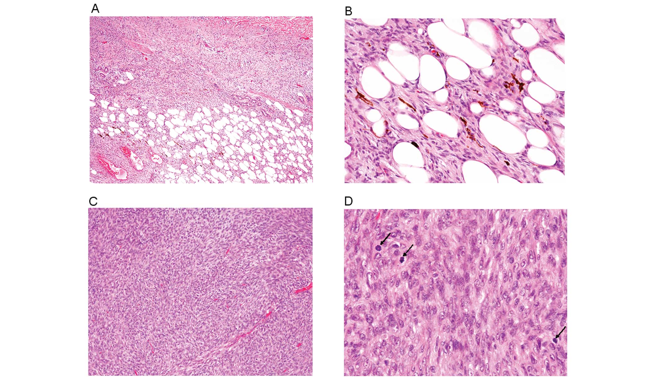
Histopathological study of the resected tumor
revealed monotonous proliferation of uniform spindle cells with
mildly enlarged slender nuclei and inconspicuous nucleoli in the
dermis and subcutis with storiform appearance (Fig. 1A and B). The epidermis was not
involved; however, these spindle cells invaded into the deep
subcutaneous tissue with a characteristic honeycomb pattern.
Mitotic figures were occasionally observed (1/10 high-power
fields). In certain areas, melanin-containing spindle cells, which
were bleached by the peroxide method, were scattered. Approximately
20% of the tumor contained the above-mentioned typical pigmented
DFSP (Bednar tumor), and in addition, further cellular spindle cell
proliferation was clearly visible. The spindle cells were arranged
in fascicles with a herringbone appearance (Fig. 1C) and had large round to short
spindle-shaped nuclei with a conspicuous nucleolus (Fig. 1D). Mitotic figures were frequently
observed in this region (23/10 high-power fields). These
histopathological features resembled fibrosarcoma, and no
melanin-containing spindle cells were present in this area.
Moreover, no pleomorphic sarcoma components or necrosis were
observed.
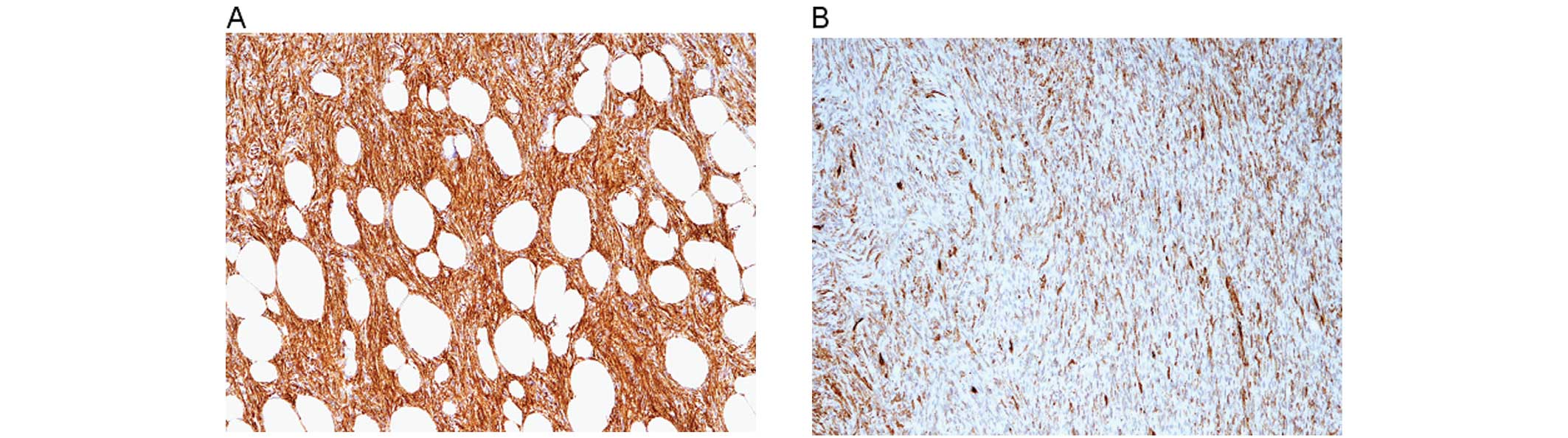
Immunohistochemically, the spindle cells in the
pigmented DFSP area were diffusely and strongly positive for CD34
(Fig. 2A), whereas those of the
fibrosarcomatous area were focally and weakly positive for this
marker (Fig. 2B). Overexpression of
p53 protein was not observed in either the pigmented DFSP or the
fibrosarcomatous components.
According to these findings, an ultimate diagnosis
of fibrosarcomatous pigmented DFSP (Bednar tumor) was made.
Discussion
FS-DFSP is a relatively rare variant of DFSP,
accounting for approximately 10% of cases (3). Although conventional DFSP has a
significant risk of local recurrence (20–50%), distant metastasis
is rare (<5%) (1,8). The local recurrence rate of FS-DFSP
(36.3%) is not significantly different from that of conventional
DFSP; however, its rate of distant metastasis is significantly
higher (13%) compared with the conventional type (8). In addition, tumor-related mortality
was observed in 5.8% of FS-DFSP cases (3). In contrast, the local recurrence rate
of pigmented DFSP (10–13%) is lower compared with conventional
DFSP, and distant metastasis is extremely rare (6).
The presence of a fibrosarcomatous area in pigmented
DFSP is extremely rare. Table I
summarizes the clinicopathological features of the five previously
reported fibrosarcomatous pigmented DFSP cases (3–7) along
with the present one. This lesion mainly affects middle-aged males
(average 49.2 years old, range 27–75 years; male to female ratio
5:1), and occurs mostly in the extremities. A fibrosarcomatous
component was present in the primary lesion in two of the 6 cases,
including the present case, and appeared in the locally recurrent
or metastatic lesions in 3 cases (Table
I). Melanin-containing spindle cells were present only in the
conventional DFSP component in all cases.
 | Table IClinicopathological features of
fibrosarcomatous pigmented dermatofibrosarcoma protuberans. |
Table I
Clinicopathological features of
fibrosarcomatous pigmented dermatofibrosarcoma protuberans.
| Case no. | Age/gender | Location | Fibrosarcomatous
component in primary lesion | Prognosis | Ref. |
|---|
| 1 | 45/M | Upper arm | − | Multiple metastases
in skin, lung, brain, thyroid, pancreas, stomach and small
intestine. DOD. | 4 |
| 2 | 75/M | Thigh | NA | Metastases in lung
and bone. DOD. | 3 |
| 3 | 46/F | Foot | − | Local recurrence | 5 |
| 4 | 51/M | Shoulder | − | Metastases in lung,
retroperitoneum and colon. DOD. | 6 |
| 5 | 27/M | Wrist | + | Multiple metastases
in thigh, chest wall, lung, supraclavicular and parapharyngeal
regions | 7 |
| Present case | 51/M | Upper arm | + | No recurrence or
metastasis 2 months after surgery | |
The prognosis of this lesion appears to be poor,
although the number of reported cases is limited; 4 of 6 cases
displayed multiple metastases and three of the patients succumbed
to the disease. The remaining case (case 3) had local recurrence,
and the present case has been free from local recurrence and
metastasis during 2 months of medical follow-up. We conclude that
the presence of a fibrosarcomatous component in pigmented DFSP is
associated with aggressive behavior; thus, careful observation and
assessment for the presence of a fibrosarcomatous component is
necessary in the diagnosis of pigmented DFSP.
References
|
1
|
Weyers W, Mentzel T, Kasper RC, et al:
Dermatofibrosarcoma protuberans. World Health Organization
Classification of Tumours. Pathology and Genetics of Skin Tumours.
LeBoit PE, Burg G, Weedon D and Sarasain A: IARC Press; Lyon: pp.
259–261. 2006
|
|
2
|
Dupress WB, Langloss JM and Weiss SW:
Pigmented dermatofibrosarcoma protuberans (Bednar tumor): A
pathologic, ultrastructural, and immunohistochemical study. Am J
Surg Pathol. 9:630–639. 1985. View Article : Google Scholar : PubMed/NCBI
|
|
3
|
Mentzel T, Beham A, Katenkamp D, Dei Tos
AP and Fletcher CDM: Fibrosarcomatous (“high-grade”)
dermatofibrosarcoma protuberans: clinicopathologic and
immunohistochemical study of a series of 41 cases with emphasis on
prognostic significance. Am J Surg Pathol. 22:576–587. 1998.
|
|
4
|
Onoda N, Tsutsumi Y, Kakudo K, et al:
Pigmented dermatofibrosarcoma protuberans (Bednar tumor). An
autopsy case with systemic metastasis. Acta Pathol Jpn. 40:935–940.
1990.PubMed/NCBI
|
|
5
|
Porter C, Vincetic A, Saleh ME and
Goldstein H: Pigmented dermatofibrosarcoma protuberans of the foot
with fibrosarcomatous changes: a review and case presentation. J
Foot Ankle Surg. 41:186–191. 2002. View Article : Google Scholar : PubMed/NCBI
|
|
6
|
Suehara Y, Yazawa Y and Hitachi K:
Metastatic Bednar tumor (pigmented dermatofibrosarcoma protuberans)
with fibrosarcomatous change: a case report. J Orthop Sci.
9:662–665. 2004. View Article : Google Scholar : PubMed/NCBI
|
|
7
|
Kini H, Raghuveer CV and Pai MR:
Fibrosarcomatous Bednar tumor with distant metastases - a case
report. Indian J Pathol Microbiol. 47:26–29. 2004.PubMed/NCBI
|
|
8
|
Voth H, Landsberg J, Hinz T, et al:
Management of dermatofibrosarcoma protuberans with fibrosarcomatous
transformation: an evidence-based review of the literature. J Eur
Acad Dermatol. 25:1385–1391. 2011. View Article : Google Scholar : PubMed/NCBI
|