Introduction
It is rare for systemic lymphoma to involve the
hypothalamus-pituitary axis. Out of 380 cases with pituitary tumor
metastasis, there were only two cases (0.5%) with malignant
lymphomas (including Hodgkin’s lymphoma). The frequency of
non-Hodgkin’s lymphoma (NHL) involving the hypothalamus-pituitary
axis is <0.5% among malignant pituitary metastases (1). Therefore, it is essential, but
difficult, to differentiate pituitary lymphoma metastasis from
other tumors to aid the selection of the appropriate therapeutic
plan. Aided by a review of the literature, the present study aimed
to report our experiences of pituitary metastasis from NHL,
summarize the common symptoms of NHL with pituitary metastasis and
emphasize the importance of the use of MRI.
Materials and methods
Cases
The medical files of two patients diagnosed with
diabetes insipidus (DI) caused by pituitary metastasis of NHL, who
were admitted in the last five years to the Second Xiangya Hospital
(Central South University, Changsha, China), were retrospectively
studied (Table I).
 | Table IPatient characteristics at
diagnosis. |
Table I
Patient characteristics at
diagnosis.
| | | | Blood measurements
|
|---|
| Patient | Gender | Age (years) | Symptoms | Routine bloods | Kidney function | Electrolyte
(mmol/l) | Sugar | Other data |
|---|
| 1 | Male | 20 | Body aches, high
fever, weight loss, polyuria, polydipsia | Hb, 95 g/l | Normal | Na, 123.8
Others normal | Normal | Albumin, 31.3
g/l
LDH, 768.2 U/l
ESR, 109 mm/h
Ferritin, 1,000 ng/ml |
| 2 | Male | 26 | Polyuria, polydipsia,
protienuria | Hb, 62 g/l | Normal | Na, 152.4
K, 2.80
Cl, 127.7
Ca, 1.16
Mg, normal
P, normal | Normal | Albumin,
normal
LDH, 674 U/l |
Bone marrow (BM) examination
For patient 1, the BM smear exhibited decreased
hyperplasia and one type of immature cells that accounted for 23%
of the cells, but whose nature was difficult to determine. The
nuclear chromatin of these cells was normal and the nucleoli were
observable. The form of the cells was similar to that of a
lymphocyte. A BM biopsy showed that one type of primitive cell with
an unclear classification was present at increased levels, with a
++ (positive) argyrophil stain. With regard to patients 2, the BM
smear showed highly active BM hyperplasia, the lymphocytes
accounted for 61.5% of the cells, the lymphoblasts and
prolymphocytes accounted for 45% and the cell bodies were large
with observable vacuoles. The smear was negative for peroxidase
staining, while CD10, CD19, CD20, CD34 and HLA-DR were positive in
the leukemic cells, according to flow cytometric detection. By
combining these results with the kidney puncture pathology and by
considering WHO standards, the diagnosis of patient 2 was that of
acute lymphocytic leukemia (ALL; L3, Burkkit’s lymphoma, stage
IV).
Pathological examination
The color doppler flow imaging (CDFI) ultrasound of
patient 1 showed that there were multiple hypoechoic nodules in the
cervical region and groin, which were identified as lymph nodes,
thus biopsies of the left inguinal and right cervical lymph nodes
were performed. The pathology of the left inguinal lymph node
revealed that the lymph node structure was not clear using
microscopy, and that sporadic abnormal cells were present.
According to the immunohistochemistry the left inguinal lymph node
samples contained CD20+ large cells, while CD45RB, CD3,
CD45RO, ALK, CD15, CD30 and CD5 were all negative with a positive
bcl-2 focal shape (+). Consequently, lymphoma could not be
completely ruled out. The pathological results from the right
cervical lymph node showed that the lymph node structure was not
visible under microscopy due to large amounts of karyopyknosis and
necrosis, with only a few slightly degenerated cells left over from
the edge, which were similar to lymphocytes with plasma cell-like
differentiation. The immunohistochemistry of the right cervical
lymph node showed that the samples were only marginally positive
for CD138 at the edge. By combining these results with those of the
inguinal lymph node section and clinical situation, the patient was
diagnosed with lymphatic plasma cell lymphoma.
Patient 2 underwent a kidney puncture biopsy as well
as computed tomography (CT) of the chest and abdomen. The pathology
report revealed that numerous diffuse infiltrations of the renal
interstitium were present with irregular forms and large nuclei.
The nuclear staining of the infiltrations was uneven, indicating
focal aggregation from multiple sites. Degeneration was also
observed among the epithelial cells of the renal tubule and the
protein tube was observed in the small lumen. Many cells, with
irregular form and large nucleus, were found to infiltrate into the
small lumen and tubular wall and nuclear staining of these
infiltrating cells were far from uniform. There were destruction
and breaking off of the residual basement membrane of the
epithelial cells in parts of the renal tubule. Segmental mild
proliferation could be found in the mesangial matrix of renal
tubule. No obvious fuchsinophilic bodies deposition was found in
the mesangium region and the basement membrane was not thick with
capillary lumens opening well. In the immunofluorescence results,
one glomerulus was observed, IgA+ staining was localized
in the mesangial area and staining for IgG, IgM, Fib, C3, C4 and
C1q was all negative. The pathological diagnosis was of
tubulointerstitial lesions that may have been caused by a tumor.
The pathology department observed that more lymphocyte infiltration
was detected in the renal tissues (mainly the medulla) and that
certain lymphocytes infiltrated the renal tubule, causing focal
destruction, although the majority of the renal tubular structures
were present. In the immunohistochemical evaluation,
CD3+ and CD20− staining were observed and the
majority of the renal tubular structures were present.
Consequently, NHL was proposed as the diagnosis of patient 2.
Water deprivation and vasopressin
response test
Urine measurements were obtained from each patient,
including the 24-h urine volume (UV), specific gravity (SG), urine
osmotic pressure (Uosm) and urine electrolyte levels. Water
deprivation and vasopressin (DDAVP) response tests were also
performed.
Imaging
The two patients underwent plain and enhanced MRI at
diagnosis and were assessed again following treatment. Other
imaging techniques, including CT and single photon emission CT
(SPECT) bone scanning were also performed if required.
Results
Diagnoses
Based on the clinical characteristics, blood
examinations and bone marrow and histological studies, patient 1
was diagnosed with lymphatic plasma cell lymphoma and patient 2 was
diagnosed with Burkitt’s ALL (L3, stage IV). The urine
measurements, including the water deprivation and DDAVP response
tests, revealed that the two patients had developed DI (Table II).
| Table IIUrine measurements. |
Table II
Urine measurements.
| At diagnosis
| After water
deprivation
| After administration
of DDAVP
|
|---|
| Patient | UV (ml) | SG | Usom (mOsm/l) | UV (ml) | SG | Usom (mOsm/l) | UV (ml) | SG | Usom (mOsm/l) |
|---|
| 1 | 10,000 | DL | DL | 8,000 | 1.007 | 268 | DL | 1.017 | 470 |
| 2 | 8,000 | 1.007 | 300 | 11,000 | 1.007 | 200 | 2,400 | 1.017 | 610 |
Imaging
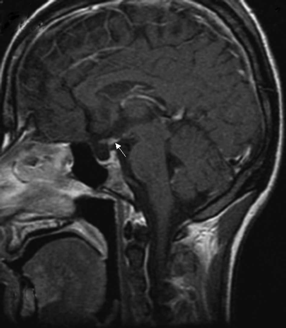
Plain and enhanced MRI examinations of the pituitary
glands were performed at diagnosis and following treatment for each
patient (patient 1, Figs. 1 and
2; patient 2, Figs. 3 and 4). With regard to patient 1, intensified
plain MRI scanning of the pituitary revealed a small nodular lesion
under the hypothalamus (tuber cinereum) and the disappearance of
the normal high signal from the posterior pituitary, suggesting
that there was infiltration and obstruction in the hypothalamic
portal system caused by lymphoma or other pathological changes.
Additionally, in the lumbar vertebral MRI of patient 1, numerous
vertebral body changes were identified as malignant lesions that
were possibly due to tumor metastasis. SPECT bone scanning also
showed bone destruction in the skull, fifth rear-right rib, third
lumbar vertebra, left sacroiliac joint and right acetabulum.
The MRI of the patient 2 revealed that the
hypophyseal fossa was small and the high signals from the posterior
pituitary had disappeared. Notably, enhanced CT of the chest and
abdomen showed bad bilateral perfusion in the kidneys that may have
been associated with diffuse and infiltrative tumors.
Treatment
The two patients were diagnosed with NHL and DI
caused by NHL pituitary metastases and were treated with DDAVP and
chemotherapy.
For patient 1, 0.1 mg DDAVP was administered twice a
day and then increased to three times per day after five days. The
patient also underwent the cyclophosphamide, epirubicin,
vincristine and prednisone (CHOP) chemotherapy regimen. After 20
days, the patient’s UV decreased, so the DDAVP dose was reduced to
0.1 mg twice a day. Subsequent to one month of chemotherapy, all
the patient’s swollen superficial lymph nodes were not palpable and
the blood index improved noticeably. A second cycle of CHOP was
subsequently administered. The patient also underwent cranial
radiation therapy using a linear accelerator five times a week for
one month. Later, the patient received the third, fourth, fifth and
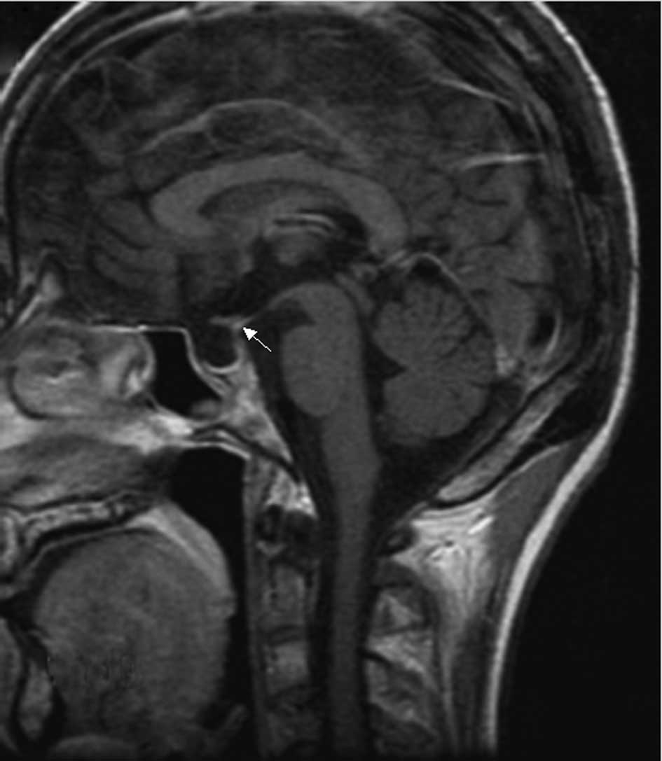
sixth CHOP regimes as monthly chemotherapy. Reexamination with
plain and enhanced MRI scanning of the pituitary gland showed that
the nodular enhanced focus in the hypothalamus (tuber cinereum) was
smaller than at diagnosis, but there was no clear change in the
other foci (Fig. 2). No enlargement
was observed in the retroperitoneal lymph node. Routine blood
tests, kidney and liver function tests, blood sedimentation rate
and lactate dehydrogenase levels were all normal. The patient
received 0.1–0.15 mg DDAVP per day to maintain a normal UV.
Patient 2 underwent the cyclophosphamide,
epirubicin, vincristine, etoposide and prednisone (EPOCH) regimen.
The patient started receiving DDAVP (0.2 mg, twice per day) two
days after the beginning of chemotherapy. Two weeks afetr
chemotherapy, the polydipsia disappeared and the patient’s UV
dropped from 7,000–8,000 ml to 2,400 ml per day. The SG was 1.018
and the Uosm was 680 mOsm/l. The patient’s blood LDH was 342.1 U/l.
DDAVP was gradually reduced to avoid withdrawal. A BM reexamination
revealed complete remission. A brain MRI showed that the pituitary
stalk had returned to normal but that the normal high signal of the
posterior pituitary remained unobservable (Fig. 4). Two months later, following the
second EPOCH course, the disease relapsed. The patient’s UV
increased to 4,000 ml per day and the SG and Uosm were 1.007 and
300 mOsm/l, respectively. Lymphoblasts and prolymphocytes in the BM
accounted for 24% of all cells. Cyclophosphamide, pirarubicin,
vinorelbine, etoposide and prednisone were administered as
chemotherapy in the third and fourth courses, in addition to 0.1 mg
oral DDAVP twice a day. The UV, SG and Uosm were controlled within
the normal range, but the lymphoblasts and prolymphocytes of the BM
continued to account for 14% of the cells. In the fifth and sixth
chemotherapy courses, itoxantrone, cytarabine and dexamethasone
were administered, as well as 100 mg oral thalidomide per day for
21 days each month. The patient remained in excellent physical
condition despite continuously presenting with 12.5% prolymphocytes
in the BM.
The present study also reviewed 17 cases from the
literature. The clinical features of these cases, as well as the
two cases described in the present study, are reviewed in Table III.
| Table IIIFeatures of patients with pituitary
metastasis of NHL. |
Table III
Features of patients with pituitary
metastasis of NHL.
| Reference | Gender | Age (years) | Type | Involvement
region | Imaging | Sequence of lymphoma
and endocrine symptoms |
|---|
| 2 | Male | 59 | DLBCL | Anterior lobe | MRI; lump in pia
mater | Headache 3 weeks
prior |
| 6 | Female | 72 | Large
B-cell
NHL | Anterior lobe | MRI; infiltration of
the pituitary and pituitary stalk | Occurred
simultaneously |
| 7 | Female | 70 | FL, transformed to
DLBCL | Anterior lobe | NR | Follicular
manifestation occurred first |
| 8 | Male | 65 | NHL | Anterior lobe | Gallium 67; pituitary
tumor | Ophthalmic signs 2
months prior |
| 9 | Male | 65 | B-cell NHL | Anterior lobe | MRI; enlargement of
the pituitary gland | Endocrine symptoms 3
months prior |
| 10 | Male | 77 | DLBCL | Anterior, posterior
lobe | MRI; the pituitary
mass | Occurred
simultaneously |
| 11 | Male | 55 | DLBCL | Anterior, posterior
lobe | CT and MRI; optic
chiasm infiltration | NR |
| 12 | Female | 50 | DLBCL | Anterior, posterior
lobe | CT and MRI;
suprasellar mass | Lymphoma in the first
half of the year |
| 2 | Male | 53 | T cell-rich
B cell NHL | Posterior lobe | MRI; the high signal
point from the pituitary disappeared | Headaches after 6
weeks of and polydipsia |
| 3 | Male | 32 | Large
B-cell
NHL | Posterior lobe | Gallium 67; cavernous
sinus infiltration | Diabetes insipidus 3
months ago |
| 4 | Female | 48 | DLBCL | Posterior lobe | MRI; no abnormality
in the pituitary with encephalitis | Lymphoma 1 month
prior |
| 13 | Male | 19 | ACTL | Posterior lobe | CT; pituitary stalk
thickening with hypothalamus involved | Lymphoma several days
ago |
| 14 | Female | 56 | ACTL | Posterior lobe | MRI; the high signal
point from the pituitary disappeared | Lymphoma 3 months
prior |
| 15 | Male | 50 | ACTL | Posterior lobe | CT; pituitary stalk
thickening, empty sella tarcica | Lymphoma 3 months
prior |
| 16 | Male | 64 | Large B-cell NHL | Posterior lobe | MRI; involvement of
the sella turcica with pituitary body and pituitary stalk
thickening | Lymphoma 2 years
prior |
| 17 | Female | 37 | DLBCL | Posterior lobe | MRI; infiltration of
the neuro-hypophyseal lymphoma | Lymphoma 3 months
ago |
| 18 | Female | 70 | B-cell NHL | Posterior lobe | MRI; a sellar mass
involving the pituitary, infundibular stalk, right cavernous sinus
and sphenoid sinus | Right palpebral
ptosis for 1 week |
| Patient 1 | Male | 20 | LPL | Posterior lobe | MRI; hypothalamic
focus with the disappearance of the high signal from the posterior
lobe | Lymphoma 9 months
prior |
| Patient 2 | Male | 26 | Burkkit’s
lymphoma | Posterior lobe | MRI; the high
signal from the posterior lobe disappeared | Polyuria and
polydipsia occurred first |
Discussion
The BM analysis, histological results of other
regions and the observed efficiency of chemotherapy aid in the
diagnosis of NHL. With regard to the two patients studied
retrospectively in the present study, the NHL diagnoses were
identified by the histology and BM examinations. However, NHL is a
systemic disease with various clinical manifestations, including
endocrine symptoms. Occasionally, the endocrine symptoms precede
the hematological diagnosis. Therefore, malignant lymphomas should
be systematically considered as a potential etiology of endocrine
disease such as DI (3). In the two
present cases, the patient’s clinical features and urine
measurements, particularly the water deprivation and DDAVP response
tests, indicated a diagnosis of DI but not NHL.
Furthermore, the two patients’ clinical conditions
were clearly improved following treatment, shown by a remission of
polyuria and polydipsia and the normalization of UV, SG and Uosm in
the urine reevaluation. This demonstrated that the diagnosis of DI
was correct. It is essential that 24-h UV, Usom, plasma
electrolytes, formal water deprivation and DDAVP response tests are
performed, as well as random plasma osmolality tests to ensure a
correct diagnosis (3). However,
among the cases from the literature, there was only one patient who
had been diagnosed with DI by determining the DDAVP in the plasma
(4). The major causes of DI,
particularly central DI, are neoplastic or infiltrative lesions of
the hypothalamus or pituitary glands, severe head injures and
pituitary or hypothalamic surgery (3), therefore, brain radiography is
required to identify the causes of DI. MRI and CT have important
roles; MRI is the modality of choice for providing multiplanar
high-contrast images, whereas CT has a complementary role in
delineating bone destruction and the visualization of
calcification. MRI is more sensitive than CT in revealing brain
lymphoma and is consequently essential (5).
In addition to the two cases reported in the present
study, the features of 17 cases from the literature were also
reviewed (Table III). The median
age of these patients was 52 years (range, 19–77 years). With
regard to the histological types, 12 cases had been diagnosed with
B-cell NHL, three with angiocentric T cell lymphoma (ACTL), one
with follicular lymphoma (FL), one with lymphoplasmacytoid lymphoma
(LPL), one with Burkkit’s lymphoma and one was not precisely
classified. Patient 1 of the present study is the first reported
case of lymphoplasmacytoid lymphoma metastasizing to the pituitary.
Among the 19 patients with pituitary function impairment, this was
manifested as DI (posterior pituitary involvement) in 11 cases
(57.9%), while five cases (26.3%) only had anterior pituitary
hypofunction and three (15.8%) had DI and anterior pituitary
hypofunction. The diagnoses of the cases were made according to the
existence of lymphoma in other parts of the body and the efficiency
of chemotherapy. Only one case underwent a biopsy of the sellar
region (12). Lymphoma symptoms
occurred first in 13 cases and the endocrine symptoms appeared
between several days and nine months later. The endocrine symptoms
appeared first in four cases and the lymphoma manifestations
appeared between six weeks and three years later. Only two cases
experienced the endocrine and lymphoma symptoms simultaneously.
Among the four patients who experienced endocrine symptoms first,
three exhibited posterior pituitary involvement and one was
diagnosed with lymphoma following a three-year anterior pituitary
function impairment. In total, three cases with ACTL exhibited
posterior pituitary involvement. Komninos et al reviewed 190
cases with systemic malignant pituitary metastases and of these, 86
cases (45.2%) developed DI due to posterior pituitary impairment,
while 45 cases (23.6%) exhibited anterior pituitary function
impairments (1). Among the 19 cases
from the present literature review, the two cases reported in the
present study and 12 other cases from the literature exhibited
posterior pituitary impairments, accounting for 73.7% of the cases
(14/19), while eight had anterior pituitary function impairment,
accounting for 42.1%. The frequency of certain symptoms in this
review did not precisely correlate with those of the published
literature (1), with the exception
of statistical differences due to the limited number of cases,
although they may be relevant in identifying the different sources
of the initial tumor (NHL vs. all malignant tumors). DI due to
posterior pituitary impairment or compression of the pituitary
stalk is the most common manifestation of systemic NHL involved
with the pituitary glands. The neurohypophysis receives its blood
supply directly from the pituitary artery, while the anterior
pituitary receives blood from the portal system and a branch
originating from the posterior pituitary. This difference in blood
supply is the reason why the posterior pituitary is more easily
impaired than the anterior pituitary.
The changes that are observable using MRI in
patients with systemic malignant pituitary metastasis have similar
presentations, including intrasellar and parasellar destructive and
nonhomogeneous enhanced impairments, often affecting adjacent
structures. Normal pituitary cells contain phospholipids or
secretory granules, so the T1-weighted imaging (WI) signal of MRI
is enhanced. In DI, during the hypofunctioning of pituitary
synthesis, transport and storage, this signal is weakened or
disappears and there is homogeneous enhancement of the pituitary
and pituitary stalk signals following the administration of
contrast agent. The MRI of patients with lymphoma shows low T1WI
and T2WI signals (5,19–21),
while for pituitary metastases from other tumors, T1WI signals are
usually low and T2WI signals are high (5). Among the 14 cases with posterior
pituitary involvement, 11 patients underwent MRI and two underwent
only CT. The disappearance of the normal higher signals in the
posterior pituitary occurred in four cases (4/11; 36.4%).
Enlargement of the pituitary gland and pituitary stalk thickening
occurred in four cases (4/13; 30.8%). There was significant
diversity in anterior pituitary involvement, including the
occurrence of suprasellar masses, destruction at the base of the
sella, optic chiasm infiltration, cavernous sinus masses, clival
damage and leptomeningeal and pituitary masses.
A number of the NHL cases first manifested as
endocrine symptoms. The NHL pituitary metastasis rate was <0.5%.
Consequently, it is difficult to identity the causes of pituitary
impairment, particularly NHL metastasis to the pituitary. Unless a
lesion biopsy of the sellar region is performed or there is
evidence of a pituitary gland tumor, it is difficult to distinguish
NHL pituitary impairment from the pituitary metastasis of other
tumors. Even an image-guided biopsy is unable to reliably avoid the
surrounding critical neurovascular structures since the sella is a
region with a small volume in close proximity to numerous complex
structures (5). In the literature
review, only one patient underwent this type of biopsy. Therefore,
it is important and necessary to perform pituitary MRI to reveal
pituitary metastases from NHL. Moreover, chemotherapy for certain
tumors that relieves pituitary impairment symptoms and improves the
imaging results is useful for an etiological diagnosis.
In summary, in our two cases, diabetes insipidus is
their main and early clinical manifestation. Thus, for patients
exhibiting endocrine symptoms, NHL should be considered as a
potential cause, particularly if hematological symptoms also exist.
It is essential to perform pituitary MRI to differentiate NHL
pituitary metastases from other tumors. The T1WI and T2WI signals
are low in patients with malignant lymphoma involving the pituitary
glands, while for pituitary metastases from other tumors, the T1WI
signals are usually low and T2WI signals are high. For patients
with NHL, the diagnosis of DI depends on water deprivation and
DDAVP tests, although plasma DDAVP tests are not necessary.
References
|
1
|
Komninos J, Vlassopoulou V, Protopapa D,
Korfias S, Kontogeorgos G, Sakas DE and Thalassinos NC: Tumors
metastatic to the pituitary gland: case report and literature
review. J Clin Endocrinol Metab. 89:574–580. 2004. View Article : Google Scholar : PubMed/NCBI
|
|
2
|
Megan Ogilvie C, Payne S, Evanson J,
Lister TA and Grossman AB: Lymphoma metastasizing to the pituitary:
an unusual presentation of a treatable disease. Pituitary.
8:139–146. 2005.PubMed/NCBI
|
|
3
|
Liozon E, Soria P, Jaccard A, et al:
Diabetes insipidus revealing primary malignant non-Hodgkin’s
lymphoma of bone. Rev Med Interne. 19:830–834. 1998.(In
French).
|
|
4
|
Scheinpflug K, Schalk E, Reschke K, Franke
A and Mohren M: Diabetes insipidus due to herpes encephalitis in a
patient with difuuse large cell lymphoma. A case report. Exp Clin
Endocrinol Diabetes. 114:31–34. 2006. View Article : Google Scholar : PubMed/NCBI
|
|
5
|
Kaltsas GA, Evanson J, Chrisoulidou A and
Grossman AB: The diagnosis and management of parasellar tumours of
the pituitary. Endocr Relat Cancer. 15:885–903. 2008. View Article : Google Scholar : PubMed/NCBI
|
|
6
|
Büchler T, Ferra C, Virgili N, Montanya E
and Grañena A: A relapsed non-Hodgkin’s lymphoma presenting as
panhypopituitarism successfully treated by chemotherapy. J
Neurooncol. 59:35–38. 2002.
|
|
7
|
Sumrall A and Herrin V: Recurrent,
transformed non-Hodgkin’s lymphoma presenting as chiasmal syndrome
with hyperprolactinemia and hypopituitarism. J Miss State Med
Assoc. 51:35–36. 2010.
|
|
8
|
Jonkhoff AR, Huijgens PC, Schreuder WO,
Teule GJ and Heimans JJ: Hypophyseal non-Hodgkin’s lymphoma
presenting with clinical panhypopituitarism successfully treated
with chemotherapy. J Neurooncol. 17:155–158. 1993.
|
|
9
|
Mathiasen RA, Jarrahy R, Cha ST, Kovacs K,
Herman VS, Ginsberg E and Shahinian HK: Pituitary lymphoma: a case
report and literature review. Pituitary. 2:283–287. 2000.
View Article : Google Scholar : PubMed/NCBI
|
|
10
|
Li JK, Chow CC, Yeung VT, Ko GT and
Cockram CS: Adrenal and hypophyseal non-Hodgkin’s lymphoma
presenting with panhypopituitarism. Int J Clin Pract. 52:513–514.
1998.
|
|
11
|
Bolanowski M, Kuliszkiewicz-Janus M and
Sokolska V: Diffuse malignant lymphoma type B with optic chiasm
infiltration, visual disturbances, hypopituitarism,
hyperprolactinaemia and diabetes insipidus. Case report and
literature review. Endokrynol Pol. 57:642–647. 2006.
|
|
12
|
Chan TW and Hoskins P: Panhypopituitarism
secondary to hypothalamic involvement in a woman with diffuse large
B-cell lymphoma. J Clin Oncol. 28:e165–e166. 2010. View Article : Google Scholar : PubMed/NCBI
|
|
13
|
Leedman PJ, Matz LR and Pullan P:
Endocrine dysfunction in lymphomatoid granulomatosis. Aust NZ J
Med. 19:97–102. 1989. View Article : Google Scholar : PubMed/NCBI
|
|
14
|
Ramsahoye BH, Griffiths DF and Whittaker
JA: Angiocentric T-cell lymphoma associated with diabetes
insipidus. Eur J Haematol. 56:100–103. 1996. View Article : Google Scholar : PubMed/NCBI
|
|
15
|
Bushunow PW, Casas V and Duggan DB:
Lymphomatoid granulomatosis causing central diabetes insipidus:
case report and review of the literature. Cancer Invest.
14:112–119. 1996. View Article : Google Scholar : PubMed/NCBI
|
|
16
|
Merlo EM, Maiolo A, Brocchieri A, Tua A
and Grignani G: Hypophyseal non-Hodgkin’s lymphoma presenting with
diabetes insipidus: a case report. J Neurooncol. 42:69–72.
1999.
|
|
17
|
Breidert M, Schimmelpfennig C, Kittner T,
Helwig A and Ehninger G: Diabetes insipidus in a patient with a
highly malignant B-cell lymphoma and stomatitis. Exp Clin
Endocrinol Diabetes. 108:54–58. 2000.PubMed/NCBI
|
|
18
|
Tamer G, Kartal I and Aral F: Pituitary
infiltration by non-Hodgkin’s lymphoma: a case report. J Med Case
Rep. 3:92932009.
|
|
19
|
Boardman JF, Rothfus WE and Dulai HS:
Lesions and pseudolesions of the cavernous sinus and petrous apex.
Otolaryngol Clin North Am. 41:195–213. 2008. View Article : Google Scholar : PubMed/NCBI
|
|
20
|
Shin JH, Lee HK, Choi CG, Suh DC, Kim CJ,
Hong SK and Na DG: MR imaging of central diabetes insipidus: a
pictorial essay. Korean J Radiol. 2:222–230. 2001. View Article : Google Scholar : PubMed/NCBI
|
|
21
|
Rennert J and Doerfler A: Imaging of
sellar and parasellar lesions. Clin Neurol Neurosurg. 109:111–124.
2007. View Article : Google Scholar
|