Introduction
Gliomas are the most common primary brain tumors in
adults and have a poor survival outcome (1). Patients with the same tumor grades
have highly variable prognoses, regardless of the therapeutic
interventions that are used (2).
Molecular behaviors have been observed to contribute to the varying
prognoses of gliomas (3). p53, one
of the most widely investigated molecules in human gliomas, has
been shown to be a prognostic marker (4,5).
Several studies have revealed that mutations in the epidermal
growth factor receptor (EGFR) gene were associated with shorter
intervals between relapses and a poorer survival rate for gliomas,
and that EGFR expression is correlated with tumor progression
(6,7). Ki-67, a marker of cell division, is a
reliable indicator of tumor cell proliferative activity that has
been associated with the histological grade and poor survival
outcome for glioma (8).
O6-methylguanine-DNA methyltransferase (MGMT) repairs
damaged DNA and renders glioma cells resistant to alkylating
agents. MGMT promoter methylation has been used as a biomarker to
predict the sensitivity of gliomas to DNA alkylating
chemotherapeutics (9).
Numerous studies have been undertaken to understand
the molecular basis of gliomagenesis (10,11).
However, to date, few studies have investigated the expression of
p53, EGFR, Ki-67 and MGMT in the same group of glioma patients,
particularly in the Chinese population. It has been reported that
certain molecular markers are associated with the ethnicity of
patients with glioma (10).
Therefore, in order to understand the molecular features of gliomas
with respect to ethnicity, the expression of these markers must be
identified. In addition, the correlation between p53, EGFR, Ki-67
and MGMT expression and the histological grade of gliomas has not
yet been established. The aim of the present study was to evaluate
the expression of these markers using the immunohistochemical
analysis of 152 tumor samples from Chinese patients with gliomas of
various grades, and also to analyze the correlation between their
expression and the tumor grades.
Materials and methods
Subjects
A total of 152 Chinese patients with gliomas (85
males and 67 females) were selected from the Nanjing Brain Hospital
and Zhongda Hospital (Nanjing, Jiangsu, China) between June 2006
and June 2008. The mean age of the patients was 40.3 years (range,
21–79 years). Approval for the study was obtained from the
institutional review board of Nanjing Brain Hospital and all
patients provided informed consent.
The tumor grades were classified by two pathologists
according to the recommendations of the World Health Organization
(WHO; 2007) (14). The pathological
grading of the cancers was as follows: grade I (5 cases of
pilocytic astrocytoma, 3 of a subependymal tumor and 1 of
subependymal giant cell astrocytoma), grade II (30 cases of
astrocytoma, 8 of oligodendroglioma, 7 of ependymoma and 11 of
small branch astrocytoma), grade III (33 cases of anaplastic
astrocytoma and 19 of anaplastic oligoastrocytoma) and grade IV (24
cases of primary glioblastoma and 11 of secondary
glioblastoma).
Immunohistochemistry
Tissue sections (5-μm thick) were obtained
from formalin-fixed and paraffin-embedded tissue blocks for
immunohistochemical staining. The samples were then incubated
overnight in primary antibodies against p53, EGFR, Ki-67 and MGMT
at 4°C. Mouse anti-human monoclonal primary antibodies against the
following antigens were used: p53 (clone Do-7; 1:50 dilution); EGFR
(clone EGFR.25; 1:50 dilution); Ki-67 (clone K-2; 1:100 dilution)
and MGMT (clone MT3.1; 1:100 dilution). All primary antibodies were
purchased from Ventana (Tucson, AZ, USA). Subsequent to the primary
antibodies being washed off, the sections were incubated with goat
anti-mouse biotin-conjugated secondary antibodies (1:1000 dilution;
Ventana) for 20 min at 37°C. The tissue sections were then
incubated with streptavidin horse-radish peroxidase for 20 min at
37°C. A 3,3′-diaminobenzidine (DAB) substrate was applied to the
section for 10 min, prior to counter-staining with hematoxylin. The
sections in which the primary antibodies were omitted were used as
negative controls.
The immunostaining was examined for the presence of
p53, EGFR, Ki-67 and MGMT, and individually observed and counted by
two independent neurosurgeons using a microscope. The presence of
p53 and EGFR was determined using the percentage of immunostained
cells per 200 cells in 5 fields. The p53 and EGFR scoring system
(based on the number of positive cells) was as follows: negative
(−), no positive cells observed in the random fields; weak positive
(+), <25% positive cells; moderately positive (++), 25–50%
positive cells; and strongly positive (+++), >50% positive
cells. The labeling index (LI) for Ki-67 was calculated as the
percentage of positive cells per 1,000 cells. The MGMT
immunostained cells were classified as negative (<10%) or
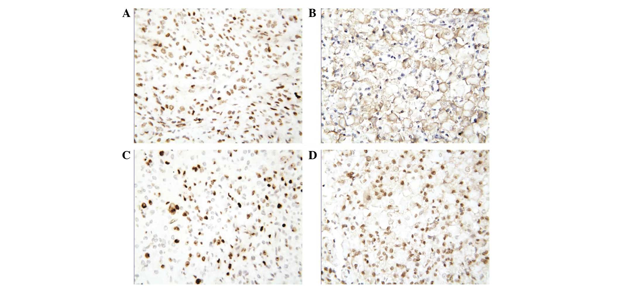
positive (≥10%; Fig. 1A–D).
Statistical analysis
Statistical analysis was performed using SPSS 12.0
(SPSS Inc., Chicago, IL, USA). The χ2 test was performed
to determine the significant differences observed in the p53, EGFR
and MGMT expression values between the various glioma grades. An
ANOVA was used to assess the significant differences observed in
the Ki-67 LI between the glioma grades. P<0.05 was considered to
indicate a statistically significant difference.
Results
Expression of p53 is lower in grade I
gliomas
p53 was expressed in 2 out of 9 cases (22.2%) of
grade I gliomas, 40 out of 56 cases (71.4%) of grade II gliomas, 38
out of 52 cases (73.1%) of grade III gliomas and 21 out of 35 cases
(60.0%) of grade IV gliomas. The frequency of p53 immunopositivity
was significantly lower in the grade I gliomas compared with the
other three categories (P<0.05). No significant differences were
identified in the frequency of p53 immunopositivity between the
grade II, III and IV tumors (P=0.074; Table I).
 | Table I.p53 immunoreactivity in human
gliomas. |
Table I.
p53 immunoreactivity in human
gliomas.
| Grade | Immunonegative cases,
n (%) | Immunopositive cases,
n (%)
|
|---|
| + | ++ | +++ | Total |
|---|
| I | 7 (77.8) | 1 (11.1) | 1 (11.1) | 0 (0.00) | 2 (22.2) |
| II | 16 (28.6) | 11 (19.6) | 12 (21.4) | 17 (30.4) | 40 (71.4) |
| III | 14 (26.9) | 11 (21.2) | 11 (21.2) | 16 (30.8) | 38 (73.1) |
| IV | 14 (40.0) | 3 (8.6) | 6 (17.1) | 12 (34.3) | 21 (60.0) |
EGFR expression is associated with glioma
grade
EGFR immmunopositive staining was observed in 2
cases (22.2%) of grade I glioma, 25 cases (44.6%) of grade II, 37
cases (71.2%) of grade III and 31 cases (88.6%) of grade IV. The
frequency of EGFR immunopositivity was significantly higher in the
grade III and IV gliomas than in the grade I and II gliomas
(P=0.021, Table II). In addition,
marked EGFR staining, ++ or +++, was observed in 40, 78.4 and 90.3%
of grade II, III and IV immunopositive gliomas, respectively. EGFR
expression was significantly higher in the higher grade gliomas
compared with the lower grade gliomas (P=0.025).
| Table II.EGFR immunoreactivity in human
gliomas |
Table II.
EGFR immunoreactivity in human
gliomas
| Grade | Immunonegative cases,
n (%) | Immunopositive cases,
n (%)
|
|---|
| + | ++ | +++ | Total |
|---|
| I | 7 (77.8) | 2 (22.2) | 0 (0.0) | 0 (0.0) | 2 (22.2) |
| II | 31 (55.4) | 15 (26.8) | 6 (10.7) | 4 (7.1) | 25 (44.6) |
| III | 15 (28.8) | 8 (15.4) | 17 (32.7) | 12 (23.1) | 37 (71.2) |
| IV | 4 (11.4) | 3 (8.6) | 10 (28.6) | 18 (51.4) | 31 (88.6) |
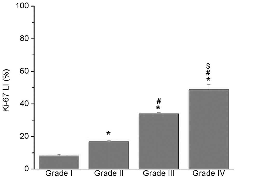
Ki-67 expression in the various glioma
grades
The mean Ki-67 LI significantly increased with the
glioma grade (Fig. 2). A
significant difference was identified in the Ki-67 LI between the
various glioma grades (P<0.05; Fig.
2), suggesting that the pathological grade was associated with
the Ki-67 LI.
MGMT expression is not associated with
glioma grade
MGMT immunoreactivity was identified in 84 (55.3%)
of the 152 glioma samples. The frequency of MGMT immunopositivity
increased with the glioma grade (Table
III). However, no significant differences were observed in the
frequency of MGMT expression between the grades. In addition, the
expression of MGMT in the grade I and II samples (30 out of 65
samples; 46.2%) was not significantly different from the expression
in the grade III and IV tumors (54 out of 87 samples; 62.1%).
| Table III.MGMT immunoreactivity in human
gliomas. |
Table III.
MGMT immunoreactivity in human
gliomas.
| Grade | Immunonegative cases,
n (%) | Immunopositive cases,
n (%) |
|---|
| I | 6 (66.7) | 3 (33.3) |
| II | 29 (51.8) | 27 (48.2) |
| III | 22 (42.3) | 30 (57.7) |
| IV | 11 (31.4) | 24 (68.6) |
Discussion
Despite developments in the diagnosis and treatment
of gliomas, due to the quick progression of malignant tumors, the
prognosis of affected patients remains poor. In order to accurately
identify the factors that affect the prognosis of glioma patients
and to select and evaluate the effectiveness of appropriate
treatments, an understanding of the molecular mechanisms and the
progression of gliomas is required. In the present study,
immunohistochemistry was used to investigate the expression of p53,
EGFR, Ki-67 and MGMT in 152 Chinese patients with gliomas, and to
analyze their correlation with the histological glioma grade. The
expression of EGFR and Ki-67 LI was observed to be significantly
correlated with the histological grade of the gliomas, while the
expression of p53 and MGMT was not associated.
p53 is one of the most frequently used molecular
markers in gliomas. Immunocytochemical experiments have
demonstrated that the overexpression of p53 is commonly considered
to be a surrogate marker for p53 mutation (12). In the present study, it was observed
that p53 expression was significantly higher in grade II gliomas
than in grade I gliomas. Furthermore, p53 expression was marginally
decreased in grade IV compared with grade II and III gliomas,
suggesting that a p53 mutation may be an early event in glioma
progression (13). The present data
also revealed that no correlation existed between the expression of
p53 and the tumor grade, suggesting that p53 expression is not a
good prognostic marker for gliomas. However, the correlation
between p53 immunoreactivity and the survival outcome of glioma
patients remains controversial. It has been suggested that p53 may
act as a weak independent prognostic marker for the clinical or
pathological features of gliomas (5). This difference may be a result of the
various methods used to detect p53 expression in glioma samples
from differing patient populations. The ethnicity of patients with
glioma appears to affect p53 expression in the various tumor grades
(10). In addition, it has been
noted that the changes in p53 overexpression in human astrocytic
gliomas are generally associated with secondary, rather than
primary glioblastomas (14).
However, the present data are not consistent with this observation
(data not shown). Furthermore, p53, a tumor suppressor gene, plays
a key role in the cellular responses to various stresses. p53 also
transfers cells with a normal p53 gene into tumor cells,
sensitizing them to chemotherapeutic drugs and/or radiotherapy and
promoting tumor cell apoptosis (15). In the present study, p53 expression
was detected in approximately two-thirds of all 152 cases,
suggesting that it may be an ideal target for glioma-targeted
therapy.
The majority of gliomas express EGFR, which is often
amplified, rearranged, mutated and/or overexpressed, particularly
in malignant tumors (16). It has
been reported that EGFR is closely correlated with tumor
proliferation, metastasis, apoptosis, angiogenesis, sensitivity to
radiotherapy and/ or chemotherapy and drug resistance (17). A previous study has shown that
glioma malignancy increases with EGFR amplification and
overexpression (18). Consistent
with this, the present study showed that EGFR overexpression was
evident in 25 cases (44.6%) of grade II, 37 cases (71.2%) of grade
III and 31 cases (88.6%) of grade IV tumors. However, the frequency
of EGFR expression shown in the present data was inconsistent with
another study stating that EGFR amplification was identified in
40–50% of glioblastomas and ∼10% of anaplastic astrocytomas, but
not in low-grade astrocytomas (19). This difference may have been a
result of the dissimilar patient populations in the two studies.
However, the two studies did each reveal that EGFR overexpression
is significantly greater in high-grade gliomas than in low-grade
tumors. In addition, EGFR activation has been shown to be a
significant indicator of glioma deterioration, as it is a vital
marker of poorly-differentiated gliomas (17–20).
In the present study, EGFR overexpression correlated with the
higher grade gliomas, suggesting that EGFR overexpression was
associated with tumor aggressiveness and invasion. However, the
role of EGFR overexpression in the prognosis of gliomas remains
controversial. It has been observed that EGFR amplification does
not significantly affect the survival of patients with glioblastoma
at any age (21). However, a
region-limited and age-limited study has observed that EGFR
amplification and overexpression is a significant predictor of
survival (7).
The levels of Ki-67, a cell proliferation nuclear
antigen, may objectively reflect the proliferation and malignancy
of tumor cells (22). Increased
Ki-67 expression has been shown to positively correlate with the
increased grade of malignancy and a poor prognosis in glioma
patients (23). In agreement with
these studies, the present data showed that a substantial increase
in Ki-67 expression was correlated with a higher tumor grade,
suggesting that Ki-67 expression is a good marker for glioma
malignancy. In addition, Ki-67 is a significant marker for
differentiating between benign and malignant tumors. When Ki-67 is
overexpressed, proliferation and invasiveness increase, resulting
in tumor recurrence and malignant changes (24).
MGMT is involved in the repair of DNA damage and the
prevention of second-level DNA damage, thus rendering glioma cells
resistant to DNA alkylating agents. The detection of MGMT
methylation does not provide useful information with regard to
prognosis, but may predict whether patients with glioblastomas are
able to benefit from temozolomide therapy (25). Methylation of the MGMT promoter may
be detected in 60% of glioblastomas, although the false-positive
rate is high (25). The expression
levels of MGMT mRNA and protein are significantly correlated with
enzyme activity. The survival time for patients who are negative
for MGMT is longer than that of patients who are positive for MGMT
(26). In the present study, it was
revealed that MGMT was expressed in 55.3% of glioma samples. The
expression of MGMT was increased in the high-grade gliomas (62.1%)
compared with the low-grade gliomas (46.2%), suggesting that the
majority of gliomas (particularly malignant gliomas) are not
sensitive to chemotherapy. The majority of studies have focused on
MGMT expression in high-grade gliomas, but few have explored MGMT
expression in the other grades. Yang et al (27) showed that MGMT expression correlates
with the glioma grade. However, the present data identified no
significant differences between MGMT expression and the various
glioma grades. This difference may have been due to the dissimilar
sample sizes and detection methods that were employed.
In summary, the expression of p53, EGFR, Ki-67 and
MGMT was investigated in gliomas in a Chinese population using
immunocytochemistry. It was identified that the expression of EGFR
and Ki-67 LI, but not p53 and MGMT, correlated with the
histological grade of the gliomas. The present study contributes to
the assessment of the invasiveness and proliferative potential of
gliomas, to predict their sensitivity to chemotherapy and/or
radiotherapy and to determine the effectiveness of
molecular-targeted therapy. Further studies are required to
understand the molecular biological behavior of malignant
gliomas.
Acknowledgements
The authors would like to thank Dr
Hai-qing Zhu and Dr Juan Wang for their technical assistance and Dr
Miao Wei for the statistical analysis.
References
|
1.
|
Kogiku M, Ohsawa I, Matsumoto K, et al:
Prognosis of glioma patients by combined immunostaining for
survivin, Ki-67 and epidermal growth factor receptor. J Clin
Neurosci. 15:1198–1203. 2008. View Article : Google Scholar : PubMed/NCBI
|
|
2.
|
van den Bent MJ and Kros JM: Predictive
and prognostic markers in neuro-oncology. J Neuropathol Exp Neurol.
66:1074–1081. 2007.PubMed/NCBI
|
|
3.
|
Noble M and Dietrich J: The complex
identity of brain tumors: emerging concerns regarding origin,
diversity and plasticity. Trends Neurosci. 27:148–154. 2004.
View Article : Google Scholar : PubMed/NCBI
|
|
4.
|
Kyritsis AP, Bondy ML, Hess KR, et al:
Prognostic significance of p53 immunoreactivity in patients with
glioma. Clin Cancer Res. 1:1617–1622. 1995.PubMed/NCBI
|
|
5.
|
Levidou G, El-Habr E, Saetta AA, et al:
P53 immunoexpression as a prognostic marker for human astrocytomas:
a meta-analysis and review of the literature. J Neurooncol.
100:363–371. 2010. View Article : Google Scholar : PubMed/NCBI
|
|
6.
|
Hoelzinger DB, Mariani L, Weis J, et al:
Gene expression profile of glioblastoma multiforme invasive
phenotype points to new therapeutic targets. Neoplasia. 7:7–16.
2005. View Article : Google Scholar : PubMed/NCBI
|
|
7.
|
Shinojima N, Tada K, Shiraishi S, et al:
Prognostic value of epidermal growth factor receptor in patients
with glioblastoma multiforme. Cancer Res. 63:6962–6970.
2003.PubMed/NCBI
|
|
8.
|
Montine TJ, Vandersteenhoven JJ, Aguzzi A,
et al: Prognostic significance of Ki-67 proliferation index in
supratentorial fibrillary astrocytic neoplasms. Neurosurgery.
34:674–679. 1994. View Article : Google Scholar : PubMed/NCBI
|
|
9.
|
von Deimling A, Korshunov A and Hartmann
C: The next generation of glioma biomarkers: MGMT methylation, BRAF
fusions and IDH1 mutations. Brain Pathol. 21:74–87. 2011.PubMed/NCBI
|
|
10.
|
Wiencke JK, Aldape K, McMillan A, et al:
Molecular features of adult glioma associated with patient
race/ethnicity, age, and a polymorphism in
O6-methylguanine-DNA-methyltransferase. Cancer Epidemiol Biomarkers
Prev. 14:1774–1783. 2005. View Article : Google Scholar : PubMed/NCBI
|
|
11.
|
Fan KJ and Pezeshkpour GH: Ethnic
distribution of primary central nervous system tumors in
Washington, DC, 1971 to 1985. J Natl Med Assoc. 84:858–863.
1992.PubMed/NCBI
|
|
12.
|
Srivastava P, Jaiswal PK, Singh V and
Mittal RD: Role of p53 gene polymorphism and bladder cancer
predisposition in northern India. Cancer Biomark. 8:21–28.
2011.PubMed/NCBI
|
|
13.
|
Pardo FS, Hsu DW, Zeheb R, Efird JT,
Okunieff PG and Malkin DM: Mutant, wild type, or overall p53
expression: freedom from clinical progression in tumours of
astrocytic lineage. Br J Cancer. 91:1678–1686. 2004.PubMed/NCBI
|
|
14.
|
Louis DN, Ohgaki H, Wiestler OD, et al:
The 2007 WHO classification of tumours of the central nervous
system. Acta Neuropathol. 114:97–109. 2007. View Article : Google Scholar : PubMed/NCBI
|
|
15.
|
Bourdon JC, Laurenzi VD, Melino G and Lane
D: p53: 25 years of research and more questions to answer. Cell
Death Differ. 10:397–399. 2003.PubMed/NCBI
|
|
16.
|
Nicholas MK, Lukas RV, Jafri NF, Faoro L
and Salgia R: Epidermal growth factor receptor - mediated signal
transduction in the development and therapy of gliomas. Clin Cancer
Res. 12:7261–7270. 2006. View Article : Google Scholar : PubMed/NCBI
|
|
17.
|
De Luca A, Carotenuto A, Rachiglio A, et
al: The role of the EGFR signaling in tumor microenvironment. J
Cell Physiol. 214:559–567. 2008.PubMed/NCBI
|
|
18.
|
Wang A, Li J and Huang Q: Preliminary
studies on the target inhibition effect of epidermal growth factor
receptor inhibitor on proliferation of glioma cells. J Int Neurol
Neurosurg. 36:189–192. 2009.(In Chinese).
|
|
19.
|
Hofer S and Lassman AB: Molecular markers
in gliomas: impact for the clinician. Target Oncol. 5:201–210.
2010. View Article : Google Scholar : PubMed/NCBI
|
|
20.
|
Ambroise MM, Khosla C, Ghosh M,
Mallikarjuna VS and Annapurneswari S: The role of
immunohistochemistry in predicting behavior of astrocytic tumors.
Asian Pac J Cancer Prev. 11:1079–1084. 2010.PubMed/NCBI
|
|
21.
|
Ohgaki H, Dessen P, Jourde B, et al:
Genetic pathways to glioblastoma: a population-based study. Cancer
Res. 64:6892–6899. 2004. View Article : Google Scholar : PubMed/NCBI
|
|
22.
|
Gerdes J, Li L, Schlueter C, et al:
Immunobiochemical and molecular biologic characterization of the
cell proliferation-associated nuclear antigen that is defined by
monoclonal antibody Ki-67. Am J Pathol. 138:867–873. 1991.
|
|
23.
|
Johannessen AL and Torp SH: The clinical
value of Ki-67/MIB-1 labeling index in human astrocytomas. Pathol
Oncol Res. 12:143–147. 2006. View Article : Google Scholar : PubMed/NCBI
|
|
24.
|
Habberstad AH, Gulati S and Torp SH:
Evaluation of the proliferation markers Ki-67/MIB-1, mitosin,
survivin, pHH3, and DNA topoisomerase IIα in human anaplastic
astrocytomas - an immunohistochemical study. Diagn Pathol.
6:432011.PubMed/NCBI
|
|
25.
|
Stupp R, Hegi ME, Mason WP, et al: Effects
of radiotherapy with concomitant and adjuvant temozolomide versus
radiotherapy alone on survival in glioblastoma in a randomised
phase III study: 5-year analysis of the EORTC-NCIC trial. Lancet
Oncol. 10:459–466. 2009.
|
|
26.
|
Nakasu S, Fukami T, Jito J and Matsuda M:
Prognostic significance of loss of O6-methylguanine-DNA
methyltransferase expression in supratentorial diffuse low-grade
astrocytoma. Surg Neurol. 68:603–609. 2007. View Article : Google Scholar : PubMed/NCBI
|
|
27.
|
Yang Z, Deng Y, Fang J, et al: Expressions
of LRP, MGMT and Topo IIα in brain glioma and normal brain tissue
and their significances. J Clin Res. 25:393–396. 2008.(In
Chinese).
|