Introduction
Glioma is the most common malignant disease of the
adult brain. The outcome of patients with glioma is poor, mainly
due to the diffusion of the tumor into the brain parenchyma
(1). Certain immunological
dysfunctions have been identified in glioma, including elevated
immunosuppressive factors (2–5),
reduced total lymphocytes (6), an
imbalance in T helper (Th) subsets (7–14) and
the infiltration of immunosuppressive microglia and macrophage
cells (15). The immunosuppressive
mechanism causes patients to be incapable of eradicating tumor
cells and results in the anergy of certain immunotherapies.
Therefore, the identification of the role of immune regulatory
factors in glioma is significant for obtaining an understanding of
the tumorigenesis mechanism and identifying a new therapeutic
strategy for this malignant disease.
IL-17 is a main effecter cytokine of Th17 cells and
has become a topic of interest following the identification of Th17
in immunology (16,17). IL-17 has been examined in
immunology, including autoimmunity, infection, transplantation,
allergy and tumors. IL-17 has been shown to promote tumorigenesis
via certain mechanisms, including the upregulation of
angiogenesis-related molecules, vascular endothelial growth factor
(VEGF) and CD31, the activation of the IL-6-STAT3 signaling
pathway, the downregulation of IL-12Rβ2, thus impairing Th1
function, and the suppression of cytotoxic T lymphocytes (CTLs),
causing them to lose their cytotoxic effect via co-operation with
CD8 (18–20).
Our recent study identified that IL-17 was expressed
at a higher level in glioma tissues compared with trauma tissues
(21). Other studies have also
demonstrated that IL-17 or Th17 are expressed at higher levels in
glioma (22,23). To further explore the role and
progress of IL-17 in glioma tumorigenesis, human IL-17 cDNA was
cloned and packed into the eukaryotic pEGFP-N1 expression vector.
The recombinant pEGFP-N1-IL-17 vector was then stably transfected
and expressed in the glioma U87MG cell line. The present study
investigated the role of IL-17 in promoting glioma
tumorigenesis.
Materials and methods
Recombinant vector and gene
amplification
The pEGFP-N1 plasmid was provided by the Institute
of Military Medicine Science (Beijing, China) and re-confirmed by
sequencing. Peripheral blood (2 ml) was drawn from the peripheral
vein from a patient with idiopathic thrombocytopenic purpura (ITP)
at the Huashan Hospital (Fudan University, Shanghai, China),
according to the Sample Manipulation Guidelines of Huashan
Hospital. This study was approved by the ethics committee of
Huashan Hospital, Fudan University. Written informed consent was
obtained from the patient. Peripheral blood mononuclear cells
(PBMCs) were separated by Ficoll centrifugation at 400 × g. The
PBMCs were cultured in RPMI-1640 medium supplemented with 100 μg/ml
penicillin, 100 μg/ml streptomycin, 2 mM glutamine and 10%
heat-inactivated fetal calf serum (Gibco, Carlsbad, CA, USA). The
PBMCs were stimulated for 4 h with 50 ng/ml phorbol myristate
acetate (PMA; Sigma-Aldrich, St Louis, MO, USA) and 1 μM ionomycin
in the presence of 10 μg/ml brefeldin A (Alexis Biochemicals, San
Diego, CA, USA). mRNA was extracted and cDNA was synthesized using
quantitative (q)PCR with primers containing enzymatic digestion
sites for BamHI and SalI, according to the
manufacturer’s instructions. The primers corresponded to NCBI
Reference Sequence (NM_002190.2) forward, 5′-CAG TCG ACG ATG ACT
CCT GGG AAG ACC TCA TTG-′3 and reverse, 5′-GG TGG ATC CCG GGC CAC
ATG GTG GAC AAT CGG-′3. The IL-17 cDNA was packed into a
pMD®19-T Simple Vector (Takara, Otsu, Japan) to form the
pMD19-T-IL-17 vector. Following the sequencing, the recombinant
segment of the correct clone was incised by BamHI and
SalI (Takara). The recombinant segment was packed into
pEGFP-N1, which was incised by the same two restriction
endonucleases. The pEGFP-N1-IL-17 clones were sequenced and the
correct clones were amplified and identified by restriction enzyme
digestion.
Cell line and transfection
The human glioma U87MG cell line was purchased from
Cell Bank (Shanghai Life Science Institute, Science Academy of
China, Shanghai, China; ATCC no. HTB-14™). The cells were cultured
in Dulbecco’s modified Eagle’s medium (DMEM) supplemented with 100
μg/ml penicillin, 100 μg/ml streptomycin, 2 mM glutamine and 10%
heat-inactivated fetal calf serum (Gibco). The cells were then
cultured at a density of 1×10⁶ cells/well in a 6-well
plate, and 20 μg pEGFP-N1-IL-17 or pEGFP-N1 plasmid were
transfected into the U87MG cells using Xfect reagent (Takara),
according to the manufacturer’s instructions. At 24 h
post-transfection, G418 was added to the culture medium (200
μg/ml). The cells were collected at 10 days post-transfection, when
the majority of the cultured cells had died. The remaining cells
were diluted to 1 cell/10 μl and 10 μl cells was added into the
96-well plate with G418. The cells were identified by fluorescence
and the positive clones were transferred into a 6-well plate.
Following amplification for 3 days, the cells were collected for
mRNA extraction and qPCR detection (Takara). The following IL-17
primer was used: Forward, 5′-CTG AAC ATC CAT AAC CGG AAT ACC A-′3
and reverse, 5′-AGC GTT GAT GCA GCC CAA G-′3. In order to determine
IL-17 secretion, 1×10⁴ cells were cultured for 3 days,
the culture supernatants were collected and IL-17 was detected
using an ELISA kit (R&D, Minneapolis, MN, USA), according to
the manufacturer’s instructions.
Mice and xenograft tumor inoculation
Nude mice were purchased and bred at the Animal
Laboratory Center, Fudan University. All the mice were handled
within a specific pathogen-free facility. All the experimental
manipulations were undertaken in accordance with the Guidelines for
the Care and Use of Laboratory Animals of Fudan University. This
study was approved by the animal ethics committee of Shanghai
Medical college, Fudan University. pEGFP-N1-U87MG,
pEGFP-N1-IL-17-U87MG and U87MG cells (5×10⁵ cells of
each type) were inoculated subcutaneously in the right flanks of
the nude mice, with 10 mice in each group. The xenograft
tumorigenesis effects were observed for the first time at 7 days
post-inoculation and monitored once every 3 days. The tumor volumes
were measured on days 32 and 35 post-inoculation. The mice were
sacrificed on day 39 and the masses and volumes of the xenograft
tumors and spleens were measured. Tumor volume (V) was calculated
using the formula V = ab2/2 (a and b are the long
and short diameters of the tumor, respectively). The RNA of the
tumor tissue was extracted and mouse (m)-CXCR2, -CD31, -matrix
metalloproteinase 3 (MMP3) and -intercellular adhesion molecule-1
(ICAM-1) were qualified using qPCR. The conditions for the qPCR
were 95°C for 30 sec, 95°C for 10 sec and 60°C for 20 sec for 40
repeats and 95°C-60°C-95°C for the melt curve observation. The
primers that were used are listed in Table I. Each sample was tested in
triplicate and the RNA of the target molecules were normalized to
glyceraldehyde 3-phosphate dehydrogenase (GAPDH). The results were
calculated as 2−ΔΔCT.
 | Table IPrimer sequences. |
Table I
Primer sequences.
| Gene | Forward primer | Reverse primer | Product size, bp |
|---|
| GAPDH |
TTCGACAGTCAGCCGCATCT |
GTGACCAGGCGCCCAATACG | 115 |
| MCP-1 |
GGCTGAGACTAACCCAGAAACATC |
TGACTGGGGCATTGATTGCAT | 158 |
| RANTES |
GCTGCTTTGCCTACATTGCCC |
ACTTGGCGGTTCTTTCGGGTG | 118 |
| CXCL1 |
GAACGTGAAGTCCCCCGGAC |
GCCACCAGTGAGCTTCCTCC | 175 |
| CXCL5 |
GCAGCGCTCTCTTGACCACT |
ACGCAACGCAGCTCTCTCAA | 169 |
| CXCL8 |
AACTTTCAGAGACAGCAGAGCACAC |
GCACTCCTTGGCAAAACTGCAC | 173 |
| CXCL10 |
TGAGCCTACAGCAGAGGAACCT |
TGCTGATGCAGGTACAGCGTAC | 139 |
| CXCL11 |
GCCTTGGCTGTGATATTGTGTGC |
CTGCTTTTACCCCAGGGCCT | 94 |
| CCL20 |
CAGTGCTGCTACTCCACCTCTG |
TGCCGTGTGAAGCCCACAATAA | 112 |
| CCR4 |
CTTCCTGAGCAAGCCTGGCA |
AGGCTCCTCAAGGCAGGTCT | 101 |
| CCR6 |
TATTGAGTCACCTCTACTTTCCT |
ACTGGAGTCGAAAACATCGCTGA | 147 |
| PD-L1 |
TGGTGGTGCCGACTACAAGC |
GGGTAGCCCTCAGCCTGACA | 129 |
| STAT3 |
AGGAGCATCCTGAAGCTGACCCA |
GAGGGTTCAGCACCTTCACCATT | 163 |
| β2-MG |
AGTATGCCTGCCGTGTGAACC |
GCGGCATCTTCAAACCTCCAT | 100 |
| PGE2 |
TGTTTTGAATGGGCGCCCG |
CGGGAACGTTTGCAGACCGT | 173 |
| IL-6 |
AAGCCAGAGCTGTGCAGATGA |
TGGTTCTGTGCCTGCAGCTT | 136 |
| TGF-β |
GCCGAGCCCTGGACACCAAC |
GCGCCCGGGTTATGCTGGTT | 220 |
| ICAM-1 |
AGTCGACGCTGAGCTCCTCT |
TGTCTGGGCATTGCCAGGTC | 137 |
| VEGF |
GGTGCCCGCTGCTGTCTAAT |
CGCCTCGGCTTGTCACATCT | 194 |
| MMP3 |
GCTAAGTAAAGCCAGTGGAAATGAA |
ACAGGACCACTGTCCTTTCTCC | 199 |
| mCXCR2a |
GTTCAACCAGCCCTGACAGCT |
TGGCAGAATAGAGGGCATGCC | 207 |
| mCD31a |
GGAAGCCAACAGCCATTACGG |
GAGCCTTCCGTTCTCTTGGTGA | 151 |
| mMMP3a |
GTGTGCTCATCCTACCCATTGC |
TAGTGTTGGAGTCCAGCTTCCCT | 211 |
| mICAM-1a |
TGGCCCTGCAATGGCTTCAA |
AGTCTCCAAGCCCAGGCTGA | 196 |
qPCR determination of mRNA expression in
vitro
In addition to using qPCR to detect the four mouse
genes being expressed in the tumor tissues, the mRNA of a panel of
molecules associated with immune and inflammatory responses in
cells cultured in vitro were also detected using this
technology. The molecules that were detected were: The chemokines,
CXCL1, CXCL5, CXCL8, CXCL10, CXCL11, monocyte chemoattractant
protein-1 (MCP-1; CCL2), regulated on activation, normal T cell
expressed and secreted (RANTES; CCL5), CCL20, CCR4 and CCR6; the
immunology regulation factors, β2-MG, PD-L1, prostaglandin E2
(PGE2), transforming growth factor (TGF)-β, IL-6 and STAT3; and the
intercellular matrix molecules, MMP3, ICAM-1 and VEGF. The primers
for the detection of these genes were human sequence-specific while
those that were used for the detection of the genes in the tumor
tissues were mouse-specific (with ‘m’ prefixing the gene name in
this study). The primer sequences are listed in Table I. qPCR was performed using the
conditions that were described previously.
Statistical analysis
The statistical analysis was performed using SPSS
11.5 (SPSS, Inc., Chicago, IL, USA). The data were analyzed by
ANOVA and logarithmic transformation was used if necessary.
P<0.05 was considered to indicate a statistically significant
difference.
Results
Human IL-17 is successfully expressed in
U87MG glioma cells
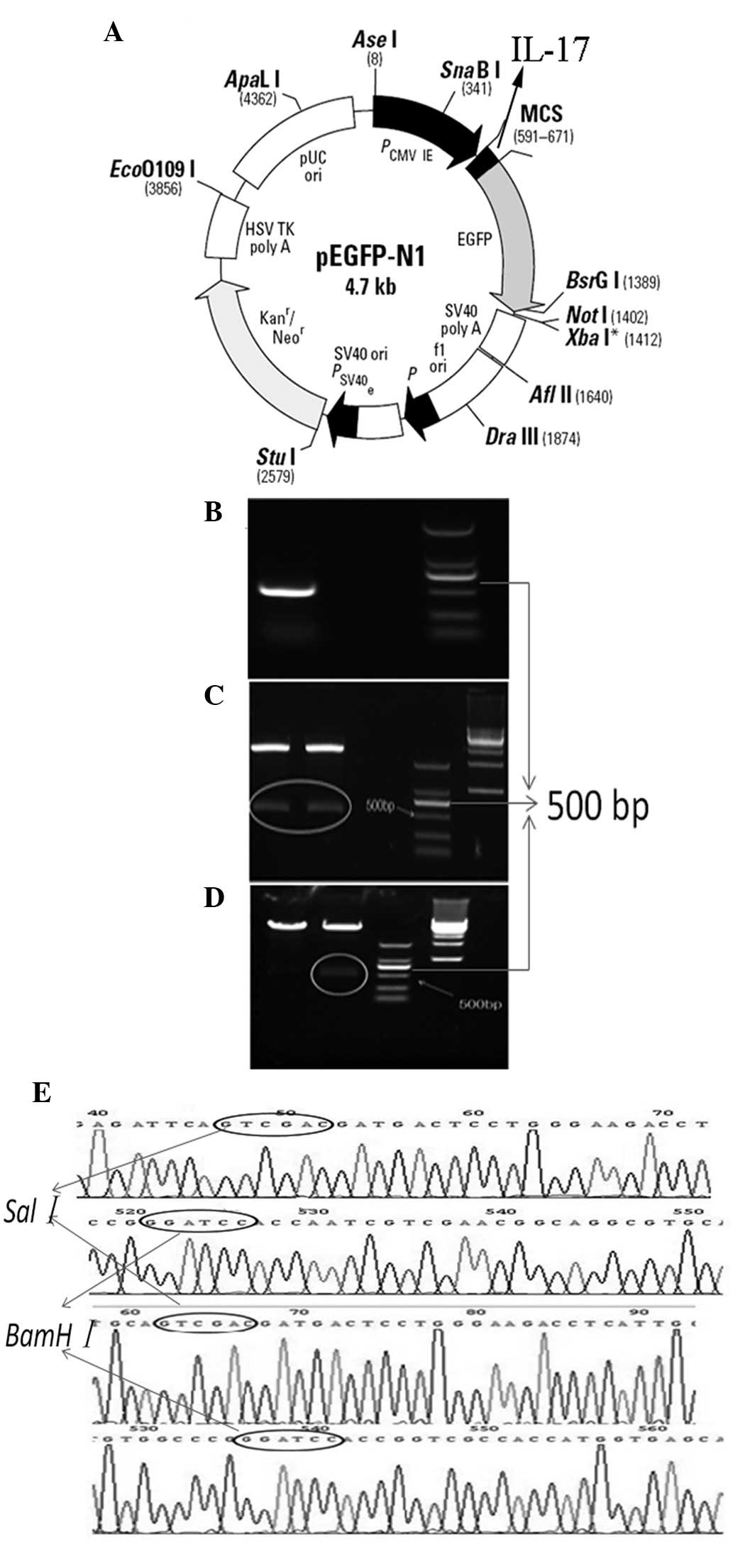
IL-17 cDNA (NCBI reference sequence, NM_002190.2)
was synthesized using RNA that was extracted from PBMCs of an ITP
patient. Sequencing and restriction enzyme digestion were used to
confirm the successful packages of IL-17 cDNA into the pMD19-T and
pEGFP-N1 vectors. The results revealed that the IL-17 cDNA was
inserted into the multiple cloning site (MCS) of the pEGFP-N1
vector. The target gene fragment (IL-17 cDNA) was 468bp and the
sequence fully corresponded to that in Genbank (Fig. 1).
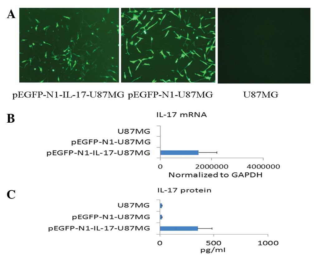
The cells that were transfected with pEGFP-N1-IL-17
and pEGFP-N1 were selected using 200 μg/ml G418. Following 10 days,
the cells were diluted to 1 cell/10 μl. Subsequent to forming an
expansive culture, the cells were identified using a fluorescence
microscope and IL-17 mRNA and protein expression was detected by
qPCR and an enzyme-linked immunosorbent assay (ELISA). The results
revealed that the U87MG cells that were transfected with
pEGFP-N1-IL-17 and pEGFP-N1 exhibited fluorescence, indicating that
the vector was expressed successfully in those cells. Notably, the
pEGFP-N1-IL-17-U87MG cells demonstrated a significantly higher
level of IL-17 mRNA and protein compared with the pEGFP-N1-U87MG
and U87MG cells (P<0.001; Fig.
2).
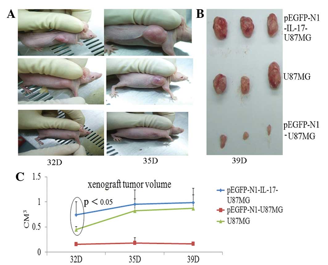
IL-17 overexpression promotes U87MG
tumorigenesis in nude mice with elevated CD31 in tumor tissues
pEGFP-N1-IL-17-U87MG, pEGFP-N1-U87MG and U87MG cells
(5×10⁵) were subcutaneously inoculated into the right
flanks of the nude mice. At 7 days post-inoculation, neoplasms
became visible and the tumor sizes were monitored every 3 days. At
32 days post-inoculation, the sizes of the neoplasms in the
pEGFP-N1-IL-17-U87MG group were larger than those of the
pEGFP-N1-U87MG (P<0.05) and U87MG (P<0.05) groups. At 35 and
39 days, the tumor volume of the former group remained larger than
the latter two groups, but had no statistical significance with the
U87MG group, indicating that IL-17 may have accelerated tumor
growth at an early stage (Fig.
3).
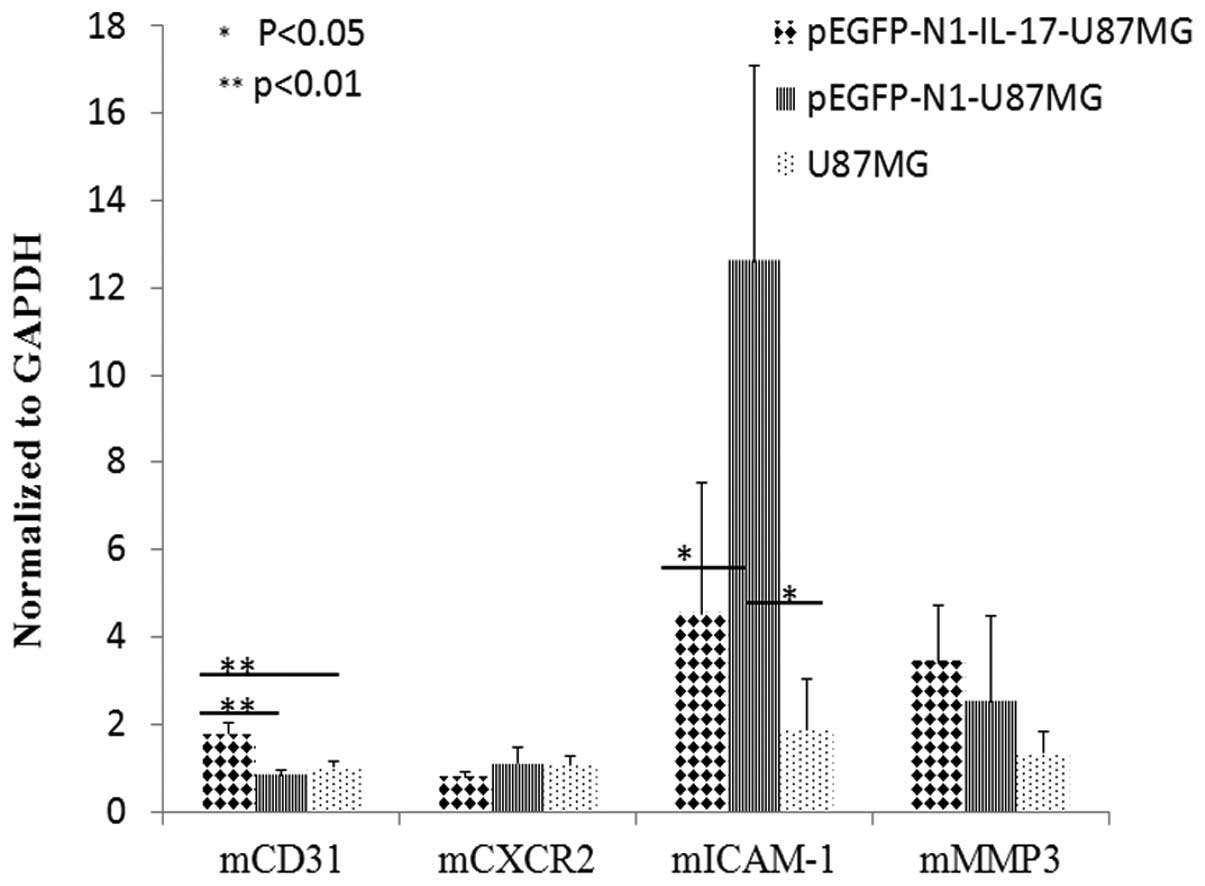
To explore the possible mechanism underlying the
differences in tumor growth among the three groups, the mRNA levels
of mCXCR2, mICAM-1, mMMP3 and mCD31 were detected in the tumor
tissues. The results revealed a higher mCD31 mRNA level in the
pEGFP-N1-IL-17-U87MG group (P<0.01) compared with the other two
groups, while mICAM-1 mRNA was higher in the pEGFP-N1-U87MG group
(P<0.05) compared with the other two groups. The levels of
mCXCR2 and mMMP3 mRNA were not significantly different among the
three groups (Fig. 4).
IL-17 transfection alters the mRNA levels
of a panel of immune/inflammation-related molecules in U87MG
To further understand the role of IL-17 in the
behavior of U87MG cells, qPCR was used to detect the mRNAs for a
panel of proteins that are associated with immune and inflammation
responses, including intercellular adhesion, the intercellular
matrix and chemokines. The expression of a series of molecules was
altered in the pEGFP-N1-IL-17-U87MG, pEGFP-N1- U87MG and U87MG
cells (Fig. 5).
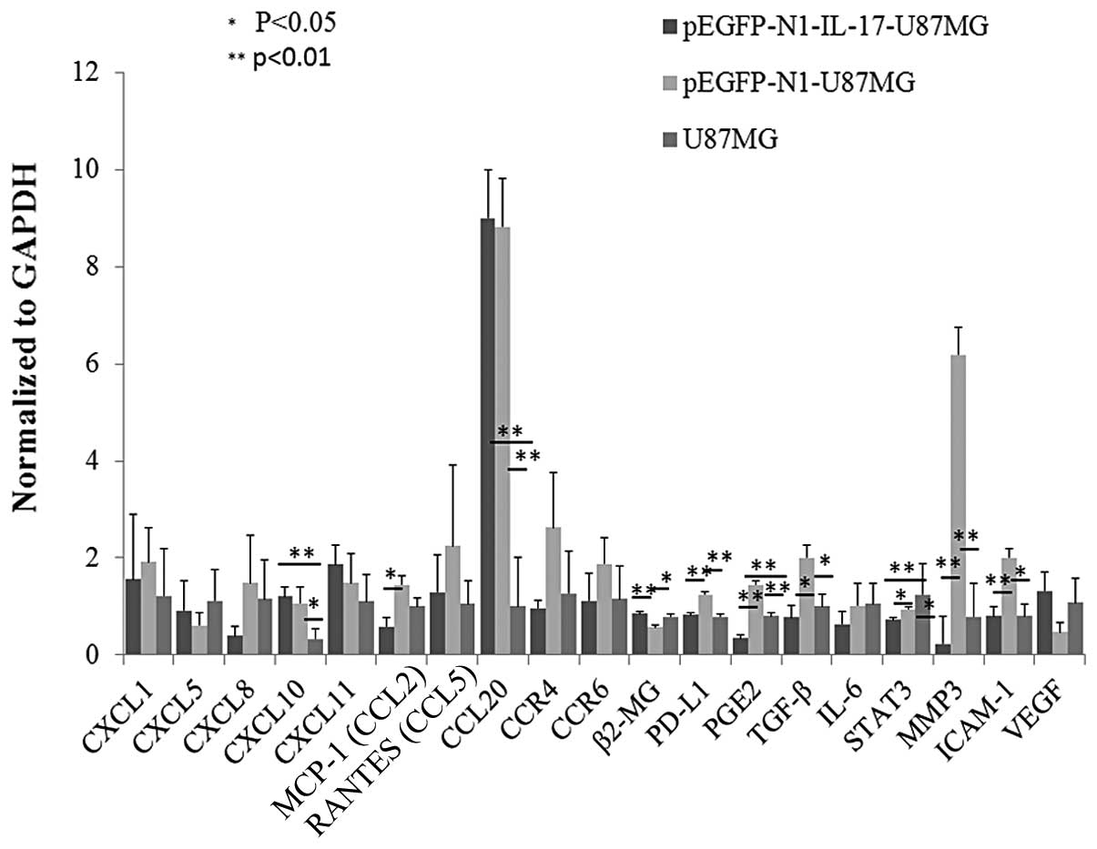 | Figure 5Variation of a panel of immune system-
and inflammation-related molecules between pEGFP-N1-IL-17-U87MG,
pEGFP-N1-U87MG and U87MG cells. The pEGPF-N1-IL-17-U87MG cells
exhibited higher CXCR10 and CCL20 mRNA levels than the U87MG cells
(P<0.01) and higher β-2MG mRNA levels than the pEGFP-N1-U87MG
cells (P<0.01). The pEGPF-N1-U87MG cells expressed higher
CXCL10, TGF-β, ICAM-1 (P<0.05), CCL20, PD-L1, PGE2 and MMP3
(P<0.01) mRNA levels than the U87MG cells and higher MCP-1,
TGF-β, STAT3 (P<0.05), PD-L1, PGE2, MMP3 and ICAM-1 levels
(P<0.01) than the pEGPF-N1-IL-17-U87MG cells. The U87MG cells
expressed higher β-2MG mRNA levels than the pEGPF-N1-U87MG cells
(P<0.05) and higher STAT3 levels than the pEGPF-N1-IL-17-U87MG
cells (P<0.01). Each group consisted of 6 independent samples,
each of which had been tested in triplicate. TGF-β, transforming
growth factor-β; ICAM-1, intercellular adhesion molecule-1; MMP3,
matrix metalloproteinase 3; MCP-1, monocyte chemoattractant
protein-1; VEGF, vascular endothelial growth factor; RANTES,
regulated on activation, normal T cell expressed and secreted
(CCL5); PGE2, prostaglandin E2. |
Discussion
IL-17, as the main regulatory element of the
emerging Th17 subset, has gained considerable interest. Our recent
study identified a higher level of IL-17 in glioma tissue (21). The present study identified that the
overexpression of IL-17 may accelerate the early-stage growth of
U87MG glioma cells in vivo. The expression of Il-17 also
altered the mRNA profile of immune/inflammation-related proteins
when transfected into a cell culture in vitro.
In the present study, human IL-17 cDNA was inserted
into the pEGFP-N1 plasmid and transfected into the glioma U87MG
cell line. The success of the procedure was confirmed using gene
sequencing, GFP detection and IL-17 mRNA and protein determination.
The U87MG, pEGFP-N1-U87MG and pEGFP-N1-IL-17-U87MG cells were
inoculated into nude mice. The pEGFP-N1-IL-17-U87MG group
demonstrated accelerated tumorigenesis compared with the other two
groups when measured at 32 days post-inoculation (P<0.05). On
days 35 and 39 post-inoculation, the implanted tumors of the
pEGFP-N1-IL-17-U87MG group were larger than those of the other two
groups. However, the difference was not statistically significant.
This result indicated that IL-17 was able to accelerate glioma
growth, particularly in the early stage of tumorigenesis.
To identify the possible mechanism behind the
accelerated tumor growth caused by IL-17 overexpression, mCXCR2,
mMMP3, mICAM-1 and mCD31 mRNA expression in the xenografted tumor
tissues were analyzed using qPCR. mCD31 expression in the tumor
tissues of the pEGFP-N1-IL-17-U87MG group was higher than in the
other groups (P<0.01). This was consistent with the results of a
study by Numasaki et al(24). The effect of early-stage
tumorigenesis acceleration caused by the overexpression of IL-17
may be associated with the promotion of angiogenesis, which is
consistent with the notion that the formation of new blood vessels
is vital for the initial growth stage for solid tumors. However, in
contrast with the results from the study by Numasaki et al,
the present data did not include an elevation in mCXCR2 in the
tumor tissues from the IL-17 overexpression group. This difference
may have been due to the different tumor types that were used in
the two studies. Alternatively, the angiogenesis-promoting effects
of IL-17 may also be active through pathways other than CXCR2, as
described by a number of studies (25–27).
The present study highlights the fact that IL-17 may be a target
for interference in tumor angiogenesis.
In addition to detecting the mRNA levels of several
molecules in the tumor tissues, the mRNA level for a panel of
molecules that are associated with the immune response and
inflammation were also analyzed in the transfected cells in order
to understand the alterations caused to the behavior of the U87MG
cells by IL-17 in vivo. IL-17 was able to increase the
levels of CXCL10, CCL20 and β2-MG. However, whether these changes
are associated with the acceleration of the early-stage growth of
the U87MG cells in vivo remains to be elucidated. Among the
molecules that were studied, β2-MG was particularly significant,
since it was the only one among the observed panel that showed an
elevation in the IL-17-transfected cells compared with those that
were mock-transfected. β2-MG was reported to be an early marker in
the plasma of hepatocellular carcinoma patients (28). However, the association between
β2-MG and glioma remains elusive and requires further
investigation. In the present study, the IL-17-transfected cells
contained elevated levels of VEGF mRNA, although they were not
increased to a statistically significant level compared with the
mock-transfected cells. This elevation of VEGF may be important,
since IL-17 tumor-promoting effects are associated with enhanced
angiogenesis, as suggested by the present study and others
(24,29,30).
Another noteworthy observation in the present study
was the effect of the vector; the pEGFP-N1 expression vector was
able to dramatically alter the behavior of the U87MG cells in
vivo and in vitro. The mock transfection group
demonstrated a strong suppression of nude mice xenograft
tumorigenesis. On days 32, 35 and 39 post-inoculation, the
pEGFP-N1-U87MG group showed a slower rate of tumorigenesis than the
other two groups (P<0.01). In addition, the tumor-inhibiting
effects were also revealed by the fact that only six out 10
pEGFP-N1-U87MG mice developed tumors. It is noteworthy that this
tumor-inhibitory effect of the vector highlights the
tumor-promoting effect of IL-17, since the mock transfection group
was a more valued control for the IL-17-transfected group compared
with the untransfected group. The in vitro culture also
demonstrated the effects of the vector, as there were relatively
more molecules showing the alterations in the mRNA levels compared
with the IL-17 transfection group. The underlying mechanism for the
effect that was observed remains unknown and thus requires further
investigation.
In the present study, human IL-17 cDNA was
successfully constructed in the glioma U87MG cell line using
eukaryotic pEGFP-N1 expression vectors. The pEGFP-N1-IL-17-U87MG
cells demonstrated accelerated growth in the early stage subsequent
to being inoculated into nude mice, which was accompanied with a
higher CD31 expression, indicating that an angiogenesis-promoting
action may be involved. IL-17 transfection may also alter the mRNA
levels of certain molecules that are associated with the immune
response and inflammation. The vector exhibited an effect in the
mock transfection group, with suppressed tumor growth and altered
mRNA levels of multiple molecules. The mechanism for this
phenomenon requires further investigation. The present study
revealed that IL-17 was able to enhance glioma growth and change
the expression of certain genes.
Acknowledgements
The authors would like to thank the Central Lab of
Huashan Hospital, Fudan University for the instrumental and
technical support. This study was supported by grants from the Open
Research Fund Program of Shanghai Key Laboratory of Female
Reproductive Endocrine-Related Diseases (no. 10dz2220300), the
National Distinguish Young Scientists Foundation Grant (no.
81025013) and the Science and Technology Development and Innovation
Fund (Pudong New Area, Shanghai, China; no. PKJ2012-Y28).
References
|
1
|
Wen PY and Kesari S: Malignant gliomas.
Curr Neurol Neurosci Rep. 4:218–227. 2004. View Article : Google Scholar
|
|
2
|
Couldwell WT, Dore-Duffy P, Apuzzo ML and
Antel JP: Malignant glioma modulation of immune function: relative
contribution of different soluble factors. J Neuroimmunol.
33:89–96. 1991. View Article : Google Scholar : PubMed/NCBI
|
|
3
|
Nitta T, Hishii M, Sato K and Okumura K:
Selective expression of interleukin-10 gene within glioblastoma
multiforme. Brain Res. 649:122–128. 1994. View Article : Google Scholar : PubMed/NCBI
|
|
4
|
Roussel E, Gingras MC, Grimm EA, Bruner JM
and Moser RP: Predominance of a type 2 intratumoural immune
response in fresh tumour-infiltrating lymphocytes from human
gliomas. Clin Exp Immunol. 105:344–352. 1996. View Article : Google Scholar : PubMed/NCBI
|
|
5
|
Bogdan C and Nathan C: Modulation of
macrophage function by transforming growth factor beta,
interleukin-4, and interleukin-10. Ann NY Acad Sci. 685:713–739.
1993. View Article : Google Scholar : PubMed/NCBI
|
|
6
|
Roszman T, Elliott L and Brooks W:
Modulation of T-cell function by gliomas. Immunol Today.
12:370–374. 1991. View Article : Google Scholar : PubMed/NCBI
|
|
7
|
Andaloussi AE, Han Y and Lesniak MS:
Progression of intracranial glioma disrupts thymic homeostasis and
induces T-cell apoptosis in vivo. Cancer Immunol Immunother.
57:1807–1816. 2008. View Article : Google Scholar : PubMed/NCBI
|
|
8
|
Heimberger AB, Abou-Ghazal M, Reina-Ortiz
C, Yang DS, Sun W, Qiao W, et al: Incidence and prognostic impact
of FoxP3+ regulatory T cells in human gliomas. Clin Cancer Res.
14:5166–5172. 2008.
|
|
9
|
Brooks WH, Latta RB, Mahaley MS, Roszman
TL, Dudka L and Skaggs C: Immunobiology of primary intracranial
tumors. Part 5: Correlation of a lymphocyte index and clinical
status. J Neurosurg. 54:331–337. 1981. View Article : Google Scholar : PubMed/NCBI
|
|
10
|
Brooks WH, Netsky MG, Normansell DE and
Horwitz DA: Depressed cell-mediated immunity in patients with
primary intracranial tumors. Characterization of a humoral
immunosuppressive factor. J Exp Med. 136:1631–1647. 1972.
View Article : Google Scholar : PubMed/NCBI
|
|
11
|
Brooks WH, Roszman TL and Rogers AS:
Impairment of rosette-forming T lymphocytes in patients with
primary intracranial tumors. Cancer. 37:1869–1873. 1976. View Article : Google Scholar : PubMed/NCBI
|
|
12
|
Elliott LH, Brooks WH and Roszman TL:
Cytokinetic basis for the impaired activation of lymphocytes from
patients with primary intracranial tumors. J Immunol.
132:1208–1215. 1984.PubMed/NCBI
|
|
13
|
Roszman TL, Brooks WH, Steele C and
Elliott LH: Pokeweed mitogen-induced immunoglobulin secretion by
peripheral blood lymphocytes from patients with primary
intracranial tumors. Characterization of T helper and B cell
function. J Immunol. 134:1545–1550. 1985.
|
|
14
|
Ashkenazi E, Deutsch M, Tirosh R, Weinreb
A, Tsukerman A and Brodie C: A selective impairment of the IL-2
system in lymphocytes of patients with glioblastomas: increased
level of soluble IL-2R and reduced protein tyrosine
phosphorylation. Neuroimmunomodulation. 4:49–56. 1997.PubMed/NCBI
|
|
15
|
Watters JJ, Schartner JM and Badie B:
Microglia function in brain tumors. J Neurosci Res. 81:447–455.
2005. View Article : Google Scholar : PubMed/NCBI
|
|
16
|
Bettelli E, Korn T and Kuchroo VK: Th17:
the third member of the effector T cell trilogy. Curr Opin Immunol.
19:652–657. 2007. View Article : Google Scholar : PubMed/NCBI
|
|
17
|
Harrington LE, Hatton RD, Mangan PR,
Turner H, Murphy TL, Murphy KM and Weaver CT: Interleukin
17-producing CD4+ effector T cells develop via a lineage distinct
from the T helper type 1 and 2 lineages. Nat Immunol. 6:1123–1132.
2005.
|
|
18
|
Nam JS, Terabe M, Kang MJ, Chae H, Voong
N, Yang YA, et al: Transforming growth factor beta subverts the
immune system into directly promoting tumor growth through
interleukin-17. Cancer Res. 68:3915–3923. 2008. View Article : Google Scholar : PubMed/NCBI
|
|
19
|
Toh ML, Kawashima M, Zrioual S, Hot A and
Miossec P and Miossec P: IL-17 inhibits human Th1 differentiation
through IL-12R beta 2 downregulation. Cytokine. 48:226–230. 2009.
View Article : Google Scholar : PubMed/NCBI
|
|
20
|
Wang L, Yi T, Kortylewski M, Pardoll DM,
Zeng D and Yu H: IL-17 can promote tumor growth through an
IL-6-Stat3 signaling pathway. J Exp Med. 206:1457–1464. 2009.
View Article : Google Scholar : PubMed/NCBI
|
|
21
|
Hu J, Mao Y, Li M and Lu Y: The profile of
Th17 subset in glioma. Int Immunopharmacol. 11:1173–1179. 2011.
View Article : Google Scholar : PubMed/NCBI
|
|
22
|
Wainwright DA, Sengupta S, Han Y, Ulasov
IV and Lesniak MS: The presence of IL-17A and T helper 17 cells in
experimental mouse brain tumors and human glioma. PLoS One.
5:e153902010. View Article : Google Scholar : PubMed/NCBI
|
|
23
|
Cantini G, Pisati F, Mastropietro A,
Frattini V, Iwakura Y, Finocchiaro G and Pellegatta S: A critical
role for regulatory T cells in driving cytokine profiles of Th17
cells and their modulation of glioma microenvironment. Cancer
Immunol Immunother. 60:1739–1750. 2011. View Article : Google Scholar : PubMed/NCBI
|
|
24
|
Numasaki M, Watanabe M, Suzuki T,
Takahashi H, Nakamura A, McAllister F, et al: IL-17 enhances the
net angiogenic activity and in vivo growth of human non-small cell
lung cancer in SCID mice through promoting CXCR-2-dependent
angiogenesis. J Immunol. 175:6177–6189. 2005. View Article : Google Scholar : PubMed/NCBI
|
|
25
|
Wakita D, Sumida K, Iwakura Y, Nishikawa
H, Ohkuri T, Chamoto K, et al: Tumor-infiltrating IL-17-producing
gammadelta T cells support the progression of tumor by promoting
angiogenesis. Eur J Immunol. 40:1927–1937. 2010. View Article : Google Scholar : PubMed/NCBI
|
|
26
|
Moran EM, Connolly M, Gao W, McCormick J,
Fearon U and Veale DJ: Interleukin-17A induction of angiogenesis,
cell migration, and cytoskeletal rearrangement. Arthritis Rheum.
63:3263–3273. 2011. View Article : Google Scholar : PubMed/NCBI
|
|
27
|
Liu J, Duan Y, Cheng X, Chen X, Xie W,
Long H, et al: IL-17 is associated with poor prognosis and promotes
angiogenesis via stimulating VEGF production of cancer cells in
colorectal carcinoma. Biochem Biophys Res Commun. 407:348–354.
2011. View Article : Google Scholar : PubMed/NCBI
|
|
28
|
Saito Y, Oba N, Nishinakagawa S, Mizuguchi
Y, Kojima T, Nomura K and Nakatsura T: Identification of
beta2-microgloblin as a candidate for early diagnosis of
imaging-invisible hepatocellular carcinoma in patient with liver
cirrhosis. Oncol Rep. 23:1325–1330. 2010.PubMed/NCBI
|
|
29
|
Rapisarda A and Melillo G: Role of the
VEGF/VEGFR axis in cancer biology and therapy. Adv Cancer Res.
114:237–267. 2012. View Article : Google Scholar : PubMed/NCBI
|
|
30
|
Murugaiyan G and Saha B: Protumor vs
antitumor functions of IL-17. J Immunol. 183:4169–4175. 2009.
View Article : Google Scholar : PubMed/NCBI
|