Introduction
Lipomatous hemangiopericytoma (LHPC) is an uncommon,
slow-growing, almost non-recurring, non-metastasizing mesenchymal
neoplasm composed of mature adipocytes and HPC-like areas (1). The majority of these tumors are
located in the deep soft tissue (2). Other commonly affected sites include
the thigh, retroperitoneum and the orbit (3). Grossly, LHPC generally presents as a
well-circumscribed, often non-encapsulated, medium-sized mass with
alternating areas of white/yellow tumor tissue in the cut surface
(2). Microscopically, these tumors
characteristically show a varying combination of patternless
cellular areas composed of round to spindle-shaped cells,
hemangiopericytoma-like vascular areas made up of small- to
medium-sized thin-walled branched vessles, and lipomatous areas
made up of mature adipocytes (2–6).
Long-term follow-up is recommended (3,5). To
date, 51 cases of LHPC have been documented in the published
literature (2–6). However, there have been no reports of
parotid gland LHPC. The current study presents the
clinicopathological features of the first case of parotid gland
LHPC in a 33-year-old male, along with a review of the literature.
Written informed consent was obtained from the patient.
Case report
A 33-year-old male presented with a four-year
history of a progressively growing, painless and fixed mass in the
left parotid gland region. The patient did not have any associated
headaches, facial weakness or numbness, or impairments in speech or
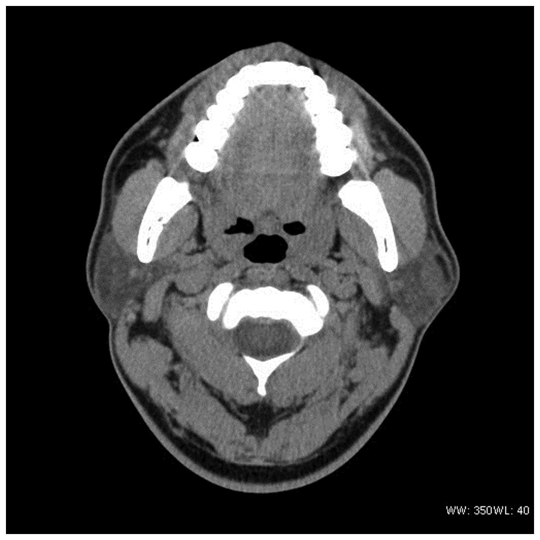
swallowing. A head and neck CT scan showed a 19.0×13.2-mm oval
heterogeneous mass within the left parotid gland (Fig. 1). A radical parotidectomy was
performed and an irregular, well-circumscribed mass compromising
the entire gland was observed during the surgery. The well-defined
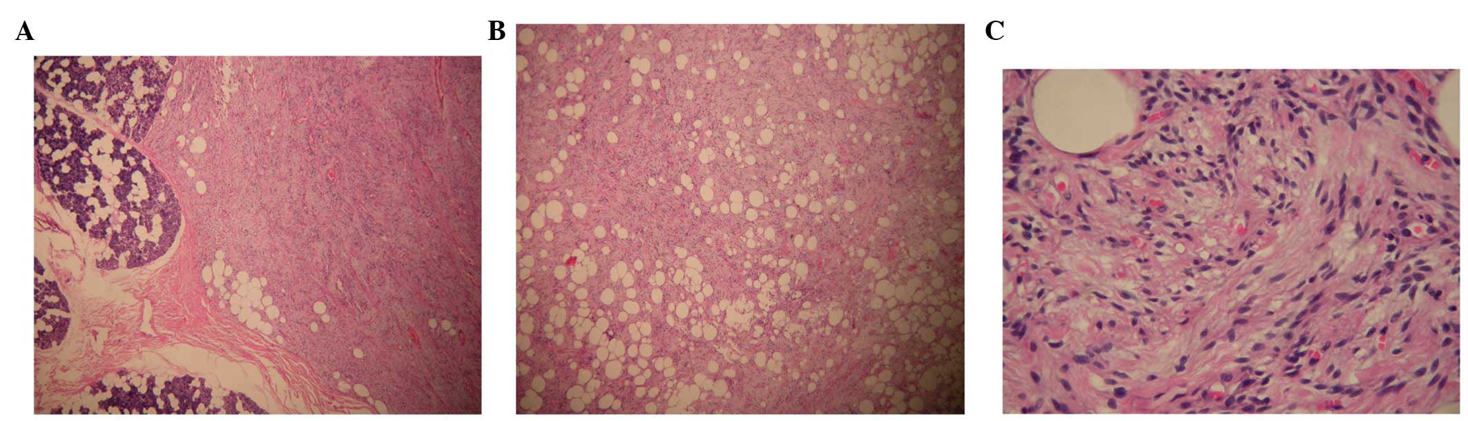
mass had a solid and tan/white cut surface. The histopathological
study identified a well-circumscribed lesion composed of cellular
nodules with the classic appearance of an HPC area admixed with
clusters and lobules of mature adipocytes. The ill-defined tumor
cells had a weakly eosinophilic cytoplasm and spindle-shaped
nuclei, with occasional small nucleoli. Nuclei atypia and mitoses
were absent, and no cellular atypia, necrosis or vascular invasion
was observed (Fig. 2).
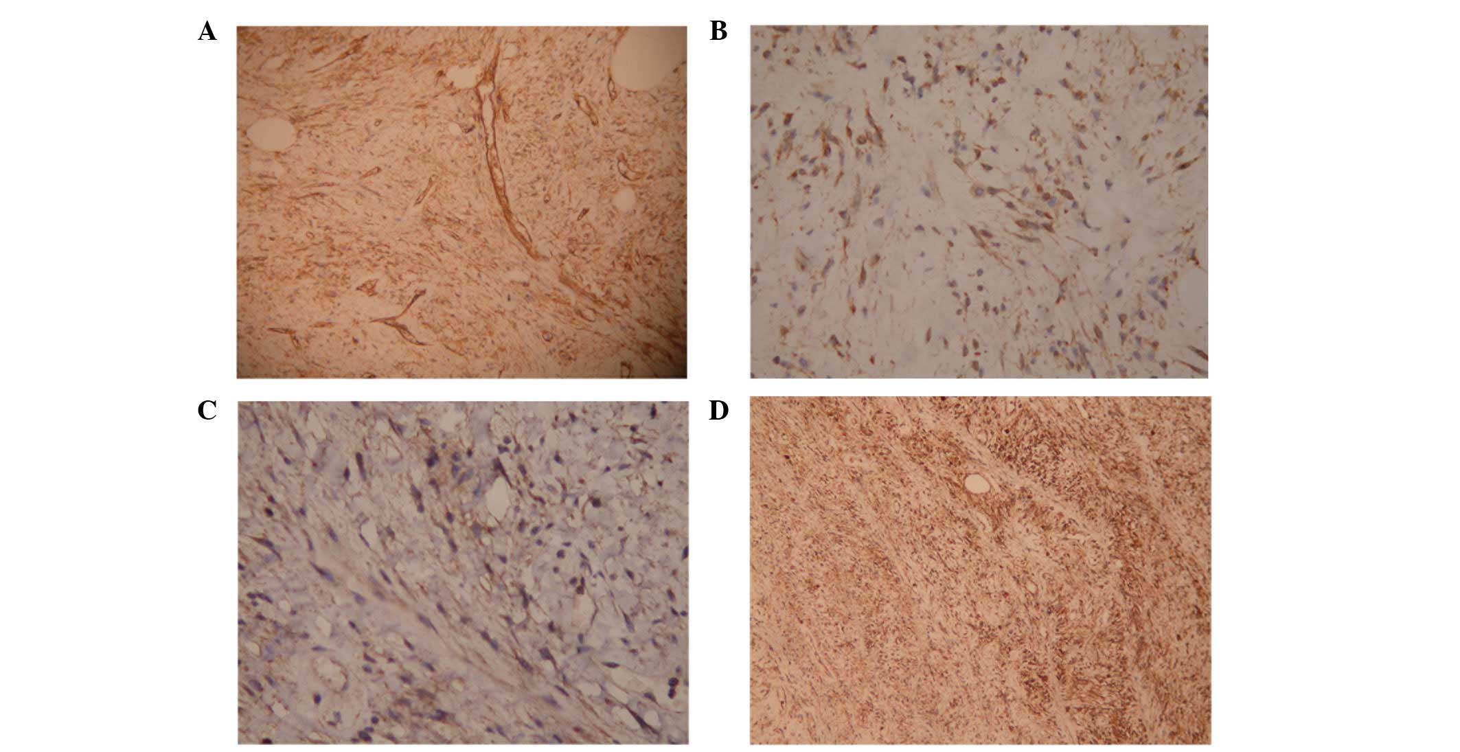
Immunohistochemistry showed that the tumor cells were diffusely
positive for CD34, CD99, Bcl-2 and vimentin (Fig. 3). Stains for cytokeratin, EMA,
HMB45, S100, CD117 and p63 were negative. The patient was managed
with surgery without any further treatment and had an uneventful
clinical course post-operatively after a follow-up of 12
months.
Discussion
LHPC was first proposed as a unique HPC variant in
1995 by Nielsen et al(4) for
tumors composed of an admixture of hemangiopericytomatous areas and
mature adipose tissue. HPC is a controversial entity. In addition
to the lack of a definite immunophenotype, the relative
non-specificity of the characteristic branching capillary pattern
and cytological features of the constituent cells has led to
uncertainty and a lack of consensus concerning this subgroup of
tumors. Due to the major histological overlap between solitary
fibrous tumors (SFTs) and HPC, and the lack of evident criteria to
judge whether a lesion should be called an SFT or HPC, pathologists
have gradually abandoned the term HPC in favor of the term SFT, so
that the majority of lesions that were called HPCs 15 years ago now
tend to be called SFTs (7).
Guillou et al(2) reviewed 100 extrapleural SFTs and
encountered 13 neoplasms that characteristically contained islands
of mature fatty tissue, and suggested that LHPC does not correspond
to a well-defined entity, but rather is representative of a
fat-containing variant of SFT. According to the 2002 edition of the
World Health Organization (WHO) Classification of Tumors of Soft
Tissue and Bone (1), there is an
overlap between SFT and both LHPC and giant cell angiofibroma.
However, the term LHPC is reserved for a variant of HPC. To date,
neither of these variant growth patterns has yet been recognized in
the parotid gland. All of the SFTs reported in the parotid gland
have been of the ‘fibrous variant’ (8).
To date, a total of 52 pathologically confirmed
cases of LHPC have been reported, including that of the present
patient (2–6). These patients consisted of 31 males
and 21 females, ranging in age from 21 to 79 years, with average
and median ages of 50.96 and 51 years, respectively. LHPC generally
presents as a well-circumscribed, often non-encapsulated,
medium-sized mass with alternating areas of whitish and yellowish
tumor tissue in the cut surface (2). The histological appearance of LHPC
consists of a varying combination of patternless cellular areas
composed of round to spindle-shaped cells, an HPC-like vasculature
made of small- to medium-sized thin-walled branched vessels and
lipomatous areas consisting of mature adipocytes.
Immunohistochemistry has shown that non-adipocytic tumor cells are
consistently positive for CD99 and, less frequently, for CD34 (75%)
and Bcl-2 (60%) (1).
Ultrastructural features are non-specific in HPC and the majority
of the lesions reported as HPC have only shown undifferentiated
spindle cell or fibroblastic features (2–6).
Convincing evidence of true pericytic differentiation has not been
observed (1).
Considering the location and histological appearance
of HPC, the main differential diagnoses considered for this type of
tumor should include pleomorphic adenoma, myoepithelioma,
schwannoma, neurofibroma and angiomyolipoma. Glial fibrillary
acidic protein (GFAP) is often positively expressed in pleomorphic
adenoma, and would be of additional assistance in excluding this
diagnosis. S100, GFAP and p63 are positively expressed in
myoepithelioma. Schwannoma and neurofibroma are strongly reactive
with S100 protein, while lacking CD34. Angiomyolipoma reacts with
HMB45, Melan-A, tyrosinase and S100 protein.
The most common treatment for LHPC is complete local
surgical excision. Although no LHPC patients developed recurrence
during the follow-up interval and even though all patients were
without disease, certain authors have recommended the use of a
long-term follow-up (3,5). In conclusion, the present study
describes the first case of a parotid gland LHPC. The
clinicopathological features of this case were similar to those of
normal LHPC. More cases of LHPC should be studied to provide
further understanding of the behavior of this rare tumor.
References
|
1
|
Guillou L, Fletcher JA, Fletcher CDM and
Mandahl N: Extrapleural solitary fibrous tumour and
hemangiopericytoma. Pathology and Genetics of Tumours of Soft
Tissue and Bone. Fletcher CDM, Unni KK and Mertens F: IARC Press;
Lyon: pp. 86–90. 2002
|
|
2
|
Guillou L, Gebhard S and Coindre JM:
Lipomatous hemangiopericytoma: a fat-containing variant of solitary
fibrous tumor? Clinicopathologic, immunohistochemical, and
ultrastructural analysis of a series in favor of a unifying
concept. Hum Pathol. 31:1108–1115. 2000. View Article : Google Scholar
|
|
3
|
Jing HB, Meng QD and Tai YH: Lipomatous
hemangiopericytoma of the stomach: a case report and a review of
literature. World J Gastroenterol. 17:4835–4838. 2011. View Article : Google Scholar : PubMed/NCBI
|
|
4
|
Nielsen GP, Dickersin GR, Provenzal JM and
Rosenberg AE: Lipomatous hemangiopericytoma. A histologic,
ultrastructural and immunohistochemical study of a unique variant
of hemangiopericytoma. Am J Surg Pathol. 19:748–756. 1995.
View Article : Google Scholar : PubMed/NCBI
|
|
5
|
Davies PE, Davis GJ, Dodd T and Selva D:
Orbital lipomatous haemangiopericytoma: an unusual variant. Clin
Experiment Ophthalmol. 30:281–283. 2002. View Article : Google Scholar : PubMed/NCBI
|
|
6
|
Barazani Y and Tareen B: Rare case of
paratesticular solitary fibrous tumour (lipomatous
hemangiopericytoma). Can Urol Assoc J. 6:E131–E133. 2012.
View Article : Google Scholar : PubMed/NCBI
|
|
7
|
Gengler C and Guillou L: Solitary fibrous
tumour and haemangiopericytoma: evolution of a concept.
Histopathology. 48:63–74. 2006. View Article : Google Scholar : PubMed/NCBI
|
|
8
|
Bauer JL, Miklos AZ and Thompson LD:
Parotid gland solitary fibrous tumor: a case report and
clinicopathologic review of 22 cases from the literature. Head Neck
Pathol. 6:21–31. 2012. View Article : Google Scholar : PubMed/NCBI
|