Introduction
Extracellular signal-regulated kinase (Erk), a
member of the mitogen-activated protein kinase (MAPK) family,
regulates cellular responses to signals that lead to cell growth,
proliferation and differentiation, as well as DNA damage (1,2).
Dysfunction of phosphatase and tensin homologue (PTEN) is one of
the main causes of cancer progression and metastasis (3,4).
Deletion of PTEN has been demonstrated to result in elevation of
phosphorylated protein kinase B (pAKT) levels, which in turn
resulted in suppression of Erk signaling, in prostate cancer
(5). Therefore, Ras/Erk signaling
may be an effective target for the treatment of advanced metastatic
prostate cancer cells. Erk signaling is associated with cancer cell
proliferation and DNA damage; however, the detailed mechanism of
Ras/Erk signaling in cancer cells remains unclear (6).
Actin cytoskeleton, the most abundant and ubiquitous
protein, is important in various signaling pathways involved in
cellular migration, adhesion, metastasis, cytokinesis, chromatin
remodeling and transcriptional processes. Malignant cancer cells
have distinct features, including modified cellular morphology and
migration machinery, and disrupted cell division. As the changes
are associated with actin dynamics, understanding the mechanism of
actin disruption in cancer cells is important for the development
of cancer chemotherapy (7). Small
molecules targeting actin microfilaments have been tested for
modulation of the rapid growth and proliferation of cancer cells
(8). The small molecule latrunculin
B (LB) inhibits actin polymerization and nucleotide exchange in
G-actin by binding to the monomer in a one to one complex, without
affecting the organization of the microtubular system (9,10).
PTEN interacts with actin cytoskeleton remodeling at
focal adhesions of the cell membrane and inhibits
phosphatidylinositide 3-kinase (PI3K) (11,12).
In addition, it regulates the repair of impaired DNA double-strand
breaks (DSBs) and nucleotide excision repair (13,14).
Thus, it was hypothesized that disruption of actin may be a
potential therapy for the treatment of highly metastatic prostate
cancer. The current study investigated the effect of disruption of
actin microfilaments on DSBs, the cell cycle and apoptotic
signaling with LB treatment, to elucidate the mechanism of actin
disruption in PTEN-null PC3M prostate cancer cells.
Materials and methods
Chemicals and antibodies
The metastatic human prostate cancer PC3M cell line
was obtained from the American Type Culture Collection (Manassas,
VA, USA). The cells were incubated in Dulbecco’s modified Eagle’s
medium supplemented with 1% antibiotic-antimycotic solution and 10%
fetal bovine serum (Gibco-BRL, Carlsbad, CA, USA) at 37°C in a
humidified atmosphere of 5% CO2. To identify the cell
signals activated upon treatment with/without LB, LY294002, U0126
or N-acetyl L cysteine (NAC), experiments were performed in the
absence of serum and exogenous growth factors. LB was purchased
from Calbiochem-Novabiochem Corp. (San Diego, CA, USA); while
LY294002, U0126, NAC, RNase A and propodium iodide (PI) were
purchased from Sigma Aldrich (St Louis, MO, USA).
Rhodamine-phalloidin and Molecular Probes® were obtained
from Invitrogen Life Technologies (Carlsbad, CA, USA), and Complete
Protease Inhibitor Cocktail tablets were purchased from Roche
(Mannheim, Germany). Protran™ nitrocellulose membranes
were purchased from Whatman International Ltd. (Maidstone, UK) and
a BCA Protein Assay kit was obtained from Pierce (Rockford, IL,
USA). Enhanced chemiluminescence (ECL) Western Blotting Detection
Reagents, and sheep anti-mouse monoclonal IgG (HRP-linked) and
donkey anti-rabbit polyclonal IgG (HRP-linked) antibodies were
purchased from GE Healthcare (Piscataway, NJ, USA); while
anti-rabbit poly (ADP-ribose) polymerase (PARP), pAkt and
phosphorylated Erk (pERK) polyclonal antibodies were purchased from
Cell Signaling Technology, Inc. (Danvers, MA, USA). Anti-Bax and
anti-Bcl-2 antibodies were purchased from Santa Cruz Biotechnology,
Inc. (Santa Cruz, CA, USA) and anti-phospho-histone H2A X antibody
(Ser139; clone, JBW301) was obtained from Upstate Biotechnology,
Inc. (Lake Placid, NY, USA).
Viability assay
Cells (2×10⁴/well) were incubated in
96-well flat bottom plates in 100 μl culture medium (Dulbecco’s
modified Eagle’s medium supplemented with 1% antibiotic-antimycotic
solution and 10% fetal bovine serum) and stabilized in a humidified
atmosphere of 5% CO2 at 37°C for >12 h. The cells
were exposed to LB (50, 75, 100, 150, 200 and 300 nM) for 24 h. The
MTT assay (Abnova, Taipei City, Taiwan) was performed according to
the manufacturer’s instructions. Briefly, 20 μl 5 μg/ml MTT was
added to each well and incubated for 4 h at 37°C. Then, the medium
was carefully removed. DMSO was added to the reactants and mixed
gently on an orbital shaker for 1 h at room temperature. The
absorbance at 570 nm was determined using a Victor3™
multilabel plate reader (model 1420; Perkin-Elmer, Waltham, MA,
USA). Triplicate wells were assayed for each condition.
Immunocytochemistry
Immunocytochemistry was performed according to the
manufacturer’s instructions (Invitrogen Life Technologies) with
certain modifications. The cells growing on the cover slip were
washed with cold phosphate-buffered saline (PBS). For
permeabilization, cells were incubated with 150 μl permeabilizing
buffer (PB; 0.15 M NaCl, 10 mM Tris, 1 mM MgCl2, 0.2 mM
dithiothreitol, 0.5 mM CaCl2 and 25% glycerol; pH 8.0)
for 4–5 min, 150 μl PB with 0.5% Triton X-100 for 3.5 min, 150 μl
PB for 30 min at 37°C and then washed with PBS with 0.2% Tween-20
(PBS-T). Cells were fixed in 4% paraformaldehyde (pH 7.2) for 15
min and washed twice with PBS-T. Before cell staining, cells were
blocked in PBS-T with 10% FBS for 20 min. Rhodamine-phalloidin
(1:200) and DAPI (0.5 mg/ml) were applied for 60 min in the dark
for staining of F-actin and DNA, respectively. Following washing
with PBS-T, phase contrast and fluorescent images of the cells were
observed using a confocal microscope (Olympus IX81; Olympus, Tokyo,
Japan).
Cell extraction
Harvested cells were incubated in a lysis buffer
consisting of one Complete Protease Inhibitor Cocktail tablet
(Roche) to 50 ml TNES buffer (50 mM Tris-HCl, pH 7.4; 1% nonidet
P-40; 2 mM EDTA; 100 mM NaCl) on ice for 30 min. Following
centrifugation at 13,000 × g for 10 min, the supernatant was stored
at −80°C until use. Nuclear extracts were prepared according to the
protocol of the Clontech Transfactor Extraction kit (Clontech
Laboratories Inc., Mountain View, CA, USA). Cells were centrifuged
at 450 × g for 5 min, washed in PBS, then 5X volume of ice-cold
Buffer A [50 mM Tris-HCl (pH 7.4), 1.5 mM MgCl2, 10 mm
KCl, 1 mM dithiothreitol, 1 mM phenylmethanesulfonylfluoride (PMSF)
and protease inhibitors] were added to the cell pellet. After 15
min incubation on ice, the cells were centrifuged at 420 × g at 4°C
for 5 min and resuspended in 2X volume of Buffer A. The mixture was
homogenized with ten strokes of a glass dounce homogenizer and then
centrifuged at 11,000 × g for 20 min. The resulting pellet
consisting of the isolated nuclei was further homogenized in two to
three volumes of 50 mM Tris-HCl (pH 7.4), 0.42 mM NaCl, 1.5 mM
MgCl2, 0.2 mM EDTA, 25% glycerol and 1 mM PMSF. The
nuclear suspension was rotated gently for 30 min at 4°C followed by
centrifugation at 13,000 × g for 30 min. The amount of protein was
determined using the Bradford protein assay (Bio-Rad Laboratories,
Inc., Hercules, CA, USA).
Western blot analysis
Total cellular extracts (30 μg) and nuclear extracts
(5 μg) were resolved by 8 and 12% sodium dodecyl
sulfate-polyacrylamide gel electrophoresis, respectively, and
transferred onto a nitrocellulose membrane by electroblotting. The
membranes were soaked in TBS-T buffer [20 mM Tris, pH 7.4; 137 mM
NaCl; 0.1% (v/v) Tween-20] containing 5% (w/v) non-fat milk, and
were probed with the primary antibodies diluted in TBS-T [PARP
(1:1000), cleaved PARP (1:10000), Bcl-2 (1:1000), Bax (1:500),
tubulin (1:500), pAkt (1:1000), pErk (1:1000) and γH2AX (1:8000)].
Detection was performed using the horseradish peroxidase-linked
secondary antibody, followed by ECL. Anti-tubulin antibody (Santa
Cruz Biotechnology, Inc.) was applied to confirm equal loading of
the proteins and successful transfer to the membranes.
Flow cytometry analysis of the cell
cycle
The cells were treated with 150 nM LB and/or 20 μM
U0126 in 6-well plates in a humidified atmosphere of 5%
CO2 at 37°C for 6–24 h. The cells were washed with
ice-cold PBS, fixed in 100% ethanol, and maintained at −20°C for up
to two weeks before analysis of DNA content. Cell cycle analysis
was performed using cells stained with PI solution, containing 50
g/ml PI, 0.1% nonidet P-40, 0.1% sodium citrate and 100 g/ml RNase
A, in the dark at 37°C for 30 min.
Statistical analysis
Data are presented as the mean ± standard error of
at least three independent experiments, and images shown are
representative of three to six independent experiments.
Results
LB induces cell growth inhibition in
PTEN-null PC3M cells
The cytotoxicity of an actin-disrupting agent is an
important encumbrance in the development of anticancer drugs based
on an actin target. Preliminary analysis of cell proliferation
under different concentrations of LB for 24 h indicated that an
IC50 value of LB was 417 nM in the PC3M cells (data not
shown). To determine an appropriate concentration of LB for the
cancer cells, the MTT assay was performed (Fig. 1). The viability of the cells
decreased in a dose-dependent manner upon treatment with various
concentrations of LB (50–300 nM) for 24 h. The cell viability
particularly decreased (to below ~65%), compared with that of the
control, with LB treatment at concentrations higher than 150
nM.
LB destabilizes actin microfilaments and
induces apoptosis via enhancement of the expression of Bax in
PTEN-null PC3M cells
To study the molecular mechanism of actin
disruption-mediated apoptosis in prostate cancer cells, PTEN-null
PC3M cells were treated with 150 nM LB (Fig. 1). Morphological analysis was
performed on the PC3M cells incubated with 150 nM LB for 24 h using
phase contrast microscopy (Fig.
2A). The cells became rounded in shape with characteristics
that included cytoplasmic shrinkage and marked convolution of the
cellular surface whilst remaining attached to the plate. A
well-developed actin cytoskeleton with numerous subcortical actin
filaments and stress fibers was observed in the control PC3M cells;
however, F-actin and irregular aggregates of phalloidin-reactive
materials were observed in cells treated with LB (Fig. 2A). Staining of the PC3M cells with
DAPI and rhodamine-phalloidin, followed by confocal microscopy
analysis, was also performed for evaluation of apoptosis. Nuclear
condensation and apoptotic bodies, hallmarks of apoptosis, were
observed in cells treated with LB (Fig.
2A). Consequently, the results indicated that a disruption of
the actin microfilament and induction of apoptosis occurred in the
cells upon treatment with LB.
 | Figure 2LB-mediated actin disruption induces
apoptosis via enhancement of Bax expression in PTEN-null PC3M
prostate cancer cell lines. (A) Disruption of actin microfilament
and induction of apoptosis. The morphology, immunostained F-actin
(rhodamine-phalloidine) and DNA (DAPI) of PC3M cells, with or
without treatment (150 nM LB for 24 h), were examined by phase
contrast microscopy and confocal microscopy. (B and C) Cleaved
PARP, as well as Bcl-2 and Bax proteins, were expressed following
LB treatment. PC3M cells were treated with either different
concentrations (0, 50, 150 and 300 nM) of LB for 24 h (B) or with
different times (0, 6, 12 and 24 h) under fixed 150 nM LB (C).
Total cell extracts (30 μg) were used for western blotting with
anti-PARP, -Bcl-2 and -Bax antibodies. LB, latrunculin B; PTEN,
phosphatase and tensin homolog; PARP, poly (ADP-ribose)
polymerase. |
Induction of apoptosis was further confirmed by the
levels of proteins that are involved in apoptotic signal
transduction pathways, using western blot analysis. This was
performed using the cells incubated with different concentrations
(0, 50, 150 and 300 nM) of LB for 24 h (Fig. 2B). The PC3M cells without LB
treatment showed expression of an uncleaved 116-kDa PARP protein
(Fig. 2B; lane 1), whilst the cells
treated with different concentrations of LB showed a progressive
decrease in expression of the 116-kDa PARP (Fig. 2B; lanes 2 to 4). Additionally, under
the same conditions, expression of the cleaved 89-kDa PARP fragment
showed a marked increase from 0–50 nM LB and from 50–150 nM LB, but
not from 150–300 nM LB. No difference was observed in the
expression levels of Bcl-2 and Bax under the conditions (Fig. 2B). Subsequently, to identify an
optimal time condition, the experiment was carried out using
different time periods (0, 6, 12 and 24 h) with 150 nM LB (Fig. 2C). The PC3M cells without incubation
time showed expression of uncleaved 116-kDa PARP protein (Fig. 2C; lane 1), which decreased with
incubation time (Fig. 2C; lanes 2
to 4). Moreover, expression of the cleaved 89-kDa PARP fragment
increased under the same conditions. The level of Bax expression,
but not of Bcl-2, increased in the cells as the incubation time
increased.
Therefore, the results supported the theory that
actin disruption causes apoptosis through activation of both PARP
and Bax signaling in PC3M cells.
LB induces DNA DSBs via the
mitogen-activated protein kinase kinase (MEK)/Erk pathway
The level of γH2AX protein expression in the PC3M
cells was measured to elucidate the effect of actin destabilization
on genome integrity (Fig. 3).
Treatment with LB resulted in an increase in the levels of γH2AX
proteins recruited to the site of DNA damage in an LB dose- and
time-dependent manner (Fig. 3A and
B). As PC3M cells have a deleted PTEN tumor suppressor gene,
LB-induced DNA damage analysis focused on Erk, one of the effectors
responding to DNA damage. Activation of Erk signaling, which
reflects LB-induced DSBs, was measured by the expression levels of
pErk in the PC3M cells treated with 0, 50, 150 and 300 nM LB for 24
h. The level of pErk expression showed a gradual increase as the LB
concentration increased (Fig. 3C).
To assess whether inhibition of MEK/Erk signaling attenuates the
production of DSBs, the levels of γH2AX expression were analyzed in
cells treated with 150 nM LB for 12 h in the presence or absence of
20 μM U0126, an MEK/ERK inhibitor. Inhibition of ERK signaling
resulted in abrogation of γH2AX protein expression activated by LB
(Fig. 3D).
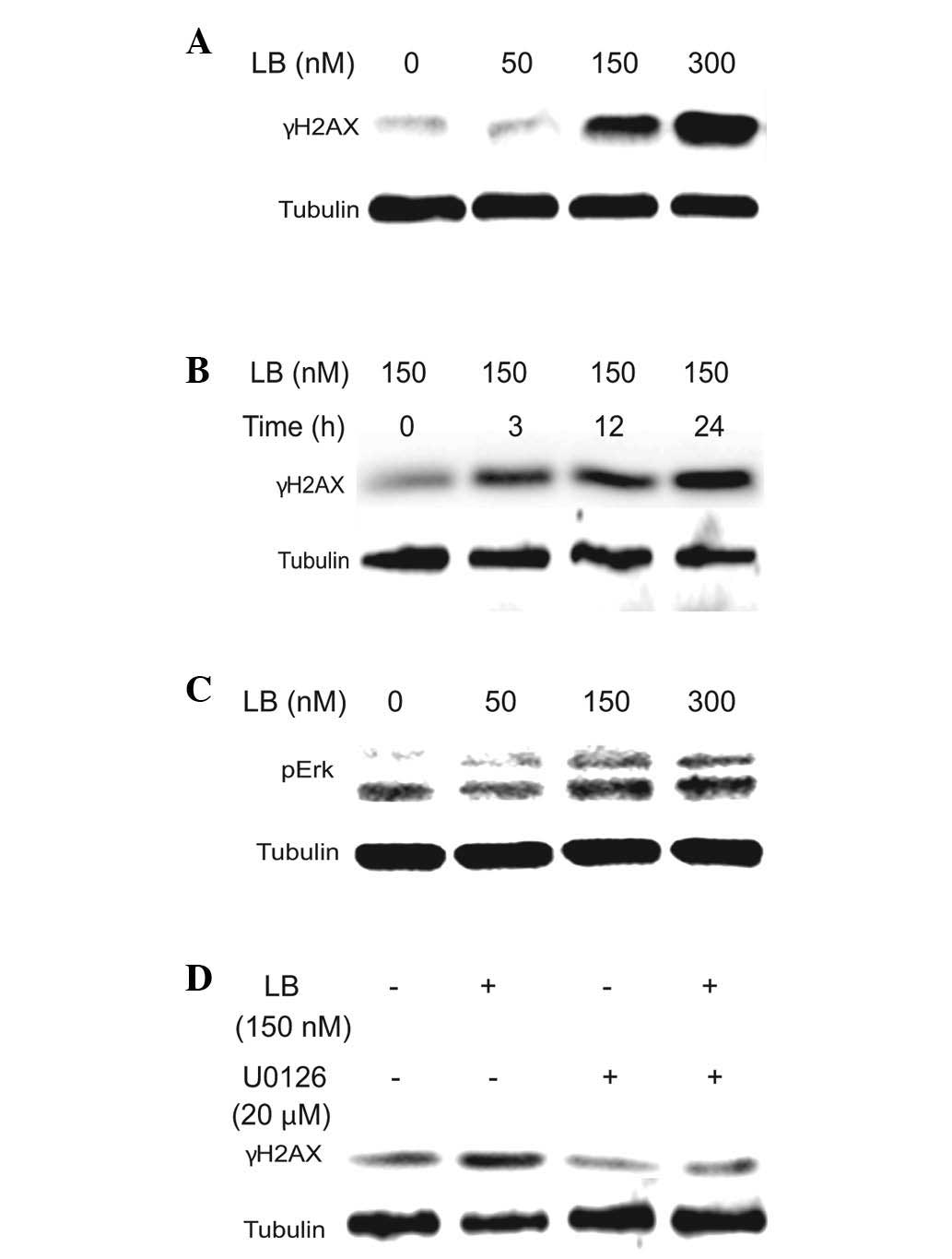 | Figure 3LB induces DSBs via the MEK/Erk
pathway in PTEN-null prostate cancer cells. (A and B) Levels of
γH2AX protein expressed following LB treatment. PC3M cells were
treated with either LB (0, 50, 150 and 300 nM) for 24 h (A) or with
150 nM LB for 0, 3, 12 and 24 h (B). (C) Levels of pErk protein
induced after LB treatment. PC3M cells were treated with LB (0, 50,
150 and 300 nM) for 24h. (D) Effect of LB treatment, with or
without U0126, on γH2AX expression. PC3M cells were treated with LB
(150 nM) in the presence or absence of U0126 (20 μM) for 24 h.
Nuclear extracts (5 μg) for γH2AX and total cell extracts (30 μg)
for pErk were used for western blotting with anti-γH2AX and
anti-pErk antibodies, respectively. LB, latrunculin B; DSBs,
double-strand breaks; MEK, mitogen-activated protein kinase kinase;
Erk, extracellular signal-regulated kinase; PTEN, phosphatase and
tensin homolog; pErk, phosphorylated Erk. |
Therefore, the results supported the theory that
actin disruption induced DSB-triggered apoptosis via the MEK/Erk
signaling pathway in PC3M cell lines.
Inhibition of MEK/Erk signaling
attenuates LB-mediated apoptosis in PC3M cell lines
To determine whether inhibition of the Ras/MEK/Erk
or the PI3K/PTEN/Akt pathway affects LB-mediated apoptosis, the
PC3M cells treated with 150 nM LB for 24 h were further incubated
with 20 μM U0126 or 10 μM LY294002, a PI3K inhibitor. Suppression
of pAkt expression indicated that LB may induce apoptosis in PC3M
cell lines (Fig. 4A; lanes 5 and
6). Inhibition of MEK/Erk by U0126, but not inhibition of PI3K
signaling by LY294002 was observed, implying that pAkt activity
confers resistance to LB-mediated apoptosis (Fig. 4A, lanes 3 and 4). To determine
whether LB-mediated apoptosis is involved in the production of
reactive oxygen species (ROS) and the activation of Erk, cells
treated with 150 nM LB for 24 h were further incubated with 10 mM
NAC (Fig. 4B and C). Typical
apoptotic morphological changes, including shrinkage of the
cytoplasm, membrane blebbing and condensation of nuclei, were
observed in PC3M cells treated with LB, with or without NAC.
However, marked suppression of LB-induced apoptotic features was
observed in cells treated with a combination of LB and U0126
(Fig. 4C).
 | Figure 4U0126 attenuates LB-mediated apoptotic
events in PTEN-null PC3M cells. (A) Levels of PARP cleavage and
pAkt after treatment with LB in the presence or absence of U0126
and LY294002. PC3M cells were treated with LB (150 nM) in the
presence or absence of U0126 (20 μM) and LY294002 (10 μM),
respectively, for 24 h. (B and C) Reduction of LB-mediated
morphological change and PARP cleavage by Erk inhibition, but not
by ROS inhibition. PC3M cells were treated with LB (150 nM) in the
presence or absence of U0126 (20 μM) and NAC (10 mM), respectively,
for 24 h. Total cell extracts (30 μg) were used for western
blotting with anti-PARP and anti-pAkt antibodies, respectively. LB,
latrunculin B; PTEN, phosphatase and tensin homolog; PARP, poly
(ADP-ribose) polymerase; pAKT, phosphorylated protein kinase B;
Erk, extracellular signal-regulated kinase; ROS, reactive oxygen
species; NAC, N-acetyl-L-cysteine. |
To confirm the results of the morphological analysis
(Fig. 4C), the level of PARP
cleavage was also investigated (Fig. 4A
and B). As expected, co-treatment with U0126 resulted in
suppression of LB-induced PARP cleavage (Fig. 4A and B; lanes 2 and 4), whereas NAC
did not affect LB-mediated apoptosis (Fig. 4B; lanes 5 and 6).
To analyze cell cycle progression in the PC3M cells
treated with 150 nM LB, measurement of the DNA content was
performed using flow cytometry. The results showed that LB arrested
the cell cycle at the G2 phase compared with that of the control
(Fig. 5A). However, addition of
U0126 to the LB-treated cells did not affect the DNA content
compared with that of the controls (Fig. 5B), indicating that inhibition of the
MAPK pathway does not influence cell cycle progression.
Therefore, the results suggested that counteracting
the activation of pAkt by MEK/Erk inhibition reduces apoptotic
events through actin disruption and not through the PI3K signaling
pathway and ROS production in PC3M cells.
Discussion
Uncontrolled growth of cancer cells is associated
with modification of actin dynamics and proteins involved in the
regulation of actin polymerization. Thus, actin dynamics have been
regarded as a principal regulator of apoptosis and as a target for
anticancer chemotherapy (15,16).
Actin-disrupting agents, such as LB, jasplakinolide and
pectenotoxin, have been used to induce apoptosis in the study of
the molecular mechanism of actin disruption and apoptosis in human
cancer cells (15,16). Upon treatment with LB, the PC3M
cells in the present study showed prominent features of apoptosis,
including cytoplasmic shrinkage, marked convolution of the cellular
surface, actin disruption, fragmentation of nuclear chromatin and
cleavage of PARP, the main substrate of caspase. These effects were
shown to be mediated through disruption of the actin cytoskeleton
and damage to the DNA, with DSBs being the most destructive,
leading to genomic instability, which is detrimental to the cell,
inducing the mitochondrial pathway of apoptosis (17). Genotoxic signals are known to
activate Bax protein release of cytochrome c from
mitochondria, resulting in inactivation and subsequent cleavage of
PARP (18). As soon as a DSB is
generated, the PI3K-related kinases phosphorylate histone H2AX to
form γH2AX at the nascent DSB sites (19). Recruitment of γH2AX serves as the
binding sites for checkpoint and repair proteins, and influences
the chromatin structure that facilitates DNA repair events
(20). These results support the
induction of apoptosis by actin dysfunction, through the formation
of DSBs in PC3M cells.
For cells with DNA damage, cell cycle arrest is
important for rendering time for repair of lesions. This study
identified that cell cycle arrest at the G2 phase occurred in the
PC3M cells treated with LB. This appeared to be the result of
LB-mediated DSBs, leading to recruitment of γH2AX around the DSB
regions and stabilization of p53 via an ATM-dependent pathway,
which in turn resulted in cell cycle arrest. However, minor changes
in the level of p21WAF1/CIF1 upon treatment with LB
(data not shown) suggested that it was not involved in G2 arrest.
Alternatively, it may implicate signaling through GADD45 or
14-3-3σ, other downstream signaling molecules of p53. The findings
of this study are novel in that phosphorylation of the histone H2AX
occurred in cells arrested at the G2 boundary, in contrast to the
role of the actin cytoskeleton undergoing early mitosis along with
microtubules in eukaryotic cells.
The PI3K/PTEN/Akt and Ras/MEK/Erk signaling pathways
have critical roles in human cancer cells (21). Inhibition of the signaling pathways
is important for cell survival and induction of apoptosis in cancer
cells. Erk1/2, a member of the MAPK kinase family, regulates the
cell cycle and progression of apoptosis (22). The effect of treatment with LB
observed in the PC3M cells was consistent with previous results
showing activation of Erk and cell cycle arrest at the G2 stage
(15,23). LB-induced cell cycle arrest at the
G2 phase was also observed in yeast and mammalian cells (15,24,25),
suggesting the involvement of Erk1/2 activation in regulation of
actin disruption for mitosis onset control. However, treatment of
cells with DNA-damaging reagents, such as hydroxyl urea, resulted
in induced phosphorylation of Erk, although the cell cycle was
arrested at S phase (2). To study
the detailed aspects of actin disruption-mediated apoptosis
signaling, cells were treated with NAC, an antioxidant, and U0126,
a highly selective inhibitor of the MEK/ERK pathway (26). While PC3M cells treated with LB
alone showed typical features of apoptosis, including cytoplasmic
shrinkage, cells treated with both LB and NAC did not show any
morphological changes associated with apoptosis. By contrast, cells
treated with LB and U0126 showed markedly suppressed apoptotic
features. Similar results were obtained for PARP cleavage, as
indicated by its induction in the cells treated with LB and U0126,
but not in cells treated with LB and NAC. The results indicated
that LB-mediated apoptosis includes induction of DSBs via the
MEK/Erk pathway, with little effect of Erk1/2 inhibition on G2
phase cell cycle arrest in PC3M cells.
Additionally, inhibition of RAS/MAPK signaling by
PD325901, an MEK inhibitor, resulted in significantly reduced
metastatic progression initiated from transplanted stem or
progenitor cells (27) ), similar
to our results showing an association between actin and Erk
signaling. The elevated expression of Akt due to PTEN gene deletion
resulted in suppression of the Raf/MEK/Erk pathway, leading to
terminal differentiation of PC3M cells (28). A markedly reduced level of pAkt
expression was observed upon co-treatment with LB and U0126,
compared with that of LB treatment alone. Previous results have
indicated that treatment with LB leads to apoptosis through
ROS-mediated signaling in MCF-7 cells (15). The current results are unique in
that they demonstrate actin disruption-mediated Erk activation and
counteracting activation of pAkt by MEK/Erk inhibition in PC3M
cells. Collectively, activation of RAS/MAPK signaling serves as a
potentiating second hit to alteration of the PTEN/PI3K/AKT axis,
and co-targeting both pathways is highly effective in preventing
development of metastatic prostate cancers.
Therefore, the findings indicate that activation of
RAS/MAPK signaling serves as a potentiating second hit for
alteration of the PTEN/PI3K/AKT axis, and co-targeting both
pathways is highly effective in preventing development of
metastatic prostate cancers. These findings will not only broaden
knowledge of the tumorigenesis mechanism in various malignant types
of cancer, but also aid the development of an anticancer
chemotherapy targeting actin disruption in highly metastatic
cancer.
Acknowledgements
This study was supported by a grant from New Growth
Engine Industry Division, Busan Met Foundation of Korea. Dr Seong-A
Ju was supported by the National Research Foundation of Korea
funded by the Ministry of Education, Science and Technology
(2009-0094050).
References
|
1
|
Basu A and Tu H: Activation of ERK during
DNA damage-induced apoptosis involves protein kinase Cdelta.
Biochem Biophys Res Commun. 334:1068–1073. 2005. View Article : Google Scholar : PubMed/NCBI
|
|
2
|
Wu D, Chen B, Parihar K, et al: ERK
activity facilitates activation of the S-phase DNA damage
checkpoint by modulating ATR function. Oncogene. 25:1153–1164.
2006. View Article : Google Scholar : PubMed/NCBI
|
|
3
|
Wang S, Gao J, Lei Q, et al:
Prostate-specific deletion of the murine Pten tumor suppressor gene
leads to metastatic prostate cancer. Cancer Cell. 4:209–221. 2003.
View Article : Google Scholar : PubMed/NCBI
|
|
4
|
Mulholland DJ, Kobayashi N, Ruscetti M, et
al: Pten loss and RAS/MAPK activation cooperate to promote EMT and
metastasis initiated from prostate cancer stem/progenitor cells.
Cancer Res. 72:1878–1889. 2012. View Article : Google Scholar : PubMed/NCBI
|
|
5
|
McCubrey JA, Steelman LS, Abrams SL, et
al: Roles of the RAF/MEK/ERK and PI3K/PTEN/AKT pathways in
malignant transformation and drug resistance. Adv Enzyme Regul.
46:249–279. 2006. View Article : Google Scholar : PubMed/NCBI
|
|
6
|
Roberts PJ and Der CJ: Targeting the
Raf-MEK-ERK mitogen-activated protein kinase cascade for the
treatment of cancer. Oncogene. 26:3291–3310. 2007. View Article : Google Scholar : PubMed/NCBI
|
|
7
|
Rao J and Li N: Microfilament actin
remodeling as a potential target for cancer drug development. Curr
Cancer Drug Targets. 4:345–354. 2004. View Article : Google Scholar : PubMed/NCBI
|
|
8
|
Giganti A and Friederich E: The actin
cytoskeleton as a therapeutic target: state of the art and future
directions. Prog Cell Cycle Res. 5:511–525. 2003.PubMed/NCBI
|
|
9
|
Coue M, Brenner SL, Spector I and Korn ED:
Inhibition of actin polymerization by latrunculin A. FEBS Lett.
213:316–318. 1987. View Article : Google Scholar : PubMed/NCBI
|
|
10
|
Ayscough KR, Stryker J, Pokala N, Sanders
M, Crews P and Drubin DG: High rates of actin filament turnover in
budding yeast and roles for actin in establishment and maintenance
of cell polarity revealed using the actin inhibitor latrunculin-A.
J Cell Biol. 137:399–416. 1997. View Article : Google Scholar
|
|
11
|
Li J, Yen C, Liaw D, et al: PTEN, a
putative protein tyrosine phosphatase gene mutated in human brain,
breast, and prostate cancer. Science. 275:1943–1947. 1997.
View Article : Google Scholar : PubMed/NCBI
|
|
12
|
Kim JS, Xu X, Li H, et al: Mechanistic
analysis of a DNA damage-induced, PTEN-dependent size checkpoint in
human cells. Mol Cell Biol. 31:2756–2771. 2011. View Article : Google Scholar : PubMed/NCBI
|
|
13
|
Ming M, Feng L, Shea CR, et al: PTEN
positively regulates UVB-induced DNA damage repair. Cancer Res.
71:5287–5295. 2011. View Article : Google Scholar : PubMed/NCBI
|
|
14
|
Ming M and He YY: PTEN in DNA damage
repair. Cancer Lett. 319:125–129. 2012. View Article : Google Scholar : PubMed/NCBI
|
|
15
|
Shin IJ, Ahn YT, Kim Y, Kim JM and An WG:
Actin disruption agents induce phosphorylation of histone H2AX in
human breast adenocarcinoma MCF-7 cells. Oncol Rep. 25:1313–1319.
2011.PubMed/NCBI
|
|
16
|
Shin IJ, Park BK, Ahn YT, Kim Y and An WG:
Actin disruption inhibits hypoxia inducible factor-1alpha
expression via inactivity of Mdm2-mediated p70S6K. Mol Med Report.
3:815–819. 2010.PubMed/NCBI
|
|
17
|
van Gent DC, Hoeijmakers JH and Kanaar R:
Chromosomal stability and the DNA double-stranded break connection.
Nat Rev Genet. 2:196–206. 2001.
|
|
18
|
Labi V, Grespi F, Baumgartner F and
Villunger A: Targeting the Bcl-2-regulated apoptosis pathway by BH3
mimetics: a breakthrough in anticancer therapy? Cell Death Differ.
15:977–987. 2008. View Article : Google Scholar : PubMed/NCBI
|
|
19
|
Rogakou EP, Pilch DR, Orr AH, Ivanova VS
and Bonner WM: DNA double-stranded breaks induce histone H2AX
phosphorylation on serine 139. J Biol Chem. 273:5858–5868. 1998.
View Article : Google Scholar : PubMed/NCBI
|
|
20
|
Downs JA, Allard S, Jobin-Robitaille O, et
al: Binding of chromatin-modifying activities to phosphorylated
histone H2A at DNA damage sites. Mol Cell. 16:979–990. 2004.
View Article : Google Scholar : PubMed/NCBI
|
|
21
|
Shaw RJ and Cantley LC: Ras, PI(3)K and
mTOR signalling controls tumour cell growth. Nature. 441:424–430.
2006. View Article : Google Scholar : PubMed/NCBI
|
|
22
|
Tang D, Wu D, Hirao A, et al: ERK
activation mediates cell cycle arrest and apoptosis after DNA
damage independently of p53. J Biol Chem. 277:12710–12717. 2002.
View Article : Google Scholar : PubMed/NCBI
|
|
23
|
Lee K and Song K: Actin dysfunction
activates ERK1/2 and delays entry into mitosis in mammalian cells.
Cell Cycle. 6:1487–1495. 2007.PubMed/NCBI
|
|
24
|
Gachet Y, Tournier S, Millar JB and Hyams
JS: A MAP kinase-dependent actin checkpoint ensures proper spindle
orientation in fission yeast. Nature. 412:352–355. 2001. View Article : Google Scholar : PubMed/NCBI
|
|
25
|
Rupes I, Webb BA, Mak A and Young PG: G2/M
arrest caused by actin disruption is a manifestation of the cell
size checkpoint in fission yeast. Mol Biol Cell. 12:3892–3903.
2001. View Article : Google Scholar : PubMed/NCBI
|
|
26
|
Favata MF, Horiuchi KY, Manos EJ, et al:
Identification of a novel inhibitor of mitogen-activated protein
kinase kinase. J Biol Chem. 273:18623–18632. 1998. View Article : Google Scholar : PubMed/NCBI
|
|
27
|
Jang K, Kim M, Seo HS and Shin I: PTEN
sensitizes MDA-MB-468 cells to inhibition of MEK/Erk signaling for
the blockade of cell proliferation. Oncol Rep. 24:787–793.
2010.PubMed/NCBI
|
|
28
|
McCubrey JA, Steelman LS, Chappell WH, et
al: Roles of the Raf/MEK/ERK pathway in cell growth, malignant
transformation and drug resistance. Biochim Biophys Acta.
1773:1263–1284. 2007. View Article : Google Scholar : PubMed/NCBI
|


