Introduction
Ovarian cancer is the fifth most common type of
cancer in females and the leading cause of mortality among
gynecological malignancies, with epithelial carcinoma being the
most frequent variety (1,2). Despite advances in surgery and
chemotherapy, the survival rate of patients with epithelial ovarian
cancer remains extremely low (3).
Furthermore, the majority of patients relapse and even become
drug-resistant (4). Increasing
evidence indicates that the initiation, progression, recurrence and
metastasis of cancer are associated with the behavior of cancer
stem cells (CSCs) (5). CSCs have
been defined as a small subpopulation of cells within the tumor
bulk mass that possess the capacity to self-renew and give rise to
all heterogeneous cancer cell lineages that comprise the tumor of
origin. CSCs are identified and isolated by the expression of
distinctive markers for enrichment (6).
Aldehyde dehydrogenase 1 (ALDH1) is a candidate
marker for cancer cells with stem cell-like properties (7). ALDH1 is from a family of intracellular
enzymes that participate in cellular detoxification,
differentiation and drug resistance through the oxidation of
intracellular aldehydes (8). It has
been demonstrated that mouse and human hematopoietic and neural
stem and progenitor cells have high ALDH1 activity (9). High ALDH1 activity associated with
poor clinical outcome has been reported in breast cancer cells
(10), ovarian cancer cells
(11) and glioblastomas (12). Therefore, the use of ALDH1 activity
as a purification strategy allows the non-toxic and efficient
isolation of human stem-like cells, based on a developmentally
conserved stem/progenitor cell function (13). The PI3K/Akt signaling pathway is
rapidly emerging as a signaling network that is important for CSC
survival (14). Therefore, the
therapeutic targeting of the PI3K/Akt axis by small molecule
inhibitors is being pursued as an option for the innovative
treatment of several types of cancer.
The present study assessed the expression of ALDH1
in human ovarian cancer cells: Wild-type A2780/WT cells and the
related paclitaxel-resistant A2780/PTX cell line. The colony
formation assay was used to characterize differences accompanied by
ALDH1 expression. It was identified that PI3K inhibition abolished
the colony formation by the A2780/PTX cells. The findings suggest
that cells with acquired paclitaxel resistance display CSC
properties, with enhanced malignant and tumor-forming
properties.
Materials and methods
Cells and reagents
The A2780/WT and A2780/PTX cell lines were obtained
from KeyGen Biotech Co., Ltd. (Nanjing, China) and maintained in
RPMI-1640 supplemented with 10% fetal bovine serum and
penicillin-streptomycin (100 U/ml penicillin and 100 μg/ml
streptomycin; Invitrogen Life Technologies, Carlsbad, CA, USA) at
37°C in a humidified atmosphere of 5% CO2. The
paclitaxel was obtained from Qilu Pharmaceutical Co., Ltd. (Jinan,
China), the LY294002 was from Selleckchem (Burlington, NC, USA) and
the agarose was from Sigma-Aldrich (St. Louis, MO, USA).
Analysis of ALDH activity
To assess ALDH activity in the two cell lines, the
Aldefluor™ kit (StemCell Technologies, Vancouver, BC, Canada) was
used according to the manufacturer’s instructions. In brief, cells
were suspended (1×10⁶ cells/ml) in the Aldefluor assay
buffer containing the ALDH substrate
(Bodipy®-aminoacetaldehyde) and incubated for 45 min at
37°C to allow substrate conversion. As a negative control for all
experiments, the cells were suspended in buffer containing the
Aldefluor substrate in the presence of diethylaminobenzaldehyde
(DEAB), a specific ALDH enzyme inhibitor. The cells were analyzed
using a FACSCalibur flow cytometer (BD Biosciences, Franklin Lakes,
NJ, USA) and the data were analyzed using FlowJo software, version
7.6.1 (Treestar Inc., Ashland, OR, USA).
In vitro soft agar for colony formation
assay
In preparation for the assay, 1% agarose in normal
growth medium was coated onto 12-well plates and allowed to cool
for 1 h at room temperature. Suspensions of each cell type
(2.0×10³ cells/well) were prepared using 0.4% agarose in
normal growth medium and plated on top of the 1% agarose base
layer. Subsequently, normal growth medium was applied on top of the
cell layer and changed every three days for two weeks. Images were
captured with a Panasonic Lumix DMC-FH3 camera (Panasonic, Osaka,
Japan). If needed, the cells were pretreated with LY294002 (10 μM,
two weeks).
Involvement of the PI3K signaling
pathway
To assess the involvement of the PI3K signaling
pathway in colony formation, LY294002 (10 μM), a specific PI3K
inhibitor, was applied to the A2780/PTX cells. DMSO (0.1%) was used
as a control.
Statistical analysis
Results are presented as the mean ± standard
deviation. Student’s t-test was used to determine the statistical
significance of the differences. P<0.05 indicated a
statistically significant difference.
Results
Acquisition of paclitaxel resistance in
the A2780/PTX cells induces the CSC phenotype
To determine the effect of the acquisition of
paclitaxel resistance on the CSC phenotype in the A2780 cells, the
proportion of ALDH1-positive A2780/PTX cells and A2780/WT cells was
measured by fluorescence-activated cell sorting analysis. The
percentage of ALDH1-positive A2780/PTX cells was significantly
greater than that of wild-type cells. These results suggested that
the acquisition of paclitaxel resistance increased the proportion
of CSCs expressing ALDH1 (Fig.
1).
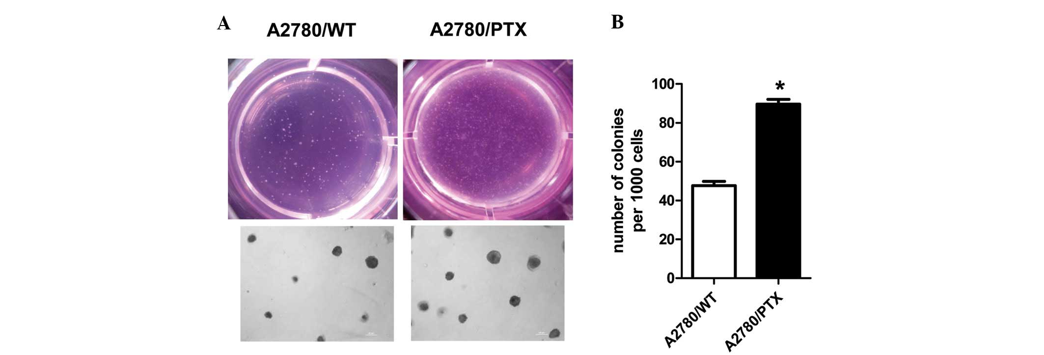
Acquisition of paclitaxel resistance in
A2780/PTX cells enhances colony formation
A characteristic of stem cells is their ability to
form clones in soft agar (15). In
order to determine whether the cancer cells form such colonies,
2,000 A2780/WT cells and A2780/PTX cells were seeded into soft agar
growth medium in 12-well plates and cultured for two weeks. The two
cell types formed colonies, but the A2780/PTX cells were more
tumorigenic than the A2780/WT cells (Fig. 2).
Inhibition of PI3K activity blocks colony
formation in A2780/PTX cells
To assess the involvement of the PI3K signaling
pathway, the PI3K inhibitor (LY294002; 10 μM) was applied to the
A2780/PTX cells. This inhibitor blocked the colony formation by the
A2780/PTX cells in soft agar (Fig.
3).
Discussion
First-line chemotherapy often leads to encouraging
responses in cancer (16), however,
in the course of the treatment, resistance frequently occurs and
ultimately limits life expectancy (17). The concept of the CSC is one
explanation for chemoresistance (18). CSCs isolated from human cancers,
including ovarian cancer (2),
glioblastoma (19) and breast
cancer (20), have reportedly
increased resistance to conventional therapies. The development of
more effective therapies may require targeting this important CSC
population. The success of this new approach is dependent on the
identification and characterization of CSCs. Recently, ALDH1
activity has been used successfully as a marker to isolate stem
cells from diverse sources (21).
The present study used wild-type (A2780/WT) and
paclitaxel-resistant (A2780/PTX) human epithelial ovarian cancer
cells to investigate the expression of ALDH1 and the associated
capacity for colony formation. The A2780/PTX cells were shown to
display high ALDH1 activity, consistent with other studies showing
that a high percentage of ALDH1-positive tumor cells is
significantly associated with a poor clinical outcome in serous
ovarian cancer (22,23). Subsequently, colony formation by the
A2780/PTX cells was examined. As predicted, the findings
demonstrated that the A2780/PTX cells displayed enhanced colony
formation in the soft agar assay. This indicated that the A2780/PTX
cells were more tumorigenic than the A2780/WT cells.
Since the PI3K/AKT axis is frequently activated in
human cancer (24), the present
study explored whether these pathways are involved in the
regulation of tumorigenicity. The PI3K inhibitor, LY294002,
abolished colony formation when added to the A2780/PTX cells.
Although further studies are required to elucidate the mechanism of
PI3K-mediated colony formation, the use of PI3K inhibitors to
reduce tumorigenicity may be a useful approach to improve the
efficacy of chemotherapy.
In conclusion, the present study showed that the
selection and isolation of stem-like ovarian cancer cells, on the
basis of ALDH1 activity, revealed enhanced colony formation.
Targeting the cells with the PI3K inhibitor, LY294002, abolished
this capacity. We believe that these findings are of importance and
support clinical trials of LY294002 in ovarian cancer, either alone
or in combination with established therapies.
Acknowledgements
This study was supported by the Program for New
Century Excellent Talents in University of The Ministry of
Education of China [NCET-12-0880 (XM)]; Fundamental Research Funds
for the Central Universities [JUSRP51311A (XM)]; The National
Natural Science Foundation of China [81100185 (XM) and 81130057
(JJ)]; and the Strategic Priority Research Program of the Chinese
Academy of Sciences [XDA01040000 (JJ)].
References
|
1
|
Aguilar-Gallardo C, Rutledge EC,
Martínez-Arroyo AM, Hidalgo JJ, Domingo S and Simón C: Overcoming
challenges of ovarian cancer stem cells: novel therapeutic
approaches. Stem Cell Rev. 8:994–1010. 2012. View Article : Google Scholar : PubMed/NCBI
|
|
2
|
Wang L, Mezencev R, Bowen NJ, Matyunina LV
and McDonald JF: Isolation and characterization of stem-like cells
from a human ovarian cancer cell line. Mol Cell Biochem.
363:257–268. 2012. View Article : Google Scholar : PubMed/NCBI
|
|
3
|
Conic I, Dimov I, Tasic-Dimov D,
Djordjevic B and Stefanovic V: Ovarian epithelial cancer stem
cells. Scientific World Journal. 11:1243–1269. 2011. View Article : Google Scholar : PubMed/NCBI
|
|
4
|
Aletti GD, Gallenberg MM, Cliby WA, Jatoi
A and Hartmann LC: Current management strategies for ovarian
cancer. Mayo Clin Proc. 82:751–770. 2007. View Article : Google Scholar : PubMed/NCBI
|
|
5
|
Diehn M, Cho RW, Lobo NA, et al:
Association of reactive oxygen species levels and radioresistance
in cancer stem cells. Nature. 458:780–783. 2009. View Article : Google Scholar : PubMed/NCBI
|
|
6
|
Clarke MF, Dick JE, Dirks PB, et al:
Cancer stem cells- perspectives on current status and future
directions: AACR Workshop on cancer stem cells. Cancer Res.
66:9339–9344. 2006. View Article : Google Scholar
|
|
7
|
Burgos-Ojeda D, Rueda BR and Buckanovich
RJ: Ovarian cancer stem cell markers: Prognostic and therapeutic
implications. Cancer Lett. 322:1–7. 2012. View Article : Google Scholar : PubMed/NCBI
|
|
8
|
Sullivan JP, Spinola M, Dodge M, et al:
Aldehyde dehydrogenase activity selects for lung adenocarcinoma
stem cells dependent on notch signaling. Cancer Res. 70:9937–9948.
2010. View Article : Google Scholar : PubMed/NCBI
|
|
9
|
Yu C, Yao Z, Dai J, et al: ALDH activity
indicates increased tumorigenic cells, but not cancer stem cells,
in prostate cancer cell lines. In Vivo. 25:69–76. 2011.PubMed/NCBI
|
|
10
|
Wicha MS, Ginestier C, Charaffe-Jauffret
E, Tarpin FI, Dontu G and Iovino F: Tumor invasion and metastasis
of human breast cancer is mediated by the ALDH-1+stem cell
component. Clin Exp Metastas. 25:11. 2008.
|
|
11
|
Steg AD, Bevis KS, Katre AA, et al: Stem
cell pathways contribute to clinical chemoresistance in ovarian
cancer. Clin Cancer Res. 18:869–881. 2012. View Article : Google Scholar : PubMed/NCBI
|
|
12
|
Liu P, Brown S, Goktug T, et al: Cytotoxic
effect of disulfiram/copper on human glioblastoma cell lines and
ALDH-positive cancer-stem-like cells. Br J of Cancer.
107:1488–1497. 2012. View Article : Google Scholar : PubMed/NCBI
|
|
13
|
Croker AK, Goodale D, Chu J, et al: High
aldehyde dehydrogenase and expression of cancer stem cell markers
selects for breast cancer cells with enhanced malignant and
metastatic ability. J Cell Mol Med. 13:2236–2252. 2009. View Article : Google Scholar : PubMed/NCBI
|
|
14
|
Martelli AM, Evangelisti C, Chiarini F, et
al: The emerging role of the phosphatidylinositol
3-kinase/Akt/mammalian target of rapamycin signaling network in
normal myelopoiesis and leukemogenesis. Biochim Biophys Acta.
1803:991–1002. 2010. View Article : Google Scholar : PubMed/NCBI
|
|
15
|
Dou J, Pan M, Wen P, et al: Isolation and
identification of cancer stem-like cells from murine melanoma cell
lines. Cell Mol Immunol. 4:467–472. 2007.PubMed/NCBI
|
|
16
|
Chang A: Chemotherapy, chemoresistance and
the changing treatment landscape for NSCLC. Lung Cancer. 71:3–10.
2011. View Article : Google Scholar : PubMed/NCBI
|
|
17
|
Yang H, Zhang Q, He J and Lu W: Regulation
of calcium signaling in lung cancer. J Thorac Dis. 2:52–56.
2010.PubMed/NCBI
|
|
18
|
Awad O, Yustein JT, Shah P, et al: High
ALDH activity identifies chemotherapy-resistant Ewings sarcoma stem
cells that retain sensitivity to EWS-FLI1 inhibition. PLoS One.
5:e13943:2010.PubMed/NCBI
|
|
19
|
Cvek B: Comment on ‘cytotoxic effect of
disulfiram/copper on human glioblastoma cell lines and
ALDH-positive cancer-stem-like cells’. Br J Cancer.
108:9932013.
|
|
20
|
Berry DA, Ueno NT, Johnson MM, et al:
High-dose chemotherapy with autologous hematopoietic stem-cell
transplantation in metastatic breast cancer: overview of six
randomized trials. J Clin Oncol. 29:3224–3231. 2011. View Article : Google Scholar
|
|
21
|
Jelski W, Kutylowska E, Laniewska-Dunaj M,
Orywal K, Laszewicz W and Szmitkowski M: Alcohol dehydrogenase
(ADH) isoenzymes and aldehyde dehydrogenase (ALDH) activity in the
sera of patients with acute and chronic pancreatitis. Exp Mol
Pathol. 91:631–635. 2011. View Article : Google Scholar : PubMed/NCBI
|
|
22
|
Deng S, Yang X, Lassus H, et al: Distinct
expression levels and patterns of stem cell marker, aldehyde
dehydrogenase isoform 1 (ALDH1), in human epithelial cancers. PLoS
One. 5:e102772010. View Article : Google Scholar
|
|
23
|
Wang YC, Yo YT, Lee HY, et al:
ALDH1-bright epithelial ovarian cancer cells are associated with
CD44 expression, drug resistance, and poor clinical outcome. Am J
Pathol. 180:1159–1169. 2012. View Article : Google Scholar : PubMed/NCBI
|
|
24
|
Amente S, Zhang J, Lavadera ML, Lania L,
Avvedimento EV and Majello B: Myc and PI3K/AKT signaling
cooperatively repress FOXO3a-dependent PUMA and GADD45a gene
expression. Nucleic Acids Res. 39:9498–9507. 2011. View Article : Google Scholar : PubMed/NCBI
|