Introduction
Urothelial carcinoma (UC) is a highly chemosensitive
disease. Cisplatin is a key drug for the treatment of advanced or
metastatic UC. To date, the combination of methotrexate,
vinblastine, doxorubicin and cisplatin (M-VAC) has been accepted as
the most effective therapy for metastatic UC (1). A randomized trial that was designed to
compare a two-drug regimen comprising gemcitabine and cisplatin
(GC) with M-VAC, revealed that GC provided a similar survival
advantage to M-VAC but with improved safety and tolerability
(2). However, the prognosis for
patients with metastatic UC of the urinary tract remains poor even
with GC treatment. From our experience with GC, the median time to
progression and the median overall survival time for
cisplatin-naïve patients were 6 and 14 months, respectively
(3). In this study, the overall
response rate to treatment for patients on this regimen was 63%,
while 37% of the patients were completely or almost resistant to
cisplatin. In addition, only 31% of the patients who relapsed >6
months after treatment with the prior cisplatin-based regimen
exhibited an objective response to cisplatin. These results suggest
that cancer cells naturally have, or eventually develop, cisplatin
resistance. Therefore, the acquisition of chemoresistance remains a
major obstacle in cancer treatment, which ultimately leads to
mortality.
We previously established a cisplatin-resistant
subline from the human HT1376 bladder cancer cell line
(HT1376-CisR) to elucidate the possible mechanisms underlying
cisplatin resistance in bladder cancer cells (4). Comparative proteomic analysis of
HT1376 and HT1376-CisR cells has revealed 36
differentially-expressed proteins, of which 21 proteins are
upregulated in HT1376-CisR cells (4). Among the differentially regulated
proteins, aldo-keto reductase family 1 member C2 (AKR1C2) was
markedly expressed in HT1376-CisR cells but not in HT1376
cells.
The AKR superfamily consists of nicotinamide adenine
dinucleotide phosphate-dependent oxidoreductases that metabolize a
wide range of endogenous and exogenous compounds. AKR
overexpression has been associated with chemotherapy resistance in
a variety of cancer cell lines (5–10). AKR
overexpression is also associated with disease progression in
bladder (11) and prostate cancer
(12). Chen et al found that
AKR overexpression, which induced resistance to chemotherapy, also
reduced reactive oxygen species (ROS) production using human
ovarian cancer cells (6). In
contrast, no correlation between AKR expression and ROS levels was
observed in lung cancer cells (8).
Thus, the importance of AKRs in the mechanism of drug resistance
remains unclear. In the present study, attempts were made to
clarify the underlying cisplatin resistance mechanisms by analyzing
the function of AKR1C2 at the cellular and molecular levels.
Materials and methods
Reagents
RPMI-1640 and fetal bovine serum (FBS) for cell
culture were supplied by Life Technologies (Carlsbad, CA, USA).
Cisplatin, 5β-cholanic acid and menadione were purchased from
Sigma-Aldrich (Tokyo, Japan). 5β-cholanic acid and menadione were
used as an AKR1C2 inhibitor and an oxidative stressor,
respectively. 2,7-Dichlorodihydrofluorescein diacetate
(H2DCFDA) was purchased from Life Technologies.
Anti-AKR1C2 and anti-β-tubulin (loading control) rabbit polyclonal
antibodies were obtained from NOVUS Biological (Littleton, CO, USA)
and Abcam (Cambridge, UK), respectively.
Cell culture
The human HT1376 bladder cancer cell line used in
this study was purchased from DS Pharma Biomedical (Osaka, Japan).
Cells were maintained in RPMI-1640 medium supplemented with 10% FBS
in a humidified incubator at 37°C and 5% CO2.
Cisplatin-resistant cells (HT1376-CisR) were obtained from the
parental HT1376 cells using an intermittent stepwise selection
protocol over 12 months, ending with exposure to 5 μM cisplatin
(4).
Western blot analysis
Cells were lysed with an ice-cold lysis buffer and
protease inhibitor cocktail mix (Sigma-Aldrich). Samples were
centrifuged at 12,000 × g for 10 min at 4°C and supernatants were
electrophoresed by SDS-PAGE and transferred to polyvinylidene
difluoride membranes (Millipore, Bedford, MA, USA). Following
blocking with 5% skimmed milk, the membranes were probed with
primary antibodies overnight at 4°C, followed by horseradish
peroxidase-conjugated secondary antibody (GE Healthcare, Chalfont
St. Giles, UK) for 1 h at room temperature. The immune complexes
were visualized with the Enhanced Chemiluminescence Plus detection
system (GE Healthcare) according to the manufacturer’s
instructions.
Drug cytotoxicity analysis
To analyze drug cytotoxicity, 1.0×10⁴
cells/well were cultured with concentrations of cisplatin graded
between 0.5×10⁻⁷ and 10−3 M cisplatin in at
least 3 replicate wells at 37°C. Following 72 h of cisplatin
treatment, the cells were counted using a Scepter 2.0 Handheld
Automated Cell Counter (Merck Millipore, MA, USA). Cell survival in
the absence of cisplatin was defined as 100% cell survival. The
drug concentration that resulted in 50% growth inhibition
(IC50) was determined from the corresponding
dose-response curve.
Intracellular ROS accumulation
Intracellular ROS accumulation was determined using
the method described by Tardito et al (13). H2DCFDA does not fluoresce
but becomes fluorescent when it is hydrolyzed to H2DCF
inside cells by nonspecific esterases. Briefly, the samples were
plated in 96-well plates at a density of 4.0×10⁴
cells/well. Following overnight incubation with or without reagent,
intracellular ROS was examined. Growth medium was removed and 100
μl prewarmed Hank’s balanced salt solution (HBSS; Life
Technologies) containing 20 μM H2DCFDA was added at 37°C
without exposure to light. The prewarmed HBSS with
H2DCFDA was prepared fresh for each assay. Following
incubation at 37°C for 30 min, the cells were washed twice with
HBSS and ROS generation was measured as fluorescence intensity
using a fluorescence multiplate reader (Flex Station 3; Molecular
Devices, Sunnyvale, CA, USA) with an excitation wavelength of 480
nm and an emission wavelength of 530 nm.
Technologies
The small interfering (si) RNA sequences were as
follows: Sense, 5′-CGGCCGGAAAAGAAAGACATT-3′ and antisense,
5′-UGUCUUUCUUUUCCGGCCGAT-3′. For the control, the following
non-targeting siRNA cocktails were used: 5′-ATCCGCGCGATAGTACGTA-3′,
5′-TTACGCTA GCGTAATACG-3′ and 5′-TATTCGCGCCTATAGCGGT-3′. The cells
were transiently transfected with AKR1C2 siRNA and control siRNA
using Lipofectamine RNAiMAX (Life Technologies) and Optimen I (Life
Technologies) at a 120 pM concentration. Following 48-h incubation,
cells were utilized for each assay.
Statistical analysis
All values are expressed as the mean ± standard
deviation of at least 3 independent experiments. The unpaired
Student’s t-test was used for statistical analysis in this study.
P<0.05 was considered to indicate a statistically significant
difference.
Results
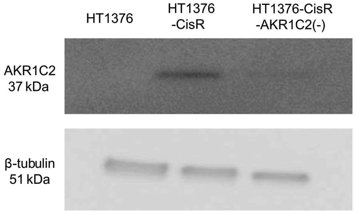
Expression levels of AKR1C2 protein levels were
examined by western blot analysis (Fig.
1). Expression was detected in the HT1376-CisR cells, but in
the parental cells. AKR1C2-siRNA reduced the AKR1C2 protein levels
by ~80% in HT1376-CisR cells.
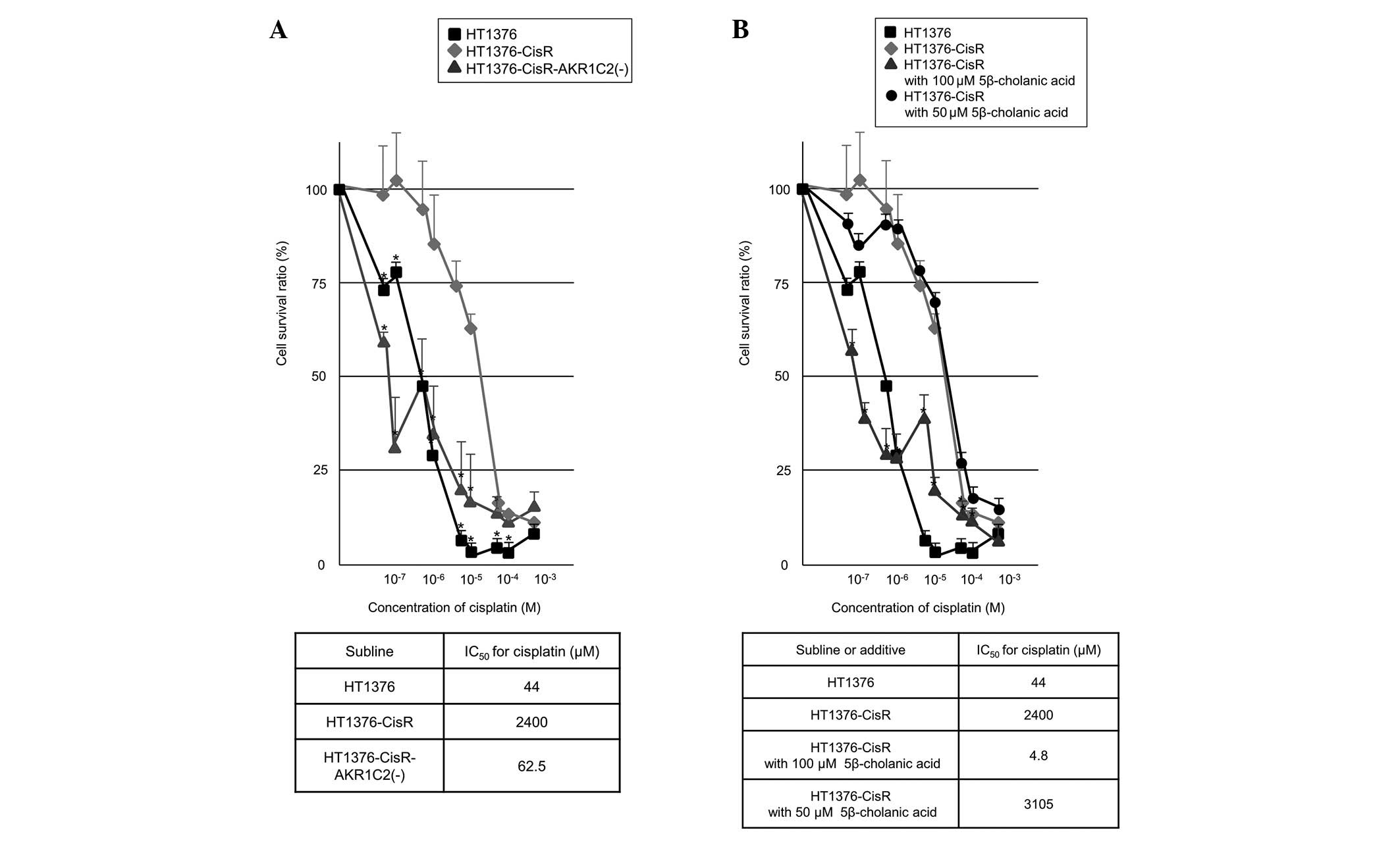
Next, the effect of AKR1C2 expression on cell
survival was examined. Fig. 2A
shows the relative number of surviving HT1376 and HT1376-CisR cells
following treatment with various concentrations of cisplatin. The
IC50 values for cisplatin treatment in HT1376 and
HT1376-CisR cells were 44 and 2,400 μM, respectively. The
IC50 for HT1376-CisR was thus 54.5-fold higher than that
of HT1376 cells, indicating that a cisplatin-resistant cell line
was successfully established. AKR1C2-siRNA markedly rescued the
cisplatin sensitivity of HT1376-CisR. The IC50 value for
cisplatin treatment in HT1376-CisR cells transiently transfected
with AKR1C2 siRNA [HT1376-CisR-AKR1C2(−)] was 62.5 μM.
Next, the inhibitory effect of 5β-cholanic acid on
cell survival was examined. Fig. 2B
shows the relative number of surviving HT1376-CisR cells following
treatment with or without 5β-cholanic acid and various
concentrations of cisplatin. All HT1376-CisR cells died following
incubation for 72 h in medium with 150 μM 5β-cholanic acid,
possibly due to its strong cytotoxicity. Addition of 100 μM
5β-cholanic acid to the medium restored the cisplatin response of
HT1376-CisR cells, whereas 50 μM 5β-cholanic acid did not. The
IC50 values for cisplatin treatment in HT1376-CisR cells
cultured at concentrations of 50 and 100 μM 5β-cholanic acid were
3,105 and 4.8 μM, respectively. These results indicate that AKR1C2
plays an important role in cisplatin resistance in HT1376
cells.
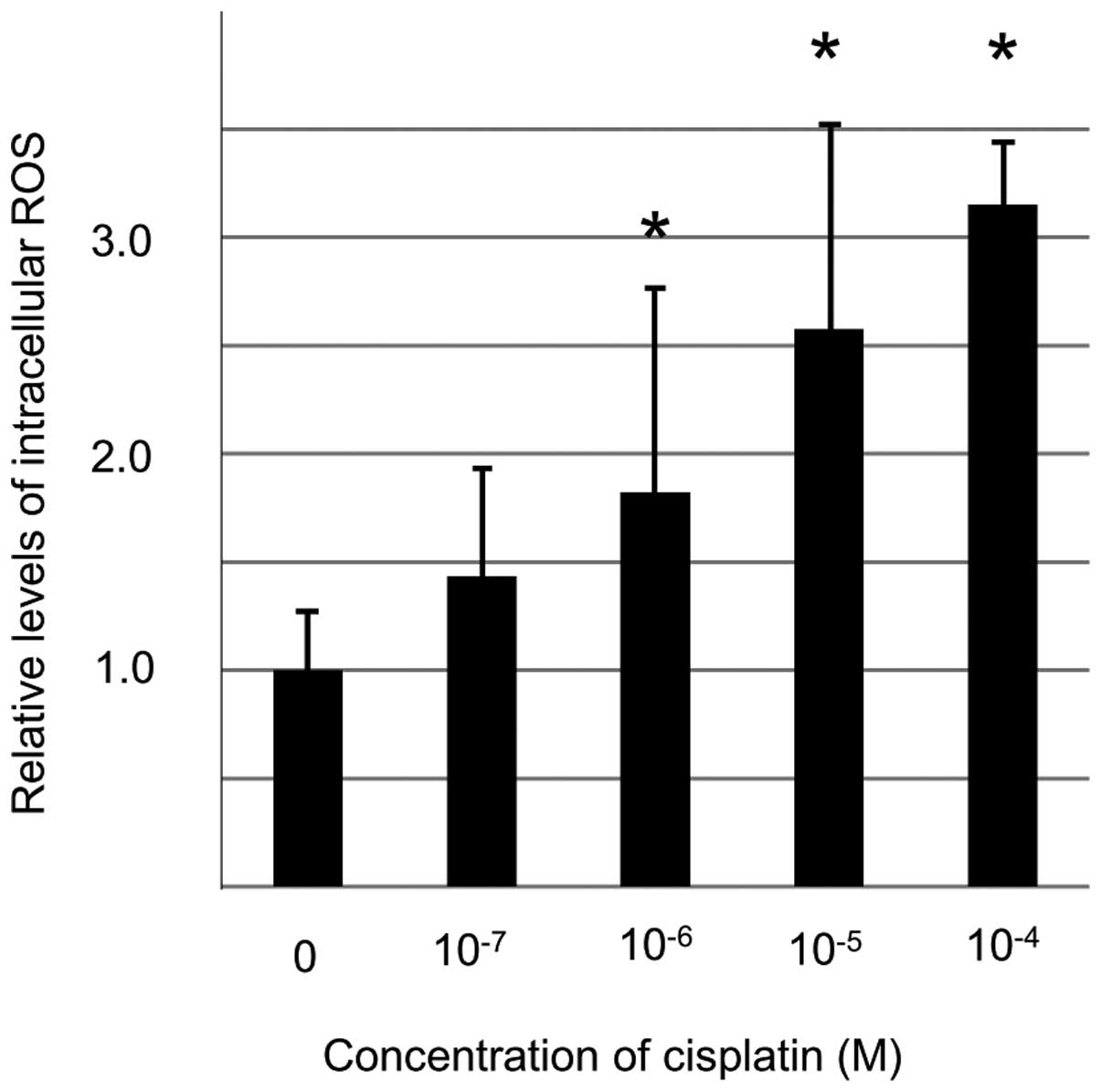
To elucidate the role of AKR1C2 in cisplatin
resistance, the levels of intracellular ROS were determined using
an H2DCFDA probe under various conditions. Exposure to
cisplatin for 2 h increased the level of intracellular ROS in
HT1376 cells in a dose-dependent manner (Fig. 3). Significant differences were
detected between the ROS levels of HT1376 cells treated without
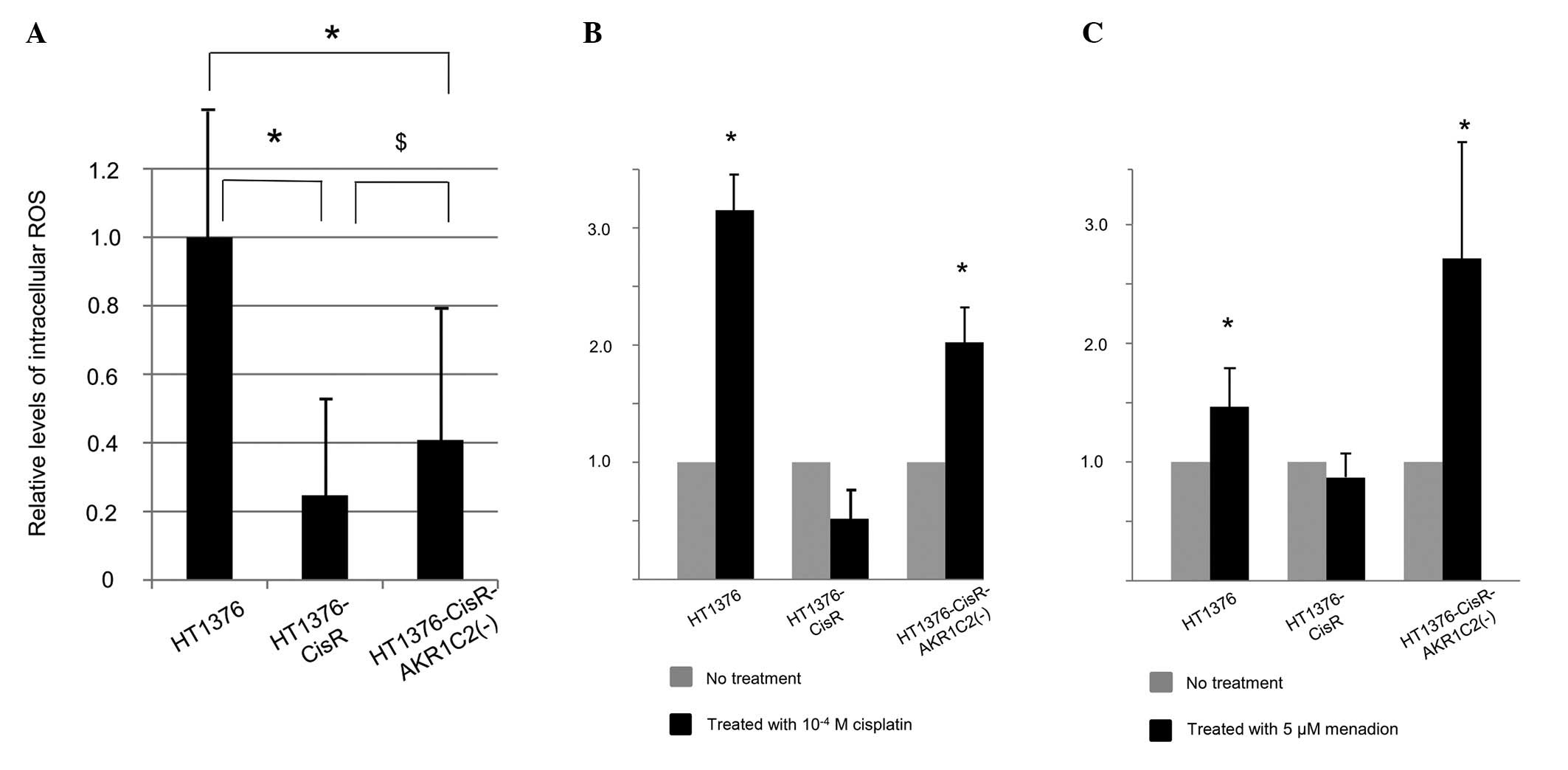
cisplatin and with >10−6 M cisplatin. Fig. 4A shows a comparison of relative
basal levels of intracellular ROS in HT1376, HT1376-CisR and
HT1376-CisR-AKR1C2(−) cells. Intracellular ROS in HT1376-CisR cells
was significantly lower than that found in HT1376 cells.
Furthermore, AKR1C2 knockdown significantly rescued intracellular
ROS levels, although these did not reach the levels found in HT1376
cells. The effects of 10−4 M cisplatin exposure for 2 h
in the respective cells are shown in Fig. 4B. Cisplatin exposure did not
increase the level of intracellular ROS in HT1376-CisR cells,
whereas exposure increased the ROS level by 3-fold in HT1376 cells.
Silencing AKR1C2 mRNA restored this ROS increase in HT1376-CisR
cells.
The effects of 5 μM menadione as an oxidative
stressor in the respective cell lines were also examined (Fig. 4C). The addition of menadione to the
media increased the ROS levels in HT1376 and HT1376-CisR-AKR1C2(−)
cells, but not in HT1376-CisR cells. These data suggest that AKR1C2
expression impairs reactivity against cisplatin-induced oxidative
stress in HT1376 cells, thus resulting in cisplatin resistance.
Discussion
In the present study, AKR1C2 expression was
identified only in the cisplatin-resistant human bladder cancer
cells. In addition, silencing or inhibition of AKR1C2 restored
cisplatin cytotoxicity in these cells, perhaps due to the increase
in cisplatin-induced intracellular ROS.
Although cisplatin is widely used for the treatment
of advanced and metastatic bladder cancer, the majority of patients
relapse with a cisplatin-resistant disease during chemotherapy. The
development of chemoresistance remains a major obstacle in the
treatment of bladder and other types of cancer (3). The cause of cisplatin resistance has
previously been investigated, and proposed mechanisms include
reduced intracellular drug accumulation, increased detoxification
of the drug by thiol-containing molecules, increased DNA damage
repair activities, escape from reactive oxygen species-mediated
cytotoxicity and the involvement of apoptosis mediators (14–16).
The general consensus is that chemoresistance is multifactorial
(i.e., several mechanisms are simultaneously encountered within the
same tumor cell) (17–20).
Cisplatin activity is known to generate ROS. For
example, cisplatin-induced hearing loss is caused by ROS generation
in the cochlea (21). ROS also
function as common mediators of apoptosis induced by anticancer
drugs. Bragado et al (22)
reported that the apoptotic activity of cisplatin requires the
onset of the p53-mediated p38α mitogen-activated protein kinase
pathway through ROS generation. Furthermore, cisplatin-induced
apoptosis of cancer cells has been found to act through
ROS-dependent Fas aggregation (23). When cancer cells are exposed to high
concentrations of ROS by cisplatin treatment, a defense mechanism
against intrinsic ROS is activated in these cells. Previous studies
have identified several important defense mechanisms that are
triggered by cisplatin treatment. The Kelch-like ECH-associated
protein 1 (Keap1)/nuclear factor erythroid 2-related factor 2
(Nrf2) system, is one of the most important cellular mechanisms
acting against oxidative stressors and electrophiles (24). Keap1 and Nrf2 are oxidative stress
sensors and transcription factors for the antioxidant responsive
element (ARE). When cells are exposed to stressors such as ROS,
Nrf2 is released from the constraint of Keap1 and activates
ARE-dependent gene expression (25). The Nrf2/ARE signaling pathway
regulates the expression of cytoprotective proteins, including
AKR1C2 (25). Although the
cytoprotective system is designed to prevent normal cells from
becoming cancerous, in a cisplatin-induced ROS-rich environment,
cancer cells may hijack the Keap1/Nrf2 system and induce AKR1C2
protein expression as an antioxidant substance. The upregulation of
antioxidant capacity in adaptation to intrinsic oxidative stress in
cancer cells can result in drug resistance (26).
Previous studies have reported an interaction
between AKRs and drug resistance in certain cancer cells. Chen
et al (6) demonstrated that
overexpression of dihydrodiol dehydrogenases (DDHs), which belong
to the AKR family, leads to resistance to platinum-based drugs in
several human cancer cell lines. These DDH levels are directly
responsible for the reduced production of ROS. Chen et al
(8) also suggested that cisplatin
sensitivity appeared to be associated with DDH levels in epithelial
lung cancer cell lines. The present study demonstrated that
induction of AKR1C2 can be found in cisplatin-resistant human
bladder cancer cells and contributes to cisplatin drug resistance.
Furthermore, inhibition of AKR1C2 was found to lead to restoration
of cisplatin drug sensitivity.
In this study, a cisplatin-resistant human bladder
cell line was established from HT1376 cells. Although the
biological characteristics of this cell line may not be universal,
AKR1C2 expression has frequently been detected in pathological
specimens of UC (11). Further
studies are required to validate the practical significance of
AKR1C2 in bladder cancer. However, we hypothesize that AKR1C2 is
one of the biomarkers that indicates cisplatin resistance. In
addition, AKR1C2 may be one of the effective molecular targets for
rescuing cisplatin sensitivity.
References
|
1
|
Logothetis CJ, Dexeus FH, Finn L, et al: A
prospective randomized trial comparing MVAC and CISCA chemotherapy
for patients with metastatic urothelial tumors. J Clin Oncol.
8:1050–1055. 1990.PubMed/NCBI
|
|
2
|
von der Maase H, Hansen SW, Roberts JT, et
al: Gemcitabine and cisplatin versus methotrexate, vinblastine,
doxorubicin, and cisplatin in advanced or metastatic bladder
cancer: results of a large, randomized, multinational, multicenter,
phase III study. J Clin Oncol. 18:3068–3077. 2000.
|
|
3
|
Tanji N, Ozawa A, Miura N, et al:
Long-term results of combined chemotherapy with gemicitabine and
cisplatin for metastatic urothelial carcinomas. Int J Clin Oncol.
15:369–375. 2010. View Article : Google Scholar : PubMed/NCBI
|
|
4
|
Miura N, Takemori N, Kikugawa T, et al:
Adseverin: A novel cisplatin-resistant marker in the human bladder
cancer cell line HT1376 identified by quantitative proteomic
proteomic analysis. Mol Oncol. 6:311–322. 2012. View Article : Google Scholar : PubMed/NCBI
|
|
5
|
Veitch ZW, Guo B, Hembruff SL, et al:
Induction of 1C aldoketoreductases and other drug dose-dependent
genes upon acquisition of anthracycline resistance. Pharmacogenet
Genomics. 19:177–188. 2009. View Article : Google Scholar : PubMed/NCBI
|
|
6
|
Chen J, Adikari M, Pallai R, et al:
Dihydrodiol dehydrogenases regulate the generation of reactive
oxygen species and the development of cisplatin resistance in human
ovarian carcinoma cells. Cancer Chemother Pharmacol. 61:979–987.
2008. View Article : Google Scholar
|
|
7
|
Matsunaga T, Yamane Y, Iida K, et al:
Involvement of the aldo-keto reductase, AKR1B10, in mitomycin-c
resistance through reactive oxygen species-dependent mechanisms.
Anticancer Drugs. 22:402–408. 2011. View Article : Google Scholar : PubMed/NCBI
|
|
8
|
Chen J, Emara N, Solomides C, et al:
Resistance to platinum-based chemotherapy in lung cancer cell
lines. Cancer Chemother Pharmacol. 66:1103–1111. 2010. View Article : Google Scholar : PubMed/NCBI
|
|
9
|
Chow KC, Lu MP and Wu MT: Expression of
dihydrodiol dehydrogenase plays important roles in apoptosis- and
drug-resistance of A431 squamous cell carcinoma. J Dermatol Sci.
41:205–212. 2006. View Article : Google Scholar : PubMed/NCBI
|
|
10
|
Wang HW, Lin CP, Chiu JH, et al: Reversal
of inflammation-associated dihydrodiol dehydrogenases (AKR1C1 and
AKR1C2) overexpression and drug resistance in nonsmall cell lung
cancer cells by wogonin and chrysin. Int J Cancer. 120:2019–2027.
2007. View Article : Google Scholar : PubMed/NCBI
|
|
11
|
Tai HL, Lin TS, Huang HH, et al:
Overexpression of aldo-keto reductase 1C2 as a high-risk factor in
bladder cancer. Oncol Rep. 17:305–311. 2007.PubMed/NCBI
|
|
12
|
Huang KH, Chiou SH, Chow KC, et al:
Overexpression of aldo-keto reductase 1C2 is associated with
disease progression in patients with prostatic cancer.
Histopathology. 57:384–394. 2010. View Article : Google Scholar : PubMed/NCBI
|
|
13
|
Tardito S, Bussolati O, Maffini M, et al:
Thioamido coordination in a thioxo-1,2,4-triazole copper(II)
complex enhances nonapoptotic programmed cell death associated with
copper accumulation and oxidative stress in human cancer cells. J
Med Chem. 50:1916–1924. 2007. View Article : Google Scholar
|
|
14
|
Siddik ZH: Cisplatin: mode of cytotoxic
action and molecular basis of resistance. Oncogene. 22:7265–7279.
2003. View Article : Google Scholar : PubMed/NCBI
|
|
15
|
Hour TC, Lai YL, Kuan CI, et al:
Transcriptional up-regulation of SOD1 by CEBPD: a potential target
for cisplatin resistant human urothelial carcinoma cells. Biochem
Pharmacol. 80:325–334. 2010. View Article : Google Scholar : PubMed/NCBI
|
|
16
|
Tsunoda T, Koga H, Yokomizo A, et al:
Inositol 1,4,5-trisphosphate (IP3) receptor type1
(IP3R1) modulates the acquisition of cisplatin
resistance in bladder cancer cell lines. Oncogene. 24:1396–1402.
2005.
|
|
17
|
Richon VM, Schulte N and Eastman A:
Multiple mechanisms of resistance to
cis-diamminedichloroplatinum(II) in murine leukemia L1210 cells.
Cancer Res. 47:2056–2061. 1987.
|
|
18
|
Eastman A and Schulte N: Enhanced DNA
repair as a mechanism of resistance to
cis-diamminedichloroplatinum(II). Biochemistry. 27:4730–4734. 1988.
View Article : Google Scholar : PubMed/NCBI
|
|
19
|
Teicher BA, Holden SA, Kelley MJ, et al:
Characterization of a human squamous carcinoma cell line resistant
to cis-diammine-dichloroplatinum(II). Cancer Res. 47:388–393.
1987.PubMed/NCBI
|
|
20
|
Rabik CA and Dolan ME: Molecular
mechanisms of resistance and toxicity associated with platinating
agents. Cancer Treat Rev. 33:9–23. 2007. View Article : Google Scholar : PubMed/NCBI
|
|
21
|
Sodhi A and Gupta P: Increased release of
hydrogen peroxide (H2O2) and superoxide anion
(O2−) by murine macrophages in vitro after
cis-platin treatment. Int J Immunopharmacol. 8:709–714. 1986.
|
|
22
|
Bragado P, Armesilla A, Silva A and Porras
A: Apoptosis by cisplatin requires p53 mediated p38alpha MAPK
activation through ROS generation. Apoptosis. 12:1733–1742. 2007.
View Article : Google Scholar : PubMed/NCBI
|
|
23
|
Huang HL, Fang LW, Lu SP, et al:
DNA-damaging reagents induce apoptosis through reactive oxygen
species-dependent Fas aggregation. Oncogene. 22:8168–8177. 2003.
View Article : Google Scholar
|
|
24
|
Nguyen T, Nioi P and Pickett CB: The
Nrf2-antioxidant response element signaling pathway and its
activation by oxidative stress. J Biol Chem. 284:13291–13295. 2009.
View Article : Google Scholar : PubMed/NCBI
|
|
25
|
Halim M, Yee DJ and Sames D: Imaging
induction of cytoprotective enzymes in intact human cells:
coumberone, a metabolic reporter for human AKR1C enzymes reveals
activation by panaxytriol, an active component of red ginseng. J Am
Chem Soc. 130:14123–14128. 2008. View Article : Google Scholar
|
|
26
|
Trachootham D, Alexandre J and Huang P:
Targeting cancer cells by ROS-mediated mechanisms: a radical
therapeutic approach? Nat Rev Drug Discov. 8:579–591. 2009.
View Article : Google Scholar : PubMed/NCBI
|