Spandidos Publications Logo
  • About
    • About Spandidos
    • Aims and Scopes
    • Abstracting and Indexing
    • Editorial Policies
    • Reprints and Permissions
    • Job Opportunities
    • Terms and Conditions
    • Contact
  • Journals
    • All Journals
    • Oncology Letters
      • Oncology Letters
      • Information for Authors
      • Editorial Policies
      • Editorial Board
      • Aims and Scope
      • Abstracting and Indexing
      • Bibliographic Information
      • Archive
    • International Journal of Oncology
      • International Journal of Oncology
      • Information for Authors
      • Editorial Policies
      • Editorial Board
      • Aims and Scope
      • Abstracting and Indexing
      • Bibliographic Information
      • Archive
    • Molecular and Clinical Oncology
      • Molecular and Clinical Oncology
      • Information for Authors
      • Editorial Policies
      • Editorial Board
      • Aims and Scope
      • Abstracting and Indexing
      • Bibliographic Information
      • Archive
    • Experimental and Therapeutic Medicine
      • Experimental and Therapeutic Medicine
      • Information for Authors
      • Editorial Policies
      • Editorial Board
      • Aims and Scope
      • Abstracting and Indexing
      • Bibliographic Information
      • Archive
    • International Journal of Molecular Medicine
      • International Journal of Molecular Medicine
      • Information for Authors
      • Editorial Policies
      • Editorial Board
      • Aims and Scope
      • Abstracting and Indexing
      • Bibliographic Information
      • Archive
    • Biomedical Reports
      • Biomedical Reports
      • Information for Authors
      • Editorial Policies
      • Editorial Board
      • Aims and Scope
      • Abstracting and Indexing
      • Bibliographic Information
      • Archive
    • Oncology Reports
      • Oncology Reports
      • Information for Authors
      • Editorial Policies
      • Editorial Board
      • Aims and Scope
      • Abstracting and Indexing
      • Bibliographic Information
      • Archive
    • Molecular Medicine Reports
      • Molecular Medicine Reports
      • Information for Authors
      • Editorial Policies
      • Editorial Board
      • Aims and Scope
      • Abstracting and Indexing
      • Bibliographic Information
      • Archive
    • World Academy of Sciences Journal
      • World Academy of Sciences Journal
      • Information for Authors
      • Editorial Policies
      • Editorial Board
      • Aims and Scope
      • Abstracting and Indexing
      • Bibliographic Information
      • Archive
    • International Journal of Functional Nutrition
      • International Journal of Functional Nutrition
      • Information for Authors
      • Editorial Policies
      • Editorial Board
      • Aims and Scope
      • Abstracting and Indexing
      • Bibliographic Information
      • Archive
    • International Journal of Epigenetics
      • International Journal of Epigenetics
      • Information for Authors
      • Editorial Policies
      • Editorial Board
      • Aims and Scope
      • Abstracting and Indexing
      • Bibliographic Information
      • Archive
    • Medicine International
      • Medicine International
      • Information for Authors
      • Editorial Policies
      • Editorial Board
      • Aims and Scope
      • Abstracting and Indexing
      • Bibliographic Information
      • Archive
  • Articles
  • Information
    • Information for Authors
    • Information for Reviewers
    • Information for Librarians
    • Information for Advertisers
    • Conferences
  • Language Editing
Spandidos Publications Logo
  • About
    • About Spandidos
    • Aims and Scopes
    • Abstracting and Indexing
    • Editorial Policies
    • Reprints and Permissions
    • Job Opportunities
    • Terms and Conditions
    • Contact
  • Journals
    • All Journals
    • Biomedical Reports
      • Information for Authors
      • Editorial Policies
      • Editorial Board
      • Aims and Scope
      • Abstracting and Indexing
      • Bibliographic Information
      • Archive
    • Experimental and Therapeutic Medicine
      • Information for Authors
      • Editorial Policies
      • Editorial Board
      • Aims and Scope
      • Abstracting and Indexing
      • Bibliographic Information
      • Archive
    • International Journal of Epigenetics
      • Information for Authors
      • Editorial Policies
      • Editorial Board
      • Aims and Scope
      • Abstracting and Indexing
      • Bibliographic Information
      • Archive
    • International Journal of Functional Nutrition
      • Information for Authors
      • Editorial Policies
      • Editorial Board
      • Aims and Scope
      • Abstracting and Indexing
      • Bibliographic Information
      • Archive
    • International Journal of Molecular Medicine
      • Information for Authors
      • Editorial Policies
      • Editorial Board
      • Aims and Scope
      • Abstracting and Indexing
      • Bibliographic Information
      • Archive
    • International Journal of Oncology
      • Information for Authors
      • Editorial Policies
      • Editorial Board
      • Aims and Scope
      • Abstracting and Indexing
      • Bibliographic Information
      • Archive
    • Medicine International
      • Information for Authors
      • Editorial Policies
      • Editorial Board
      • Aims and Scope
      • Abstracting and Indexing
      • Bibliographic Information
      • Archive
    • Molecular and Clinical Oncology
      • Information for Authors
      • Editorial Policies
      • Editorial Board
      • Aims and Scope
      • Abstracting and Indexing
      • Bibliographic Information
      • Archive
    • Molecular Medicine Reports
      • Information for Authors
      • Editorial Policies
      • Editorial Board
      • Aims and Scope
      • Abstracting and Indexing
      • Bibliographic Information
      • Archive
    • Oncology Letters
      • Information for Authors
      • Editorial Policies
      • Editorial Board
      • Aims and Scope
      • Abstracting and Indexing
      • Bibliographic Information
      • Archive
    • Oncology Reports
      • Information for Authors
      • Editorial Policies
      • Editorial Board
      • Aims and Scope
      • Abstracting and Indexing
      • Bibliographic Information
      • Archive
    • World Academy of Sciences Journal
      • Information for Authors
      • Editorial Policies
      • Editorial Board
      • Aims and Scope
      • Abstracting and Indexing
      • Bibliographic Information
      • Archive
  • Articles
  • Information
    • For Authors
    • For Reviewers
    • For Librarians
    • For Advertisers
    • Conferences
  • Language Editing
Login Register Submit
  • This site uses cookies
  • You can change your cookie settings at any time by following the instructions in our Cookie Policy. To find out more, you may read our Privacy Policy.

    I agree
Search articles by DOI, keyword, author or affiliation
Search
Advanced Search
presentation
Oncology Letters
Join Editorial Board Propose a Special Issue
Print ISSN: 1792-1074 Online ISSN: 1792-1082
Journal Cover
May-2014 Volume 7 Issue 5

Full Size Image

Sign up for eToc alerts
Recommend to Library

Journals

International Journal of Molecular Medicine

International Journal of Molecular Medicine

International Journal of Molecular Medicine is an international journal devoted to molecular mechanisms of human disease.

International Journal of Oncology

International Journal of Oncology

International Journal of Oncology is an international journal devoted to oncology research and cancer treatment.

Molecular Medicine Reports

Molecular Medicine Reports

Covers molecular medicine topics such as pharmacology, pathology, genetics, neuroscience, infectious diseases, molecular cardiology, and molecular surgery.

Oncology Reports

Oncology Reports

Oncology Reports is an international journal devoted to fundamental and applied research in Oncology.

Experimental and Therapeutic Medicine

Experimental and Therapeutic Medicine

Experimental and Therapeutic Medicine is an international journal devoted to laboratory and clinical medicine.

Oncology Letters

Oncology Letters

Oncology Letters is an international journal devoted to Experimental and Clinical Oncology.

Biomedical Reports

Biomedical Reports

Explores a wide range of biological and medical fields, including pharmacology, genetics, microbiology, neuroscience, and molecular cardiology.

Molecular and Clinical Oncology

Molecular and Clinical Oncology

International journal addressing all aspects of oncology research, from tumorigenesis and oncogenes to chemotherapy and metastasis.

World Academy of Sciences Journal

World Academy of Sciences Journal

Multidisciplinary open-access journal spanning biochemistry, genetics, neuroscience, environmental health, and synthetic biology.

International Journal of Functional Nutrition

International Journal of Functional Nutrition

Open-access journal combining biochemistry, pharmacology, immunology, and genetics to advance health through functional nutrition.

International Journal of Epigenetics

International Journal of Epigenetics

Publishes open-access research on using epigenetics to advance understanding and treatment of human disease.

Medicine International

Medicine International

An International Open Access Journal Devoted to General Medicine.

Journal Cover
May-2014 Volume 7 Issue 5

Full Size Image

Sign up for eToc alerts
Recommend to Library

  • Article
  • Citations
    • Cite This Article
    • Download Citation
    • Create Citation Alert
    • Remove Citation Alert
    • Cited By
  • Similar Articles
    • Related Articles (in Spandidos Publications)
    • Similar Articles (Google Scholar)
    • Similar Articles (PubMed)
  • Download PDF
  • Download XML
  • View XML
Article

Adenomatoid tumors of the testis: A report of two cases and review of the literature

  • Authors:
    • Duqun Chen
    • Zuhu Yu
    • Liangchao Ni
    • Yaoting Gui
    • Shangqi Yang
    • Bentao Shi
    • Yongqing Lai
  • View Affiliations / Copyright

    Affiliations: Department of Urology, Peking University Shenzhen Hospital, Shenzhen PKU‑HKUST Medical Center, Shenzhen 518036, P.R. China
  • Pages: 1718-1720
    |
    Published online on: March 5, 2014
       https://doi.org/10.3892/ol.2014.1938
  • Expand metrics +
Metrics: Total Views: 0 (Spandidos Publications: | PMC Statistics: )
Metrics: Total PDF Downloads: 0 (Spandidos Publications: | PMC Statistics: )
Cited By (CrossRef): 0 citations Loading Articles...

This article is mentioned in:


Abstract

Adenomatoid tumors are rare benign neoplasms that normally occur in the scrotum. The clinical symptoms and routine examinations mean that it is difficult to distinguish adenomatoid tumors from malignant intratesticular solid tumors, which may result in unnecessary orchidectomies. The present report describes two adenomatoid tumor patients treated between 2006 and 2013 at the Peking University Shenzhen Hospital who presented with an asymptomatic mass in the scrotum. Based on thorough analysis of clinical features, blood, radiological images and intra‑operative findings, limited local excisions were performed, revealing adenomatoid tumors of the testis on pathological examination. The patients were followed up and exhibit no recurrence at the time of writing. The present report also summarizes the morphological and immunohistochemical features of paratesticular tumors and reviews the literature to improve understanding of these rare lesions and assist in accurate diagnosis.

Introduction

Adenomatoid tumors are benign masses usually located within the genital tract organs of males and females (1). The majority involve the epididymis, but sites may also include the testicular tunica, spermatic cord and, in rare cases, the testicular parenchyma (2–4). Extragenital sites of involvement include the heart, lymph nodes, adrenal glands, intestinal mesentery, omentum and retroperitoneum (5). The majority present as small, firm, asymptomatic intrascrotal masses which can be mistaken for a large variety of diseases, including benign lesions and intratesticular malignancies. In numerous cases, diagnosis of adenomatoid tumors results from incidental pathological findings following orchidectomy. Therefore, it is imperative that urologists be aware of the features of this rare lesion to avoid erroneous diagnosis and unnecessary extensive surgical excision. The patients provided their written informed consent.

Case reports

Between 2006 and 2013, two patients with a one-year history of an asymptomatic mass in the scrotum (Table I) were admitted to Peking University Shenzhen Hospital (Shenzhen, China).

Table I

Physical characteristics and clinical and radiological features of the patients.

Table I

Physical characteristics and clinical and radiological features of the patients.

No.Age, yearsChief complaintDate of presentationAFPβ-HCGUltrasonographyCT
140Asymptomatic mass in scrotum2006NormalNormalWell-defined heterogeneous noduleNo metastasis
239Asymptomatic mass in scrotum2013NormalNormalHypoechoic homogeneous massNot performed

[i] AFP, α-fetoprotein; β-HCG, β-human chorionic gonadotrophin; CT, computed tomography.

Case 1

A 40-year-old patient presented with a palpable and painless swelling with a gradual increase in testicular size over one year. The patient had no history of local trauma or surgery to the scrotum. Local examination revealed a firm and painless mass of 1×1 cm fixed to the testis in the left side of the scrotum. The overlying skin appeared normal and the inguinal lymph nodes were not palpable. Serum levels of α-fetoprotein (AFP) and β-human chorionic gonadotrphin (β-HCG) were within normal range. Ultrasonography revealed a well-defined, heterogeneous, round nodule with no capsule at the lower pole of the testis. Computed tomography scan of the pelvis revealed no significant intra-abdominal lymphadenopathy.

Under suspicion of malignancy, radical orchidectomy was prescribed. During the surgery, paratesticular nodular masses were identified between the tunica vaginalis and the head of the epididymis, firm in consistency with no evidence of invasion which is indicates that there is a high possibility of a benign lesion.

Case two

A 39-year-old male presented with a palpable mass in the right side of the scrotum. There was no history suggestive of urinary infection, genito-urinary tuberculosis or epididymo-orchitis. On local examination, a mass was identified in the right testis which was 2×1.5 cm in size, firm in consistency and well-marginated with smooth swelling. The overlying skin and surrounding adenexa appeared normal. Serum levels of AFP and β-HCG were within normal limits. Scrotal ultrasound revealed a solid mass in the epididymal body of the right scrotum that was hypoechoic compared with the testicular parenchyma.

Management

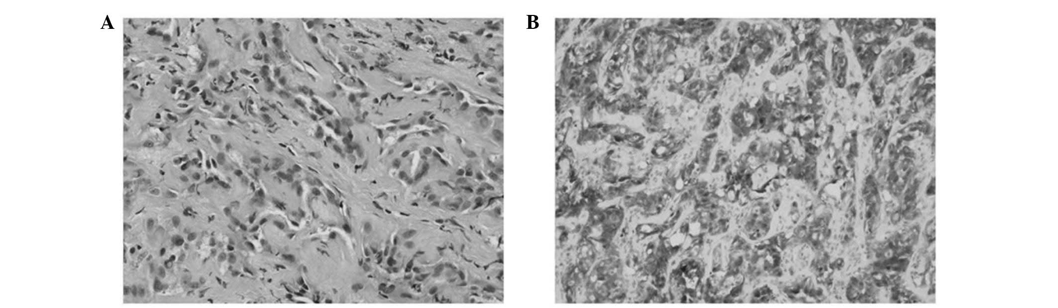
Limited local excisions were performed in the two cases and the morphological and immunohistochemical features are presented in Table II and Fig. 1. At the time of writing, follow-ups have revealed no recurrence.

Figure 1

Adenomatoid tumor of the testis. (A) Hematoxylin-eosin stain revealing tubules of cuboidal cells with occasional cytoplasmic vacuolation. Nuclei are bland with dispersed chromatin and surrounded by fibrous stroma (magnification, ×200). (B) Immunohistochemical staining revealing marked Wilms’ tumor gene 1 expression by neoplastic cells (magnification, ×100).

Table II

Morphological and pathological features of the masses.

Table II

Morphological and pathological features of the masses.

No.Size, cmDensityLocationMicroscopic features Immunohistochemistry
11×1FirmTunica vaginalisChannels lined by flattened cells with cytoplasmic vacuolation, myxoid changeCK+, D2–40+, CR+, CD68−
22×1.5FirmEpididiymisTubules of cuboidal cells with cytoplasmic vacuolation, bland nuclei, fibrous stromaWT1+, calretinin+, D2–40+, CK5/6−

[i] CK, cytokine; CR, complement receptor; CD, cluster of differentiation; WT1, Wilms’ tumor gene 1.

Discussion

Adenomatoid tumors are relatively uncommon benign tumors of mesothelial origin frequently located within the genital tract organs (6). In the female genital tract, adenomatoid tumors may be located in the uterus, fallopian tubes and ovarian hilus, and the epididymis, spermatic cord, prostate and ejaculatory duct in the male (1). Adenomatoid tumors account for ~30% of all paratesticular tumors and are are therefore the most common type of paratesticular tissues (7). In contrast to testicular neoplasms, which are 95% malignant, paratesticular tumors are more frequently benign and may be cured by local excision (7). However, it is not always easy to differentiate between the two masses.

Adenomatoid tumors present either as an incidental finding or a slow growing scrotal mass, most often in patients between 30 and 50 years of age. Enlargement is usually painless and the scrotal skin and surrounding adenexa remain normal. The majority of adenomatoid tumors are present asymptomatically for several years and are uniformly benign. Rarely, patients have presented with testicular pain. In the majority of cases, adenomatoid tumors present as painless, firm, intrascrotal masses of <2 cm diameter.

The ultrasound features of adenomatoid tumors may vary but usually include a well-defined, homogeneous, round nodule with variable echogenicity ranging from hypoechoic to hyperechoic. On magnetic resonance imaging, such masses are slightly hypointense compared with the testicular parenchyma on T2-weighted images and, on post-contrast images, exhibit similar enhancement to the testis (8).

Microscopically, they contain a number of histological patterns: adenoid or tubular glandular, angiomatoid, solid, cystic or transitional (9), which may pose a range of diagnostic problems, particularly for differentiating adenomatoid tumors from other paratesticular tumors (10). An understanding of the pathological features of paratesticular tumors is crucial for accurate diagnosis (Table III) (9,10).

Table III

Morphological and immunohistochemical features of paratesticular tumors.

Table III

Morphological and immunohistochemical features of paratesticular tumors.

LesionMicroscopic features Immunohistochemistry
Adenomatoid tumorCords and tubules of cuboidal to columnar cells with vacuolated cytoplasm and fibrous stromaWT1+, D2–40+, calretinin+, CK5+, vimentin+
Fibrous pseudotumorDense fibrous tissue with interspersed fibroblasts and mixed inflammatory cellsNot clear
CystadenomaEpithelial-like tumor cells with a sertoliform growth pattern and cystic dilatationsInhibin+, S-100+, CD99+
LeiomyomasInterlacing and whorling bundles of smooth muscle cells Desmin+
Serous borderline tumorCystic with numerous intracystic, blunt papillae lined by stratified epithelial cells having minimal to mild cytologic atypiaER+, PR+, CD15+, MOC-31+, calretinin−
MesotheliomaEpithelioid cells arising from the tunica vaginalis with papillary, tubulopapillary or solid architectural patterns Calretinin+, WT1+, D2–40+, CK7+, CK5/6+

[i] WT1, Wilms’ tumor gene 1; CK, cytokine; CD, cluster of differentiation; ER, estrogen receptor; PR, progesterone receptor.

An asymptomatic mass in the scrotum is a nonspecific symptom that can be caused by benign lesions and malignances. Ultrasound has become a first-line imaging technique, used to assess acute and nonacute conditions of the scrotum, and in many cases it is the only modality required (8). When ultrasound confirms that the mass is extratesticular (particularly if located in the epididymis), circumscribed and homogeneous with no evidence of invasion to adjacent tissue, in addition to typical clinical features and normal levels of serum tumor makers, a diagnosis of paratesticular mass can be made, as in case two.

In many cases, the similar imaging studies and clinical signs make it hard to distinguish adenomatoid tumors from malignant intratesticular tumors, leading to unnecessary extensive surgical excision of the whole testis (11). The present study is the first reported case to avoid extensive surgical excision based on intra-operative evaluation when this dilemma was encountered.

In summary, adenomatoid tumors in the testis are rare disorders that may pose difficulty for diagnosis, particularly when occurring in unusual locations where the aim is to preserve endogenous testicular function. This report presents two cases of adenomatoid tumors of the testis with the symptom of asymptomatic mass in the scrotum. Local excisions were performed based on thorough analysis of the clinical features, tumor makers, radiological images and intra-operative findings. A complete understanding of the pathological features of paratesticular tumors is critical in making accurate diagnoses.

Acknowledgements

This study was supported by the National Natural Science Foundation of China (grant no. 81101922), the Medical Scientific Research Foundation of Guangdong Province of China (grant nos. A2012584 and A2013606) and the Science and Technology Development Fund Project of Shenzhen (grant no. JCYJ20130402114702124).

References

1 

Schwartz EJ and Longacre TA: Adenomatoid tumors of the female and male genital tracts express WT1. Int J Gynecol Pathol. 23:123–128. 2004. View Article : Google Scholar : PubMed/NCBI

2 

Alexiev BA, Xu LF, Heath JE, et al: Adenomatoid tumor of the testis with intratesticular growth: a case report and review of the literature. Int J Surg Pathol. 19:838–842. 2011. View Article : Google Scholar : PubMed/NCBI

3 

Monappa V, Rao AC, Krishnanand G, et al: Adenomatoid tumor of tunica albuginea mimicking seminoma on fine needle aspiration cytology: a case report. Acta Cytol. 53:349–352. 2009. View Article : Google Scholar

4 

Di Pierro GB, Sciarra A, Innocenzi M and Cristini C: Rare case of multiple adenomatoid tumors arising from tunica vaginalis of testis and epididymis. Actas Urol Esp. 34:560–561. 2010.(In Spanish).

5 

Makkar M, Dayal P, Gupta C and Mahajan N: Adenomatoid tumor of testis: A rare cytological diagnosis. J Cytol. 30:65–67. 2013. View Article : Google Scholar : PubMed/NCBI

6 

Alam K, Maheshwari V, Vashney M, et al: Adenomatoid tumour of testis. BMJ Case Rep. 2011.2011:

7 

Goel A, Jain A and Dalela D: Can radical orchiectomy be avoided for paratesticular adenomatoid tumor? Indian J Urol. 27:556–557. 2011. View Article : Google Scholar : PubMed/NCBI

8 

Aganovic L and Cassidy F: Imaging of the scrotum. Radiol Clin North Am. 50:1145–1165. 2012. View Article : Google Scholar

9 

Amin MB: Selected other problematic testicular and paratesticular lesions: rete testis neoplasms and pseudotumors, mesothelial lesions and secondary tumors. Mod Pathol. 18(Suppl 2): S131–S145. 2005. View Article : Google Scholar

10 

Rubenstein RA, Dogra VS, Seftel AD and Resnick MI: Benign intrascrotal lesions. J Urol. 171:1765–1772. 2004. View Article : Google Scholar : PubMed/NCBI

11 

Pacheco AJ, Torres JL, de la Guardia FV, Arrabal Polo MA and Gómez AZ: Intraparenchymatous adenomatoid tumor dependent on the rete testis: A case report and review of literature. Indian J Urol. 25:126–128. 2009. View Article : Google Scholar : PubMed/NCBI

Related Articles

  • Abstract
  • View
  • Download
  • Twitter
Copy and paste a formatted citation
Spandidos Publications style
Chen D, Yu Z, Ni L, Gui Y, Yang S, Shi B and Lai Y: Adenomatoid tumors of the testis: A report of two cases and review of the literature. Oncol Lett 7: 1718-1720, 2014.
APA
Chen, D., Yu, Z., Ni, L., Gui, Y., Yang, S., Shi, B., & Lai, Y. (2014). Adenomatoid tumors of the testis: A report of two cases and review of the literature. Oncology Letters, 7, 1718-1720. https://doi.org/10.3892/ol.2014.1938
MLA
Chen, D., Yu, Z., Ni, L., Gui, Y., Yang, S., Shi, B., Lai, Y."Adenomatoid tumors of the testis: A report of two cases and review of the literature". Oncology Letters 7.5 (2014): 1718-1720.
Chicago
Chen, D., Yu, Z., Ni, L., Gui, Y., Yang, S., Shi, B., Lai, Y."Adenomatoid tumors of the testis: A report of two cases and review of the literature". Oncology Letters 7, no. 5 (2014): 1718-1720. https://doi.org/10.3892/ol.2014.1938
Copy and paste a formatted citation
x
Spandidos Publications style
Chen D, Yu Z, Ni L, Gui Y, Yang S, Shi B and Lai Y: Adenomatoid tumors of the testis: A report of two cases and review of the literature. Oncol Lett 7: 1718-1720, 2014.
APA
Chen, D., Yu, Z., Ni, L., Gui, Y., Yang, S., Shi, B., & Lai, Y. (2014). Adenomatoid tumors of the testis: A report of two cases and review of the literature. Oncology Letters, 7, 1718-1720. https://doi.org/10.3892/ol.2014.1938
MLA
Chen, D., Yu, Z., Ni, L., Gui, Y., Yang, S., Shi, B., Lai, Y."Adenomatoid tumors of the testis: A report of two cases and review of the literature". Oncology Letters 7.5 (2014): 1718-1720.
Chicago
Chen, D., Yu, Z., Ni, L., Gui, Y., Yang, S., Shi, B., Lai, Y."Adenomatoid tumors of the testis: A report of two cases and review of the literature". Oncology Letters 7, no. 5 (2014): 1718-1720. https://doi.org/10.3892/ol.2014.1938
Follow us
  • Twitter
  • LinkedIn
  • Facebook
About
  • Spandidos Publications
  • Careers
  • Cookie Policy
  • Privacy Policy
How can we help?
  • Help
  • Live Chat
  • Contact
  • Email to our Support Team