Introduction
Adenosquamous carcinoma (ASC) is a rare form of
malignancy which consists of two types of cell, including squamous
cells and glandular-like cells (1,2). It
has been reported that ASC can be found as an isolated tumor in
various systemic organs, including the stomach, intestines, uterus,
lungs, esophagus, anus and vagina (3–8).
However, ASC may also exist with other types of malignancy
(3), which suggests that an
accurate diagnosis is required in each case. Several studies have
demonstrated that computed tomography scanning is particularly
useful in the diagnosis of ASC (6,8), as
radiographic findings differ between organs. ASC is known to show
aggressive behavior in addition to metastatic spread (4). Imaoka et al recently analyzed
clinical features and prognosis using a large number of pancreatic
cancers including 28 cases with ASC, in which the median overall
survival rate was unfavorable for ASC compared with that for ductal
adenocarcinoma (7). Due to its
aggressive nature, surgical excision is considered one of the major
treatment options for ASC (4).
The conjunctiva contains columnar epithelium
together with goblet cells, which tends to show squamous metaplasia
in adults. Squamous cell carcinoma is one of the commonest
conjunctival epithelial malignancies (9); however, ASC has yet to be reported.
The present study reports the first known case of ASC in the
conjunctiva and analyzes the histological findings.
Case report
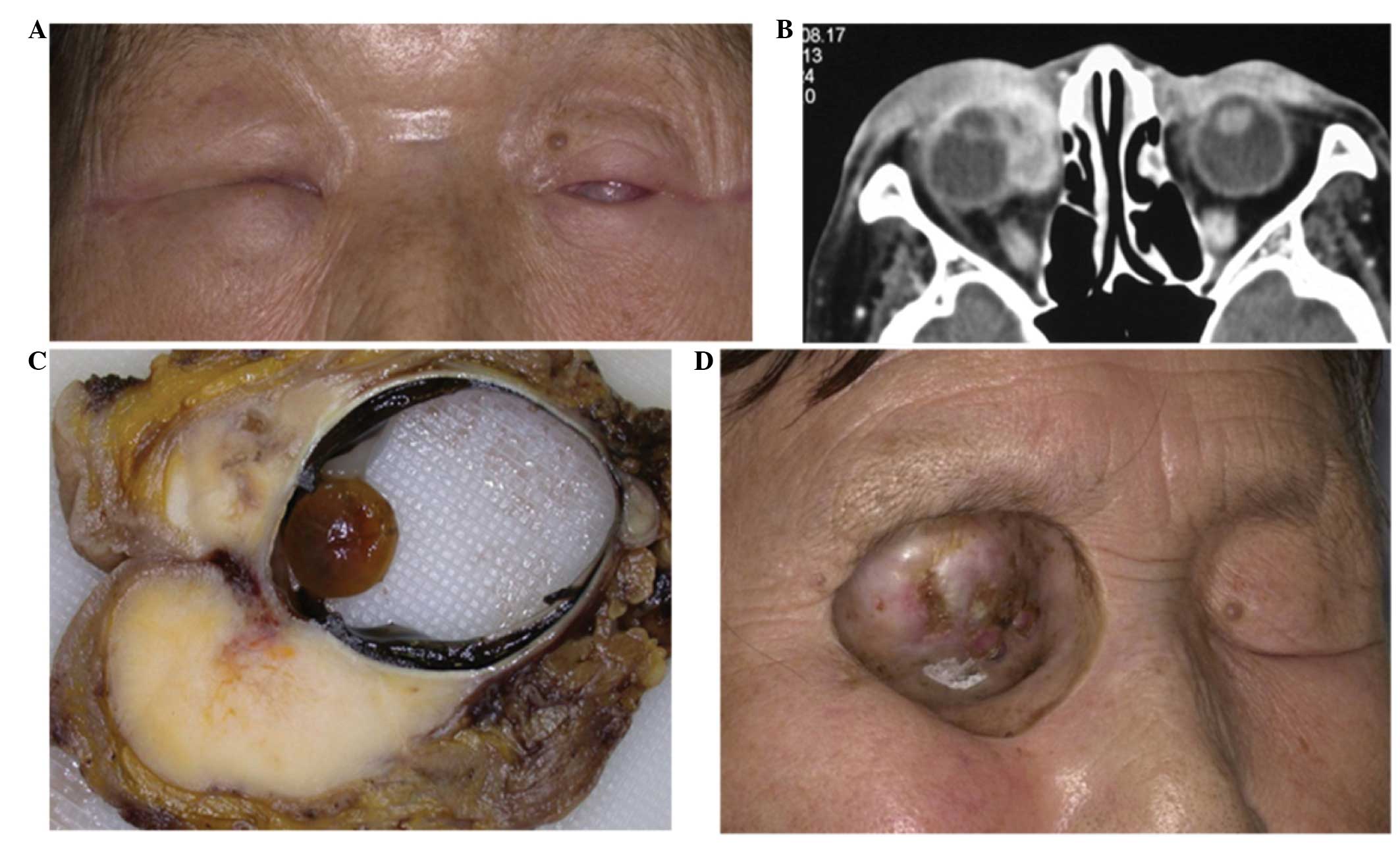
A 76-year-old female presented with right eyelid
swelling in 2001 (Fig. 1A). The
patient had a medical history of conjunctival infection with
Chlamydia trachomatis in both eyes. On initial clinical
examination, a right conjunctival tumor was noted and a biopsy was
performed. Histologically, the tumor was diagnosed as a squamous
cell carcinoma. The patient underwent radiotherapy, but the tumor
rapidly relapsed. Computed tomography demonstrated massive
high-intensity eyelid and orbital mass lesions (Fig. 1B). The patient underwent orbital
exenteration on March 7, 2003. The excised tissues revealed marked
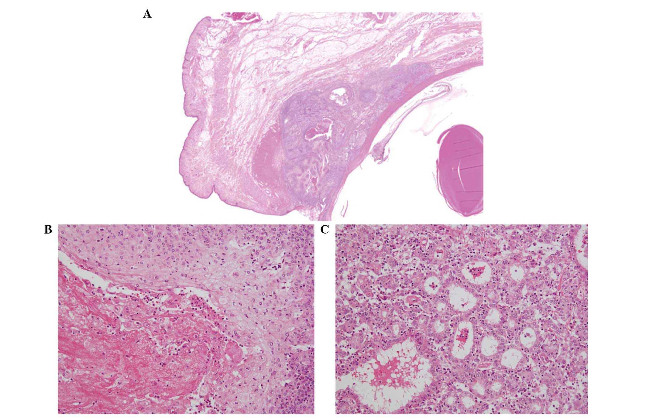
whitish nodules adjacent to the eyeball (Fig. 1C). Histologically, the excised
tissue exhibited a collection of tumor cells localized in the
conjunctiva with intermingled cystic change (Fig. 2A, arrows). At high magnification,
the conjunctival tissues were replaced with invasive tumor cells.
The majority of the tumor cells exhibited a high nucleus/cytoplasm
ratio and severe nuclear atypia with frequent mitoses. Numerous
tumor cells demonstrated squamous differentiation with a
keratinizing tendency (Fig. 2B).
Other tumor cells exhibited glandular formation (Fig. 2C). Tumor cells had invaded the
tarsal plate, extraocular muscles and cornea. The conjunctival
tumor was diagnosed as an ASC. The surgical margins were free of
tumor cells and, at the time of writing, the patient is well
without local recurrence or distant metastases (Fig. 1D).
Discussion
The occurrence of ASC is more common in organs where
adenocarcinomas arise frequently, including the stomach, intestines
and uterus. ASC has also been identified in the esophagus, anus and
vagina, where squamous cell carcinomas predominate (5). Squamous cell carcinoma is a common
malignant epithelial tumor of the conjunctiva which occurs in
predominantly male and immunosuppressed patients (9). Metastatic ASC involving the orbit has
also been reported (10), but while
it is known that primary ASC can arise from the lacrimal gland
(11), there are no reports of ASC
arising in the conjunctiva.
Although the pathogenesis of ASC remains largely
unknown, the following four hypotheses have been proposed (5): i) Malignant transformation of both
squamous and glandular-like cells originating from pleiotropic
epithelial stem cells, ii) tumorigenesis of squamous metaplasia in
the columnar epithelium, iii) transdifferentiation of
adenocarcinoma to squamous cell carcinoma, and iv) the coexistence
of both carcinomas. It is likely that the conjunctival epithelium
can exhibit squamous metaplasia, from which squamous cell carcinoma
arises. In addition, the conjunctiva contain microscopic pockets,
called the crypts of Henle, around the eyeball which are
responsible for secreting mucin, a proteinaceous substance that
makes up the inner layer of tears (12). Therefore, the ASC in the present
report may have arisen from malignant transformation of the crypts
of Henle, including squamous and glandular cells. A second
possibility is that a recurrent squamous cell carcinoma
subsequently transformed into adenocarcinoma, since the patient had
a medical history of eyelid squamous cell carcinoma.
ASC exhibits aggressive biological behavior, and is
typically associated with worse prognosis than conventional
adenocarcinoma. Therefore, surgical excision is considered a key
treatment option for ASC. The extent of surgery is dependent on the
location of the tumor (4); in a
previous case, although surgical intervention was successfully
conducted, the prognosis remained unfavorable (5). Thus, prognosis following surgical
intervention is not always clear. In the present case, orbital
exenteration was performed to eliminate the tumor cells completely.
Histologically, the surgical margin was free of tumor cells, and
the patient has remained well, without local recurrence or distant
metastases, for 10 years. Additional observation is required to
manage this rare aggressive tumor in the conjunctiva.
References
|
1
|
Straus R, Heschel S and Fortmann DJ:
Primary adenosquamous carcinoma of the stomach. A case report and
review. Cancer. 24:985–995. 1969. View Article : Google Scholar : PubMed/NCBI
|
|
2
|
Mori M, Iwashita A and Enjoji M:
Adenosquamous carcinoma of the stomach. A clinicopathologic
analysis of 28 cases. Cancer. 57:333–339. 1986. View Article : Google Scholar : PubMed/NCBI
|
|
3
|
He JJ, Ding KF, Zheng L, Xu JH, Li J, Wu
YL, Sun LF, Zhou DE and Zheng S: Adenosquamous carcinoma of the
uncinate process of the pancreas with synchronous gastrointestinal
stromal tumor of the stomach: Case report and review of the
literature. Oncol Lett. 4:1191–1194. 2012.PubMed/NCBI
|
|
4
|
Shafaghi A, Askari K, Ashoobi MT and
Mansour-Ghanaei F: Adenosquamous carcinoma of the sigmoid colon: a
case report and review of literature. Int J Clin Exp Med.
6:390–392. 2013.PubMed/NCBI
|
|
5
|
Yang SJ, Ooyang CH, Wang SY, Liu YY, Kuo
IM, Liao CH and Wu TJ: Adenosquamous carcinoma of the ampulla of
Vater - a rare disease at unusual location. World J Surg Oncol.
11:1242013. View Article : Google Scholar : PubMed/NCBI
|
|
6
|
Yin Q, Wang C, Wu Z, Wang M, Cheng K, Zhao
X, Yuan F, Tang Y and Miao F: Adenosquamous carcinoma of the
pancreas: multidetector-row computed tomographic manifestations and
tumor characteristics. J Comput Assist Tomogr. 37:125–133. 2013.
View Article : Google Scholar : PubMed/NCBI
|
|
7
|
Imaoka H, Shimizu Y, Mizuno N, Hara K,
Hijioka S, Tajika M, Kondo S, Tanaka T, Ogura T, Obayashi T,
Hasegawa T, Niwa Y and Yamao K: Clinical characteristics of
adenosquamous carcinoma of the pancreas: a matched case-control
study. Pancreas. 43:287–290. 2014. View Article : Google Scholar : PubMed/NCBI
|
|
8
|
Watanabe Y, Tsuta K, Kusumoto M, Yoshida
A, Suzuki K, Asamura H and Tsuda H: Clinicopathologic features and
computed tomographic findings of 52 surgically resected
adenosquamous carcinomas of the lung. Ann Thorac Surg. 97:245–251.
2014. View Article : Google Scholar
|
|
9
|
Kao AA, Galor A, Karp CL, Abdelaziz A,
Feuer WJ and Dubovy SR: Clinicopathologic correlation of ocular
surface squamous neoplasms at Bascom Palmer Eye Institute: 2001 to
2010. Ophthalmology. 119:1773–1776. 2012. View Article : Google Scholar : PubMed/NCBI
|
|
10
|
Som PM, Silvers AR, Catalano PJ, Brandwein
M and Khorsandi AS: Adenosquamous carcinoma of the facial bones,
skull base, and calvaria: CT and MR manifestations. AJNR Am J
Neuroradiol. 18:173–175. 1997.PubMed/NCBI
|
|
11
|
Cherian I, Shrestha SP, Panhani ML, Talwar
OP, Yogi N, Rai S, Lalchan S and Fernandes C: Adenosquamous
carcinoma of the lacrimal gland. BMJ Case Rep. 2010:2010.
|
|
12
|
Hase K, Kase S, Noda M, Ohashi T, Shinkuma
S and Ishida S: Ectopic cilia: a histopathological study. Case Rep
Dermatol. 4:37–40. 2012. View Article : Google Scholar : PubMed/NCBI
|