Introduction
Prostate cancer is the second leading type of
malignancy in males in North America with an estimated 186,320 new
cases and 28,660 mortalities reported in 2008 (1). The number of patients with prostate
cancer has also been increasing in Japan (2). Alterations in nuclear morphometry,
gene and protein expression, gene promoter methylation and
angiogenesis are known to be involved in prostate carcinogenesis
and contribute to field cancerization in the prostate (3).
Our understanding of carcinogenesis has been
enhanced by the recently revived cancer stem cell (CSC) theory.
CSCs have been reported in multiple solid tumors in different
tissues, including the prostate (4–6). CSCs
are endowed with high tumorigenic capacity and may drive tumor
formation, maintain tumor homeostasis and mediate tumor metastasis.
A number of primary non-malignant and malignant tumor-derived human
prostate epithelial cell lines have been developed using a
retroviral vector encoding human telomerase reverse transcriptase.
These cell lines exhibit the characteristics of stem cells and
express embryonic stem (ES) cell markers, such as NANOG, octamer 4
(OCT4) and SRY-box 2 (Sox-2), as well as the early progenitor cell
markers, cluster of differentiation 133 (CD133), CD44 and NESTIN
(7,8).
The multipotent stem cell marker NANOG was
identified in 2003 (9,10). NANOG is specifically expressed in
the human embryonic pluripotent stem cells of embryos prior to or
following implantation, primordial germ cells, ES cells cultured
in vitro, embryonic germ cells and embryonic carcinoma
cells, and functions in the promotion of cell proliferation. NANOG
is expressed in dysgerminoma and embryonic carcinoma, but not in
immature teratoma, endodermal sinus tumors or choriocarcinoma
(11). NANOG can be used to
distinguish between germ cell tumors and non-germ cell tumors
(11). NANOG has also been found to
be a sensitive and specific marker of metastatic germ cell tumors
(11,12). With regard to prostate cancer,
several studies have recently suggested the positive reaction of
adenocarcinoma (ADC) cells against NANOG (13,14).
Therefore, NANOG is an emerging focus in developmental biology, due
to its importance in the maintenance of self-renewal and
multipotential capacity in a variety of malignancies, including
prostate cancer. Octamer 4 (OCT4) belongs to the family of
Pit-Oct-Unc-domain transcription factors and has been found in ES
and germ cells (15). A number of
reports have shown that OCT4 is pivotal in maintaining the
self-renewal and pluripotency of ES cells (16). Recently, it has also been shown that
cancer cells expressing OCT4 and Sox2 may be crucial in cancer
development (17). The two genes,
Sox2 and OCT4, are part of an important gene regulatory network,
and are essential for embryogenesis and the pluripotency and
self-renewal of cells (16).
Previous studies have also suggested that certain cancers,
including prostate cancer (14,18),
express Sox2 and OCT4 simultaneously (19,20),
and their expression has been associated with the differentiation
of tumors (21). These two genes
are significant for cancer cell survival. CD133 is a transmembrane
glycoprotein that is originally expressed in a subset of stem cells
in the hematopoietic system as well as in the solid tumors of other
tissues (22), including the
prostate (23). CD133-positive
cancer cells have cancer stem/progenitor cells that exhibit
resistance to cancer therapies (including radiation and
chemotherapy), a greater invasion ability and metastasis in various
malignancies. Thus, the utility of CD133 expression as a prognostic
marker has been suggested (22), as
well as in the prostate (23).
NESTIN is an intermediate filament protein that is known to be
important as a neural stem cell marker (24). However, the expression of NESTIN has
recently been reported to be associated with the proliferation of
progenitor cell populations within neoplasms (25). In addition, the upregulation of
NESTIN has been found to closely correlate with the malignancy and
metastasis of a variety of malignancies (25), including prostate cancer (26).
The expression of NANOG, OCT4, Sox, NESTIN and CD44
has been observed in human prostate ADC cells (7), which suggests the importance of cancer
stem and progenitor cells in prostate carcinogenesis. However,
OCT4A-expressing cells have rarely been identified in human benign
and malignant prostate glands (27). The number of OCT4A-expressing cells
has been shown to increase in prostate ADC with high Gleason scores
(27). OCT4A-expressing cancer
cells have also been shown to coexpress Sox2, an ES cell marker,
but did not express other putative stem cell markers, such as NANOG
and CD133 (27). The neuroendocrine
differentiation markers, chromogranin A and synaptophysin, are also
coexpressed by the majority of OCT4A-expressing cells (27). Thus, discrepancies exist in reports
investigating the role and expression of certain stem and
progenitor cell markers in prostate cancer cells.
In the current study, in order to determine whether
certain stem cell markers may be used for the diagnosis of prostate
cancer, the immunohistochemical expression of NANOG, OCT4, CD133
and NESTIN, which are well-known stem cell markers, were
investigated in 38 cases from a total of 114 biopsy specimens
obtained from Japanese patients with prostate cancer between
January 2011 and December 2011. In addition, the correlation
between the expression of these stem cell markers in prostate
cancer and non-cancerous tissues was evaluated. Hypoxia has been
associated with an aggressive course and poor clinical outcome of
cancer (28,29); low oxygen may promote the
self-renewal of CSCs (14,30–32).
Therefore, the immunohistochemical expression of hypoxia-inducible
factor (HIF)-1α was also examined.
Materials and methods
Study samples
Between October 2010 and September 2011, a total of
114 patients with elevated serum prostate-specific antigen levels
of >4 ng/ml and/or abnormal digital rectal examinations were
referred to the Kanazawa Medical University Hospital (Uchinada,
Japan) and underwent transrectal ultrasound sonography-guided
eight-core biopsies. Histopathological diagnosis was re-evaluated
by a certified pathologist on hematoxylin and eosin-stained
sections from the biopsy samples. Prior to this study, written
informed consent was obtained from all patients. The study was
approved by the Ethics Committee of Kanazawa Medical University
(Uchinada, Japan), and the Declaration of Helsinki regarding the
use of human tissue was strictly followed.
Immunohistochemistry
Serial sections, 4 μm in thickness, prepared from
formalin-fixed, paraffin-embedded specimens, were available for
immunohistochemical analysis. Sections were deparaffinized and
rehydrated following standard methods. Briefly, the sections were
deparaffinized three times with xylene for 5 mins, and rehydrated
in graded ethanol (80–100%) for 5 mins. A microwave antigen
retrieval procedure was performed for 20 min in citrate buffer (pH
6.0) and hydrogen peroxide was used to block non-specific
peroxidase reactions. Following washing with phosphate-buffered
saline (PBS, pH 7.4), sections were incubated with rabbit
polyclonal anti-human NANOG (ab21624; 1:30 dilution; Abcam,
Cambridge, MA, USA), OCT4 (ab18976; 1:100 dilution; Abcam), CD133
(ab19898; 1:200 dilution; Abcam) and NESTIN (ab93666; 1/120
dilution; Abcam), as well as mouse monoclonal anti-human HIF-1α
(ab10625; 1:200 dilution; Abcam). Following washing three times
with PBS, sections were incubated at 37°C with biotin-conjugated
goat anti-rabbit polyclonal antibody (ab6720; Abcam) for 20 min.
Visualization was achieved by incubation with diaminobenzidine for
10 min and slides were counterstained with Mayer hematoxylin.
Following hydration in graded alcohol and clearing with xylene, the
slides were mounted with neutral gum. Seminomas obtained from
testicular cancer specimens of two patients (Kanazawa Medical
University Hospital) who had undergone surgical resection, which
had been confirmed to overexpress NANOG and OCT4, were selected as
the appropriate positive controls (33), and paraffin-embedded Caco-2 cells
(cat. no. CRL-2102; American Type Culture Collection, Manassas, VA,
USA) and endothelial cells in ADC obtained from colorectal cancer
specimens of two patients (Kanazawa Medical University Hospital)
who had undergone surgical resection were used as internal positive
controls for CD133 and NESTIN (34,35).
Negative controls were prepared by incubating samples without the
primary antibody. The intensity of immunoreactivity against all the
primary antibodies used was assessed using a microscope (Olympus
BX41; Olympus Optical, Tokyo, Japan). Indices were determined by
counting the number of positive nuclei among ≥300 cells in
high-power fields, and were indicated as percentages. Positive
cells were evaluated for their intensity of immunoreactivity on a 0
or 3+ scale. The overall intensity of the staining reaction was
scored as follows: 0, no immunoreactivity and no positive cells; 1
(+/−), weak expression in <50% cells; 2 (+), moderate expression
in ≥50% cells; 3 (++), moderate to strong expression in 51–75%
cells; and 4 (+++), strong and diffuse expression in >76% cells.
Slides were reviewed by one pathologist blinded to the clinical
data.
Statistical analysis
Incidences among the groups were compared using a
two-tailed unpaired t-test and Bonferroni multiple comparison test
(GraphPad InStat version 3.05; GraphPad Software, San Diego, CA,
USA). P<0.05 was considered to indicate a statistically
significant difference between the groups.
Results
General observations
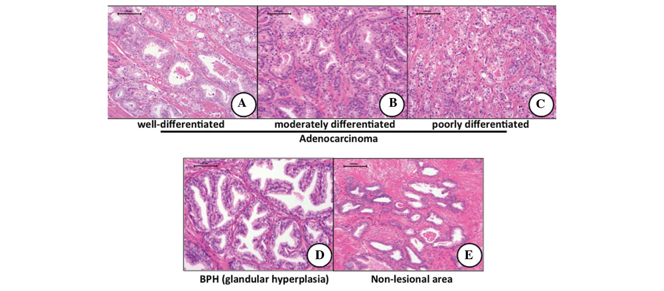
Prostate cancer was found in 38 (33.3%) of 114 males
who underwent eight core biopsy and were divided into two subgroups
according to the following Gleason scores: 30 cases with <6
(3+3) and eight cases with >7 (3+4), as shown in Fig. 1A–C. Other specimens were diagnosed
as benign prostate hyperplasia with marginal prostatitis (Fig. 1D) or normal prostate glands
(Fig. 1E).
Immunohistochemical findings
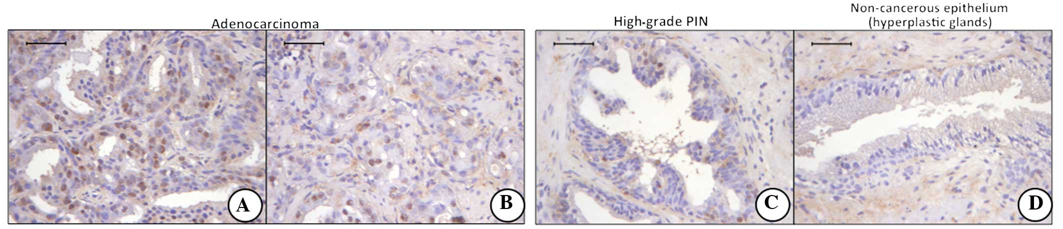
Of the four stem cell markers, ADC cells in all
specimens of the 38 cases of prostate ADC were found to positively
express the NANOG (Fig. 2A) and
OCT4 (Fig. 2B) proteins. However,
the immunohistochemical expression of CD133 (Fig. 2C) and NESTIN (Fig. 2D) was extremely weak or absent in
the cancer cells of prostate ADC and those of non-cancerous cells.
High-grade prostate intraepithelial neoplasia (PIN) was positive
for NANOG (Fig. 2E) and OCT4
(Fig. 2F); however, the number of
positive cells was fewer than that of prostate cancer. The majority
of hyperplastic glands were negative for NANOG (Fig. 2I) and OCT4 (Fig. 2J) staining. The cells of
hyperplastic glands were completely negative for CD133 (Fig. 2K) and NESTIN (Fig. 2L). Positive reactions for NANOG and
OCT4 were predominantly localized in the nuclei of cancer cells and
the cell nuclei of PIN. The staining intensity of NANOG was
stronger than that of OCT4.
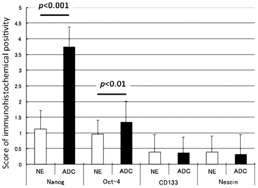
Fig. 3 shows the
scorings for the immunohistochemical expression of the four
different stem cell markers in prostate cancer and non-cancerous
cells. The immunoreactivities of NANOG (P<0.001) and OCT4
(P<0.01) in prostate cancer was significantly greater than those
in the non-cancerous cells. No significant differences were
identified between the immunoreactivities of CD133 and NESTIN in
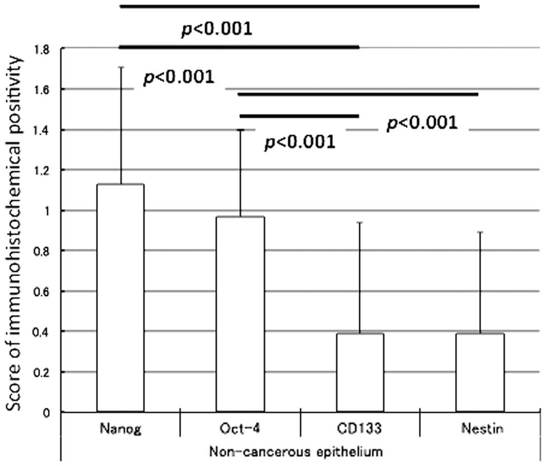
the prostate cancer and non-cancerous cells. Based on the detailed
analysis, the scoring data for the four different stem cell markers
in the non-cancerous and prostate cancer cells are also illustrated
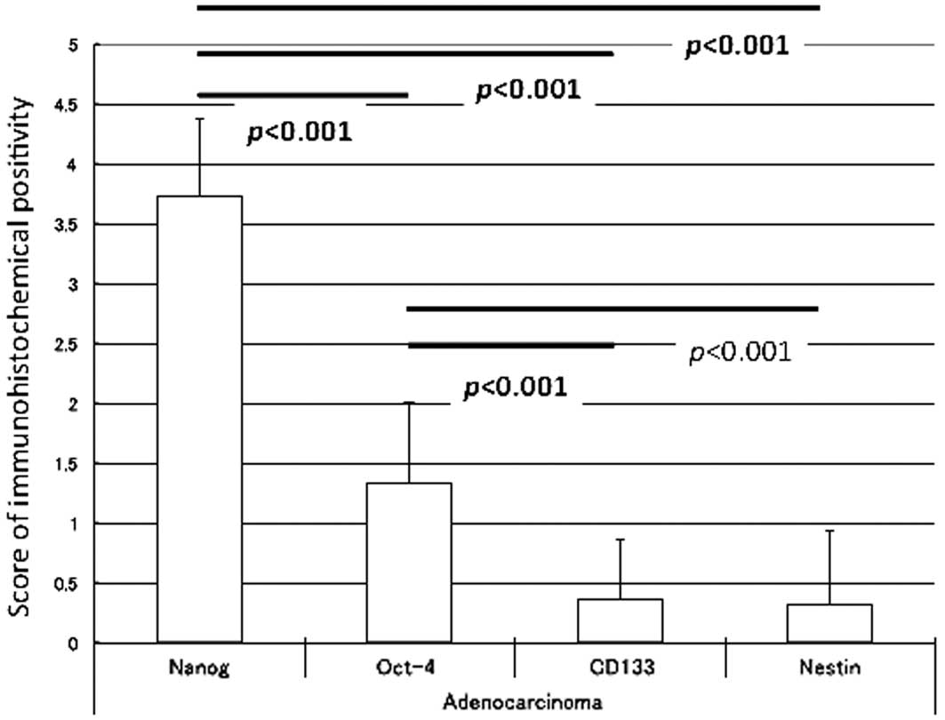
in Figs. 4 and 5, respectively. The immunohistochemical
intensity of prostate cancer was weakest for NANOG followed by
OCT4, with the strongest staining for CD133 and NESTIN. In the
non-cancerous tissue, as shown in Fig.
4, the immunoreactivities of NANOG (P<0.001) and OCT4
(P<0.001) were significantly greater than those of CD133 and
NESTIN. In prostate cancer, NANOG (P<0.001) immunoreactivity was
the strongest among the four stem cell markers (Fig. 5).
The expression score for NANOG in the prostate
cancer cells was significantly greater than that of cells in
high-grade PIN and the hyperplastic glands (P<0.001 for each
comparison; Fig. 6). The number of
atypical cells in high-grade PIN was also higher than that in the
hyperplastic glands (P<0.001; Fig.
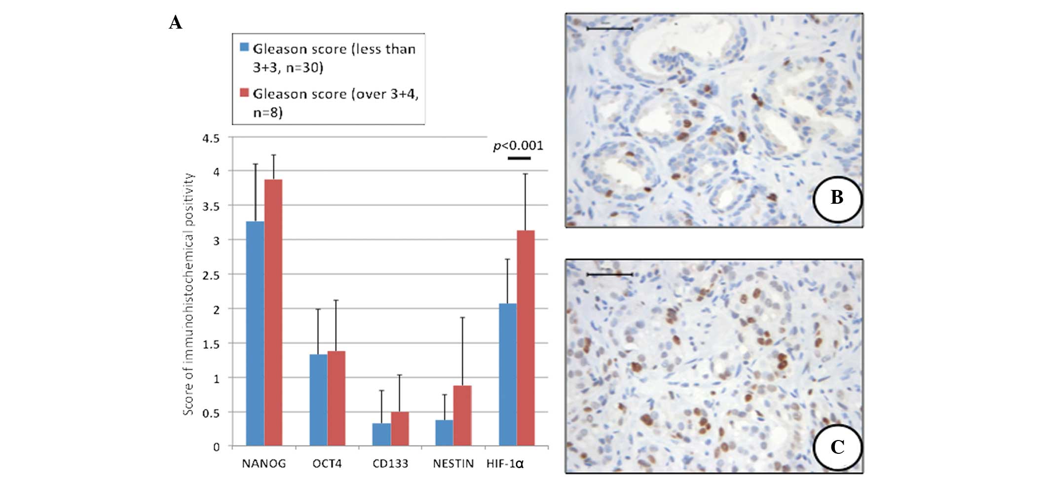
6). The expression of NANOG, OCT4, CD133 and NESTIN in prostate
ADCs with high Gleason scores (>3+4) was greater than that in
prostate cancers with low Gleason scores (<3+3), although this
difference was not significant (Fig.
7).
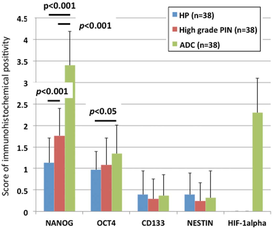 | Figure 6Immunohistochemical scores of four
stem cell markers and HIF-1α in the hyperplastic glands (benign
prostate hyperplasia), high-grade PIN and prostatic ADC. The score
of NANOG in ADC was significantly greater than that of the
hyperplastic glands (P<0.001) and high-grade PIN (P<0.001);
the value of high-grade PIN was significantly higher than that of
hyperplastic glands (P<0.001). The score of OCT4 in ADC was
significantly higher than that in the hyperplastic glands
(P<0.05), while the scores of CD133 and NESTIN of three lesions
(hyperplastic glands, high-grade PIN and ADC) were almost similar.
HIF-1α was expressed in the nuclei of ADC, but not in the
hyperplastic glands and high-grade PIN. HP, hyperplastic; ADC,
adenocarcinoma; HIF-1α, hypoxia-inducible factor-1α; PIN, prostate
intraepithelial neoplasia; OCT4, octamer 4; CD133, cluster of
differentiation 133. |
HIF-1α immunohistochemistry revealed that specific
cancer cell nuclei (Fig. 8A and B),
corresponding to their Gleason score, as well as a few cell nuclei
in high-grade PIN showed a positive reaction for HIF-1α (Fig. 8C). However, hyperplastic and normal
glandular cells were negative for HIF-1α (Fig. 8D). The mean score for HIF-1α with a
high Gleason score was significantly greater than that of HIF-1α
with a low Gleason score (P<0.001; Fig. 7).
Discussion
Pluripotency-associated transcription factors,
including NANOG, Sox2 and OCT4, are known as regulators of cellular
identity in ES cells (36) and have
recently been identified in the epithelial malignancies of a
variety of tissues (33,37), including prostate cancer (13,14,18).
CD133 (23) and NESTIN (26) have also been reported to be
expressed in prostate cancer. However, these reports were
predominantly from human prostate cancer cell lines. Consistent
with their role in sustaining the stemness of ES cells,
pluripotency-related factors have been suggested to be expressed at
a higher frequency in cancer exhibiting lower degrees of
differentiation (37). In this
study, the immunohistochemical expression of NANOG was markedly
higher than that of the other stem cell markers, OCT4, CD133 and
NESTIN. The reason for the discrepancy between the findings of the
current study and those reported by others is not known; however,
differences between prostate cancer obtained from biopsy specimens
and human prostate cancer cell lines may have influenced the
stainability of the four different stem cell markers, NANOG, OCT4,
CD133 and NESTIN. Although Miki et al (8) observed tumor compartments and
high-grade PIN with higher CD133 and an inverse correlation with
androgen receptor staining, a CD133-positive reaction was not
detected in the prostate cancer cells, PIN or hyperplastic
glandular cells in this study.
CSCs comprise of ~0.01% of the tumor cell
population. In this study, a large number of strongly positive
NANOG and/or OCT4 cancer cells were observed. This high level of
expression is not necessarily associated with stem cell behavior,
but rather to the deregulation proteins that provide some type of
growth advantage to cancer cells (38–41).
Androgen deprivation-induced atrophy of the prostate
gland and subsequent regeneration following androgen replacement
have indicated that the stem cell population may reside in the
adult prostate gland in rodents (42). The origin of prostate cancer remains
unknown and has given rise to a series of hypotheses (43). Prostate ADCs are frequently
multifocal, show the same immunohistochemical profile as benign
glandular cells, and lack basal cell markers, such as p63 and
cytokeratin 34β. This indicates that prostate cancer may develop
from altered benign glandular cells. However, multiple pluripotency
markers, such as CD44, CD117 and Oct3/4, have been shown to be
expressed in prostate cancer, indicating that prostate cancer may
develop from common stem cell-like or intermediate cells (8,44). The
findings on the expression of NANOG described in the current study
may also support this hypothesis.
NANOG (9,10) is one of the four factors known to
reprogram adult cells into germline-competent induced pluripotent
stem cells (45). NANOG is also
critical in maintaining the self-renewal and pluripotency of ES
cells by regulating the cell fate of the pluripotent inner cell
mass (46–48). Notably, elevated NANOG protein
expression in several types of human cancer has been reported,
predominantly in germ cell tumors, as well as the malignancies of
non-germ cells (38), suggesting
the involvement of NANOG in tumorigenesis and progression. Non-germ
cell tumors, including breast (38)
and oral cancer (49), also express
NANOG. A systematic study using animal models and in vitro
cell systems has provided substantial evidence for the key function
of NANOG in human tumor development (50). A recent study has shown that the
transforming growth factor (TGF)-β pathway is involved in the
regulation of NANOG gene expression via binding with the NANOG
proximal promoter (51). TGF-β
functions as a key tumor suppressor of the prostate and can also
promote malignant progression and metastasis of the advanced
disease (52). Human cultured
prostate cancer cells, prostate cancer xenografts and primary
prostate cancer cells express a functional variant of NANOG, NANOG
mRNA, in cancer cells (50). This
expression is derived predominantly from a retrogene locus termed
NANOGp8 (50). In this study, the
NANOG protein was detected in the nucleus of cancer cells, but was
not expressed in hyperplastic glandular cells. These findings
suggest that NANOG has a particular function in prostate cancer
development. In addition, a significant correlation has been
reported between NANOG-, OCT4- and HIF-1α-positive regions
(31). Low oxygen levels promote
self-renewal in stem cells and hypoxia has been associated with an
aggressive disease course and poor clinical outcomes in
malignancies, including prostate cancer (28,29).
Furthermore, a number of aggressive neoplasms exhibit gene
expression signatures characteristic of human ES cells. Thus, HIF
may act as a key inducer of a dynamic state of stemness in
pathological conditions.
OCT4 maintains pluripotency in embryogenesis; the
upregulation of OCT4 results in differentiation to the primitive
endoderm and mesoderm, while downregulation induces a loss of
pluripotency and dedifferentiation into the trophectoderm (53). A recent report questioned the
function of OCT4 as a pure stem cell marker by showing its
expression in differentiated cells (54). Ugolkov et al (18) reported that OCT4 nuclear expression
was markedly associated with benign prostatic lesions, but not
prostate cancer. In the present study, OCT4 expression was found in
the prostate cancer and non-cancerous glandular cells; however,
differences were observed in its expression between prostate cancer
and non-cancerous glands. Although, these differences were not as
marked as those observed in NANOG expression.
One notable observation in the current study was
that prostate cancer cells expressing NANOG and OCT4 were also
positive for HIF-1α reactivity. Hypoxia-regulated genes are
mediated by the HIF-1 complex composed of a heterodimeric pair of
HIF-1α and -1β (28,29), and HIF-1α is an important
transcription factor in prostate carcinogenesis, which suggests
that HIF-1α may be a potential prognostic biomarker in the
proteomic assessments of prostate cancers (55,56).
Additionally, HIF-1α induces human ES cell markers, such as NANOG
(14,30,31),
OCT4 (14,30,31)
and CD133 (32), in cancer cells.
The findings of the current study showing the coexpression of
NANOG, OCT4 and HIF-1α support these studies. However, a slightly
positive reaction or null of CD133 in cancer cells was observed.
The reason for this was unknown; although, a strong correlation was
identified between NANOG and HIF-1α expression, which may suggest
that NANOG and HIF-1α co-operate in prostate carcinogenesis.
The results of this study showed that of the four
CSC markers examined (NANOG, OCT4, CD133 and NESTIN), NANOG was
intensively expressed in prostate cancer. In addition, HIF-1α was
coexpressed in cancer cells. These findings suggest that NANOG, in
conjunction with HIF-1α, may be important in prostate
carcinogenesis. In addition, the immunohistochemical expression of
NANOG may present as a biomarker for investigating the pathobiology
of prostate cancer.
Acknowledgements
The authors would like to thank Dr Kohei Kawaguci,
Dr Osamu Ueki and Mr. Hideaki Nishida for providing the biopsy
samples. This study was supported, in part, by a grant (2010) from
the Hokkoku Cancer Research Promotion Foundation and a Grant for
Alumni Research from the Kanazawa Medical University (grant no.
AR2012-01).
References
|
1
|
Jemal A, Siegel R, Ward E, Hao Y, Xu J,
Murray T and Thun M: Cancer statistics, 2008. CA Cancer J Clin.
58:71–96. 2008.
|
|
2
|
Matsuda T, Marugame T, Kamo K, Katanoda K,
Ajiki W and Sobue T: Cancer incidence and incidence rates in Japan
in 2002: based on data from 11 population-based cancer registries.
Jpn J Clin Oncol. 38:641–648. 2008.
|
|
3
|
Nonn L, Ananthanarayanan V and Gann PH:
Evidence for field cancerization of the prostate. Prostate.
69:1470–1479. 2009.
|
|
4
|
Al-Hajj M, Wicha MS, Benito-Hernandez A,
Morrison SJ and Clarke MF: Prospective identification of
tumorigenic breast cancer cells. Proc Natl Acad Sci USA.
100:3983–3988. 2003.
|
|
5
|
Collins AT, Berry PA, Hyde C, Stower MJ
and Maitland NJ: Prospective identification of tumorigenic prostate
cancer stem cells. Cancer Res. 65:10946–10951. 2005.
|
|
6
|
O’Brien CA, Pollett A, Gallinger S and
Dick JE: A human colon cancer cell capable of initiating tumour
growth in immunodeficient mice. Nature. 445:106–110. 2007.
|
|
7
|
Gu G, Yuan J, Wills M and Kasper S:
Prostate cancer cells with stem cell characteristics reconstitute
the original human tumor in vivo. Cancer Res. 67:4807–4815.
2007.
|
|
8
|
Miki J, Furusato B, Li H, Gu Y, Takahashi
H, Egawa S, Sesterhenn IA, McLeod DG, Srivastava S and Rhim JS:
Identification of putative stem cell markers, CD133 and CXCR4, in
hTERT-immortalized primary nonmalignant and malignant tumor-derived
human prostate epithelial cell lines and in prostate cancer
specimens. Cancer Res. 67:3153–3161. 2007.
|
|
9
|
Chambers I, Colby D, Robertson M, Nichols
J, Lee S, Tweedie S and Smith A: Functional expression cloning of
Nanog, a pluripotency sustaining factor in embryonic stem cells.
Cell. 113:643–655. 2003.
|
|
10
|
Mitsui K, Tokuzawa Y, Itoh H, Segawa K,
Murakami M, Takahashi K, Maruyama M, Maeda M and Yamanaka S: The
homeoprotein Nanog is required for maintenance of pluripotency in
mouse epiblast and ES cells. Cell. 113:631–642. 2003.
|
|
11
|
Zhang S, Balch C, Chan MW, Lai HC, Matei
D, Schilder JM, Yan PS, Huang TH and Nephew KP: Identification and
characterization of ovarian cancer-initiating cells from primary
human tumors. Cancer Res. 68:4311–4320. 2008.
|
|
12
|
Santagata S, Ligon KL and Hornick JL:
Embryonic stem cell transcription factor signatures in the
diagnosis of primary and metastatic germ cell tumors. Am J Surg
Pathol. 31:836–845. 2007.
|
|
13
|
Gong C, Liao H, Guo F, Qin L and Qi J:
Implication of expression of Nanog in prostate cancer cells and
their stem cells. J Huazhong Univ Sci Technolog Med Sci.
32:242–246. 2012.
|
|
14
|
Ma Y, Liang D, Liu J, Axcrona K, Kvalheim
G, Stokke T, Nesland JM and Suo Z: Prostate cancer cell lines under
hypoxia exhibit greater stem-like properties. PLoS One.
6:e291702011.
|
|
15
|
Scholer HR, Hatzopoulos AK, Balling R,
Suzuki N and Gruss P: A family of octamer-specific proteins present
during mouse embryogenesis: evidence for germline-specific
expression of an Oct factor. EMBO J. 8:2543–2550. 1989.
|
|
16
|
Boiani M and Scholer HR: Regulatory
networks in embryo-derived pluripotent stem cells. Nat Rev Mol Cell
Biol. 6:872–884. 2005.
|
|
17
|
Monk M and Holding C: Human embryonic
genes re-expressed in cancer cells. Oncogene. 20:8085–8091.
2001.
|
|
18
|
Ugolkov AV, Eisengart LJ, Luan C and Yang
XJ: Expression analysis of putative stem cell markers in human
benign and malignant prostate. Prostate. 71:18–25. 2011.
|
|
19
|
Mallanna SK and Rizzino A: Systems biology
provides new insights into the molecular mechanisms that control
the fate of embryonic stem cells. J Cell Physiol. 227:27–34.
2012.
|
|
20
|
Tysnes BB: Tumor-initiating and
-propagating cells: cells that we would like to identify and
control. Neoplasia. 12:506–515. 2010.
|
|
21
|
Till JE: Stem cells in differentiation and
neoplasia. J Cell Physiol Suppl. 1:3–11. 1982.
|
|
22
|
Grosse-Gehling P, Fargeas CA, Dittfeld C,
Garbe Y, Alison MR, Corbeil D and Kunz-Schughart LA: CD133 as a
biomarker for putative cancer stem cells in solid tumours:
limitations, problems and challenges. J Pathol. 229:355–378.
2013.
|
|
23
|
Missol-Kolka E, Karbanova J, Janich P,
Haase M, Fargeas CA, Huttner WB and Corbeil D: Prominin-1 (CD133)
is not restricted to stem cells located in the basal compartment of
murine and human prostate. Prostate. 71:254–267. 2011.
|
|
24
|
Lendahl U, Zimmerman LB and McKay RD: CNS
stem cells express a new class of intermediate filament protein.
Cell. 60:585–595. 1990.
|
|
25
|
Ishiwata T, Matsuda Y and Naito Z: Nestin
in gastrointestinal and other cancers: effects on cells and tumor
angiogenesis. World J Gastroenterol. 17:409–418. 2011.
|
|
26
|
Kleeberger W, Bova GS, Nielsen ME, Herawi
M, Chuang AY, Epstein JI and Berman DM: Roles for the stem cell
associated intermediate filament Nestin in prostate cancer
migration and metastasis. Cancer Res. 67:9199–9206. 2007.
|
|
27
|
Sotomayor P, Godoy A, Smith GJ and Huss
WJ: Oct4A is expressed by a subpopulation of prostate
neuroendocrine cells. Prostate. 69:401–410. 2009.
|
|
28
|
Kimbro KS and Simons JW: Hypoxia-inducible
factor-1 in human breast and prostate cancer. Endocr Relat Cancer.
13:739–749. 2006.
|
|
29
|
Mabjeesh NJ and Amir S: Hypoxia-inducible
factor (HIF) in human tumorigenesis. Histol Histopathol.
22:559–572. 2007.
|
|
30
|
Bao B, Ahmad A, Kong D, Ali S, Azmi AS, Li
Y, Banerjee S, Padhye S and Sarkar FH: Hypoxia induced
aggressiveness of prostate cancer cells is linked with deregulated
expression of VEGF, IL-6 and miRNAs that are attenuated by CDF.
PLoS One. 7:e437262012.
|
|
31
|
Mathieu J, Zhang Z, Zhou W, Wang AJ,
Heddleston JM, Pinna CM, Hubaud A, Stadler B, Choi M, Bar M, et al:
HIF induces human embryonic stem cell markers in cancer cells.
Cancer Res. 71:4640–4652. 2011.
|
|
32
|
Salnikov AV, Liu L, Platen M, Gladkich J,
Salnikova O, Ryschich E, Mattern J, Moldenhauer G, Werner J,
Schemme P, et al: Hypoxia induces EMT in low and highly aggressive
pancreatic tumor cells but only cells with cancer stem cell
characteristics acquire pronounced migratory potential. PLoS One.
7:e463912012.
|
|
33
|
Clark AT: The stem cell identity of
testicular cancer. Stem Cell Rev. 3:49–59. 2007.
|
|
34
|
Cizkova D, Soukup T and Mokry J: Nestin
expression reflects formation, revascularization and reinnervation
of new myofibers in regenerating rat hind limb skeletal muscles.
Cells Tissues Organs. 189:338–347. 2009.
|
|
35
|
Horst D, Kriegl L, Engel J, Kirchner T and
Jung A: CD133 expression is an independent prognostic marker for
low survival in colorectal cancer. Br J Cancer. 99:1285–1289.
2008.
|
|
36
|
Chambers I and Tomlinson SR: The
transcriptional foundation of pluripotency. Development.
136:2311–2322. 2009.
|
|
37
|
Ben-Porath I, Thomson MW, Carey VJ, Ge R,
Bell GW, Regev A and Weinberg RA: An embryonic stem cell-like gene
expression signature in poorly differentiated aggressive human
tumors. Nat Genet. 40:499–507. 2008.
|
|
38
|
Ezeh UI, Turek PJ, Reijo RA and Clark AT:
Human embryonic stem cell genes OCT4, NANOG, STELLAR, and GDF3 are
expressed in both seminoma and breast carcinoma. Cancer.
104:2255–2265. 2005.
|
|
39
|
Li L, Yu H, Wang X, Zeng J, Li D, Lu J,
Wang C, Wang J, Wei J, Jiang M and Mo B: Expression of seven
stem-cell-associated markers in human airway biopsy specimens
obtained via fiberoptic bronchoscopy. J Exp Clin Cancer Res.
32:282013.
|
|
40
|
Luo W, Li S, Peng B, Ye Y, Deng X and Yao
K: Embryonic stem cells markers SOX2, OCT4 and Nanog expression and
their correlations with epithelial-mesenchymal transition in
nasopharyngeal carcinoma. PLoS One. 8:e563242013.
|
|
41
|
Yin X, Li YW, Zhang BH, Ren ZG, Qiu SJ, Yi
Y and Fan J: Coexpression of stemness factors Oct4 and Nanog
predict liver resection. Ann Surg Oncol. 19:2877–2887. 2012.
|
|
42
|
English HF, Santen RJ and Isaacs JT:
Response of glandular versus basal rat ventral prostatic epithelial
cells to androgen withdrawal and replacement. Prostate. 11:229–242.
1987.
|
|
43
|
Lawson DA and Witte ON: Stem cells in
prostate cancer initiation and progression. J Clin Invest.
117:2044–2050. 2007.
|
|
44
|
Hurt EM, Kawasaki BT, Klarmann GJ, Thomas
SB and Farrar WL: CD44+ CD24(−) prostate cells are early cancer
progenitor/stem cells that provide a model for patients with poor
prognosis. Br J Cancer. 98:756–765. 2008.
|
|
45
|
Yu J, Vodyanik MA, Smuga-Otto K,
Antosiewicz-Bourget J, Frane JL, Tian S, Nie J, Jonsdottir GA,
Ruotti V, Stewart R, et al: Induced pluripotent stem cell lines
derived from human somatic cells. Science. 318:1917–1920. 2007.
|
|
46
|
Pan G and Thomson JA: Nanog and
transcriptional networks in embryonic stem cell pluripotency. Cell
Res. 17:42–49. 2007.
|
|
47
|
Pereira L, Yi F and Merrill BJ: Repression
of Nanog gene transcription by Tcf3 limits embryonic stem cell
self-renewal. Mol Cell Biol. 26:7479–7491. 2006.
|
|
48
|
Suzuki A, Raya A, Kawakami Y, Morita M,
Matsui T, Nakashima K, Gage FH, Rodríguez-Esteban C and Izpisúa
Belmonte JC: Maintenance of embryonic stem cell pluripotency by
Nanog-mediated reversal of mesoderm specification. Nat Clin Pract
Cardiovasc Med. 3(Suppl 1): S114–S122. 2006.
|
|
49
|
Chiou SH, Yu CC, Huang CY, Lin SC, Liu CJ,
Tsai TH, Chou SH, Chien CS, Ku HH and Lo JF: Positive correlations
of Oct-4 and Nanog in oral cancer stem-like cells and high-grade
oral squamous cell carcinoma. Clin Cancer Res. 14:4085–4095.
2008.
|
|
50
|
Jeter CR, Badeaux M, Choy G, Chandra D,
Patrawala L, Liu C, Calhoun-Davis T, Zaehres H, Daley GQ and Tang
DG: Functional evidence that the self-renewal gene NANOG regulates
human tumor development. Stem Cells. 27:993–1005. 2009.
|
|
51
|
Xu RH, Sampsell-Barron TL, Gu F, Root S,
Peck RM, Pan G, Yu J, Antosiewicz-Bourget J, Tian S, Stewart R and
Thomson JA: NANOG is a direct target of TGFbeta/activin-mediated
SMAD signaling in human ESCs. Cell Stem Cell. 3:196–206. 2008.
|
|
52
|
Danielpour D: Functions and regulation of
transforming growth factor-beta (TGF-beta) in the prostate. Eur J
Cancer. 41:846–857. 2005.
|
|
53
|
Pesce M and Scholer HR: Oct-4: control of
totipotency and germline determination. Mol Reprod Dev. 55:452–457.
2000.
|
|
54
|
Zangrossi S, Marabese M, Broggini M,
Giordano R, D’Erasmo M, Montelatici E, Intini D, Neri A, Pesce M,
Rebulla P and Lazzari L: Oct-4 expression in adult human
differentiated cells challenges its role as a pure stem cell
marker. Stem Cells. 25:1675–1680. 2007.
|
|
55
|
Lekas A, Lazaris AC, Deliveliotis C,
Chrisofos M, Zoubouli C, Lapas D, Papathomas T, Fokitis I and
Nakopoulou L: The expression of hypoxia-inducible factor-1alpha
(HIF-1alpha) and angiogenesis markers in hyperplastic and malignant
prostate tissue. Anticancer Res. 26:2989–2993. 2006.
|
|
56
|
Makarewicz R, Zyromska A and Andrusewicz
H: Comparative analysis of biological profiles of benign prostate
hyperplasia and prostate cancer as potential diagnostic, prognostic
and predictive indicators. Folia Histochem Cytobiol. 49:452–457.
2011.
|