Introduction
Breast cancer is the most common type of cancer in
the female population (1). Despite
significant progress in the treatment of breast cancer over the
past decades, breast cancer remains the primary cause of
cancer-related mortality in females worldwide (1,2). The
development and progression of this chronic disease involves the
deregulation and activation of multiple signaling pathways at
various stages in carcinogenesis. This complexity associated with
breast cancer causes limitations in designing high-efficacy
therapeutic strategies. Tamoxifen (TAM) is the mainline drug
prescribed for patients with metastatic breast cancer (3). TAM has been reported to reduce the
risk of recurrence and mortality in patients with breast cancer
when administered as an adjuvant therapy (4). TAM is a non-steroidal selective
estrogen receptor modulator, which is particularly effective in
post-menopausal women who have a significant risk of developing
estrogen receptor-positive breast cancer (5,6). The
antitumor activity of TAM has been proposed to be cytostatic and
cytotoxic for breast cancer. More specifically, TAM has been shown
to interact with the mitochondrial estrogen receptor and increase
the reactive oxygen species concentrations from the mitochondria
required for the cytotoxicity (7,8).
However, there are serious side effects associated with the
prolonged use of high-dose TAM, particularly in the uterus, which
manifests as abnormal proliferation and an increase in the risk of
endometrial cancer (9–12). Therefore, novel therapeutic
approaches that could increase TAM sensitivity are required, so
that lower doses may be used without compromising TAM efficacy. One
potential strategy may be the combination of TAM with other agents
that increase the efficacy and decrease the toxicity of TAM.
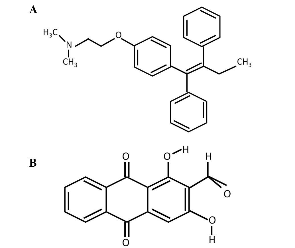
Nordamnacanthal (NDAM), also known as
2-formyl-1,3-dihydroxyanthraquinone, is an anthraquinone extracted
from the roots of Morinda elliptica (13). NDAM has been reported to have a
number of biological properties, including antitumor effects
(14,15). A previous study has shown that
depending upon the concentration/dose, NDAM is capable of
triggering and blocking cell death signaling in tumor cells
(16). Since both TAM and NDAM have
antitumor properties, a combination of these drugs may be a
therapeutic option for patients with breast cancer. However, their
potential additive effects have yet to be elucidated. Therefore,
the present study aimed to investigate the effect of TAM and NDAM
on apoptosis, cell cycle arrest, mitochondrial membrane potential
(Δψm) and oxidative stress in MCF-7 human breast cancer
cells.
Materials and methods
Cells and cell culture
The estrogen-sensitive MCF-7 human breast cancer
cell line was obtained from the American Type Culture Collection
(Rockville, MD, USA). Cells were cultured as monolayers in
RPMI-1640 (Sigma-Aldrich, , St. Louis, MO, USA) medium supplemented
with 10% heat-inactivated fetal bovine serum (Gibco-BRL, Carlsbad,
CA, USA), 100 U/ml penicillin and 100 μg/ml streptomycin (both
Sigma-Aldrich) at 37°C in a humidified environment containing 5%
CO2.
Drugs and drug treatment
NDAM was isolated from the roots of M.
elliptica using solvent fractionation and was purified using
high-performance liquid chromatography techniques. The structure
was identified by comparing spectroscopic data as reported in our
previous study (13). NDAM was
dissolved in dimethyl sulfoxide (DMSO; Sigma-Aldrich) and stored at
−20°C as a 10-mg/ml stock solution. TAM (Sigma-Aldrich) was
dissolved in DMSO at a concentration of 10 mg/ml. In the control
experiments, equal quantities of DMSO were added. The structure of
TAM and NDAM are shown in Fig.
1.
Cytotoxicity assay
Cells were seeded at a density of 5000 cells/well in
96-well microtiter culture plates and incubated overnight. The
culture medium was replaced with fresh medium containing various
concentrations of NDAM (0–30 μg/ml) and TAM (0–30 μg/ml) alone or
in combination for 24, 48 and 72 h. A total of 20 μl 3-(4,
5-dimethylthaiazol-2-yl)-2-5-diphenyltetrazolium bromide (MTT;
Calbiochem, Darmstadt, Germany) solution (0.5 mg/ml) was added to
each well and cell proliferation was analyzed as described
previously (14).
Cell viability assay
Cell death was quantified in the MCF-7 cells using
propidium iodide (PI; Sigma-Aldrich) and acridine orange (AO;
Sigma-Aldrich) double-staining as described previously, using a
fluorescence microscope (Diaphot, Nikon Inc., Melville, NY, USA)
(14). The MCF-7 cells were seeded
in a 25-ml culture flask at a concentration of 1×10⁶
cells/ml and treated with NDAM and TAM alone or in combination and
were incubated at 37°C with 5% CO2 for 72 h. The cells
were washed with phosphate-buffered saline (PBS) and stained with
10 μl AO (10 μg/ml) and PI (10 μg/ml). Slides were analyzed under
UV-fluorescence microscopy (Nikon Inc.) and the number of viable,
apoptotic and necrotic cells was calculated.
Annexin V binding assay
MCF-7 cells were treated with NDAM and TAM alone or
in combination for 72 h. Cells were resuspended in Annexin V
binding buffer (BD Pharmingen, San Diego, CA, USA) to a
concentration of 1×10⁶ cells/ml. Annexin V-fluorescein
isothiocyanate (FITC; BD Biosciences, San Diego, CA, USA) was
incubated for 15 min in the dark in 100 μl cell suspension. PI was
then spiked into 400 μl Annexin V binding buffer and added
immediately to the cell suspension, and subsequently analyzed using
a FACSCaliber™ system with CellQuest™ software (both BD
Biosciences). Care was taken to collect trypsinized cells and cells
which may have been floating prior to trypsinization to ensure that
apoptotic cells, if present, were detected.
Cell cycle analysis
MCF-7 cells (5,000 cells/ml) were seeded onto 25
cm2 flasks with NDAM and TAM either alone or combined
for 72 h. The cells were trypsinized and washed with PBS then
centrifuged at 2,000 × g for 5 min. The cell pellet was resuspended
in 1 ml 0.1% sodium citrate containing 0.05 mg PI and 50 μg RNase
(Sigma-Aldrich) for 30 min at room temperature in the dark. Flow
cytometric analysis was performed using a FACScan system (BD
Biosciences) and CellQuest software.
Assessment of Δψm
MCF-7 cells (1×10⁶) were grown in 25-ml
culture flasks for 24 h, followed by incubation with NDAM and TAM
alone or in combination in culture medium for 72 h at 37°C. The
Δψm was assessed using a BD™ MitoScreen kit (BD
Biosciences) according to the manufacturer’s instructions, and
analyzed using a FACSCaliber system (BD Biosciences). The ratio of
Δψm/mitochondrial mass was calculated to correct the
Δψm for differences in mitochondrial mass.
Assessment of lipid peroxidation
Lipid peroxidation was assessed through analyzing
the lipid peroxidation marker malondialdehyde in the cell lysates
(17,18). Cells were treated with NDAM and TAM
alone or in combination at 37°C with 5% CO2 for 72 h.
The treated MCF-7 cells were washed in ice-cold PBS and lysed in
260 μl solubilization buffer [10 mM Tris (pH 7.4), 9 g/l NP40, 1
g/l SDS and 250 U/ml benzonase; Sigma-Aldrich). Approximately 200
μl cell lysate or malondialdehyde standards (Sigma-Aldrich) were
mixed with 10 μl butylated hydroxytoluene (50 mg/ml ethanol) and
200 μl orthophosphoric acid (0.2 mM). The reaction mixture was
incubated on ice for 30 min and centrifuged at 2000 × g for 15 min
at 25°C. The supernatant was separated and added to 25 μl
2-thiobarbituric acid reagent (800 mg 2-thiobarbituric acid
dissolved in 50 ml 0.1 M NaOH) and incubated at 90°C for 45 min.
Formed malondialdehyde equivalents, thiobarbituric acid-reactive
substances (TBARS), were extracted and measured using a plate
reader (Bio-Rad Laboratories, Inc.) with excitation at 532 nm and
600 nm. Malondialdehyde standard solution was used for qualitative
determination of TBARS. The Bradford assay was performed in order
to measure the protein content.
Statistical analysis
All experiments were performed in triplicate. The
statistical significance of the differences was determined using
one-way analysis of variance followed by Dunnett’s multiple
comparison test. Values are presented as the mean ± standard
deviation and P<0.05 was considered to indicate a statistically
significant difference.
Results
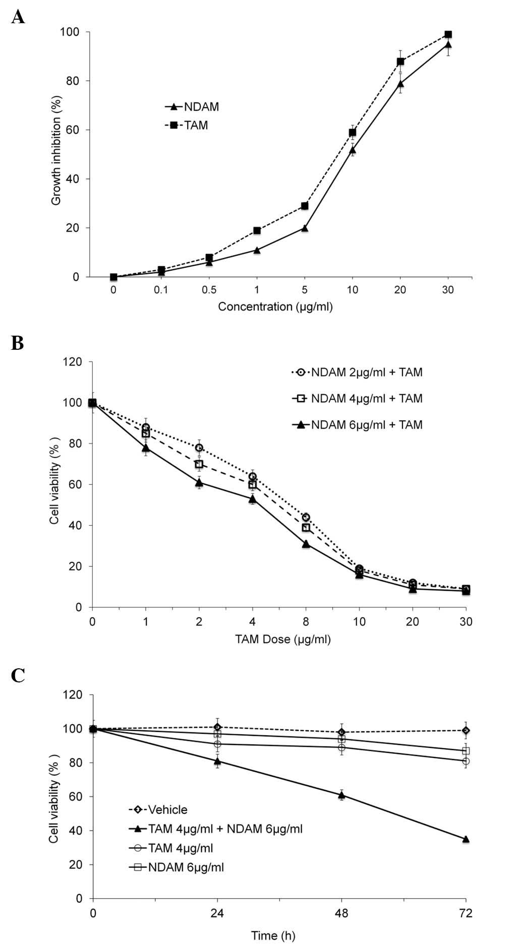
NDAM enhances the cytotoxic effect of
TAM
In the present study, it was hypothesized that TAM
in combination with NDAM may be a superior therapeutic strategy for
breast cancer. In order to test this hypothesis, the effect of
incremental doses of TAM and NDAM, alone or combined, was analyzed
on the growth of MCF-7 human breast cancer cells. Cellular growth,
determined using MTT assay, revealed that TAM and/or NDAM were
effective in inhibiting the growth of the MCF-7 cells in a
dose-dependent manner (Fig. 2A).
Low doses of TAM (4 μg/ml) were observed to reduce the
proliferation of breast cancer cells and complete cell death was
achieved at a concentration of 23 μg/ml following treatment for 72
h (Fig. 2B). At a concentration of
6 μg/ml, NDAM was found to reduce cell viability by 24.5% and
complete cell death was achieved at a concentration of 28 μg/ml
NDAM (Fig. 2B). Upon combining NDAM
with TAM, a markedly enhanced induction of cell death was observed,
even at lower TAM concentrations (Fig.
2B). While treatment with 4 μg/ml TAM alone did not
significantly reduce MCF-7 cell viability (12%), the combination of
NDAM (6 μg/ml) and TAM (4 μg/ml) was found to significantly reduce
MCF-7 cell viability by up to 77.0% (Fig. 2C; P<0.05). The same
concentrations of TAM and NDAM were not sufficient to induce
apoptosis when used alone.
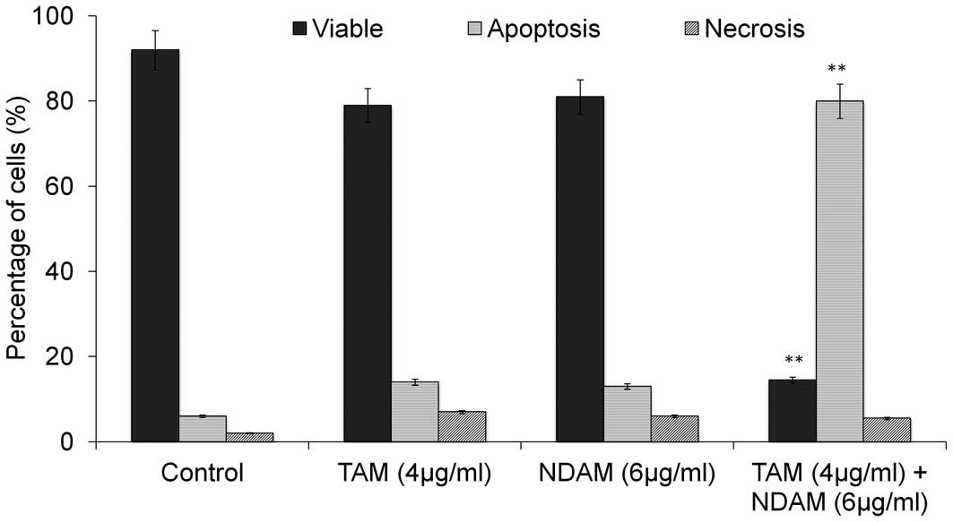
NDAM enhances TAM-induced apoptosis
Morphological changes were observed in the cells
treated with the TAM/NDAM combination. Co-incubation of MCF-7 cells
with TAM and NDAM resulted in significantly increased levels of
apoptosis (Fig. 3) compared with
the control cells and those treated with TAM or NDAM alone, with
the proportion of the viable cells accounting for only 14.5% of the
total cell population and 79% undergoing apoptosis. Necrotic cells
were also observed in the treatment group, but the numbers were
insignificant. Treatment with NDAM (6 μg/ml) and TAM (4 μg/ml)
alone was not found to induce significant levels of apoptosis in
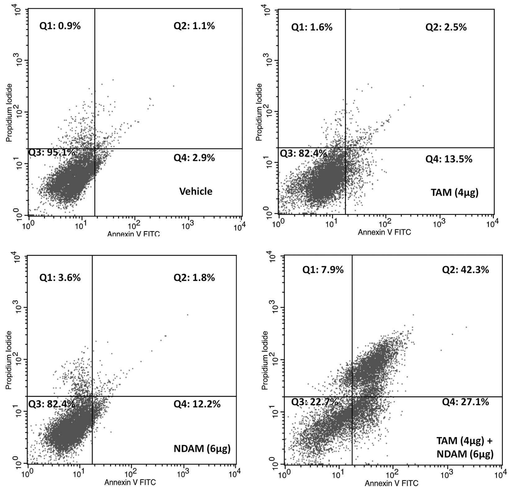
the MCF-7 cells (Fig. 4). Annexin
V-FITC analysis was performed to confirm the induction of apoptosis
in the MCF-7 cells upon cotreatment with TAM and NDAM.
Fluorescence-activated cell sorting revealed a significant increase
in apoptotic cells (69%) upon cotreatment with TAM and NDAM for 72
h compared with the vehicle-treated cells (4%) or those treated
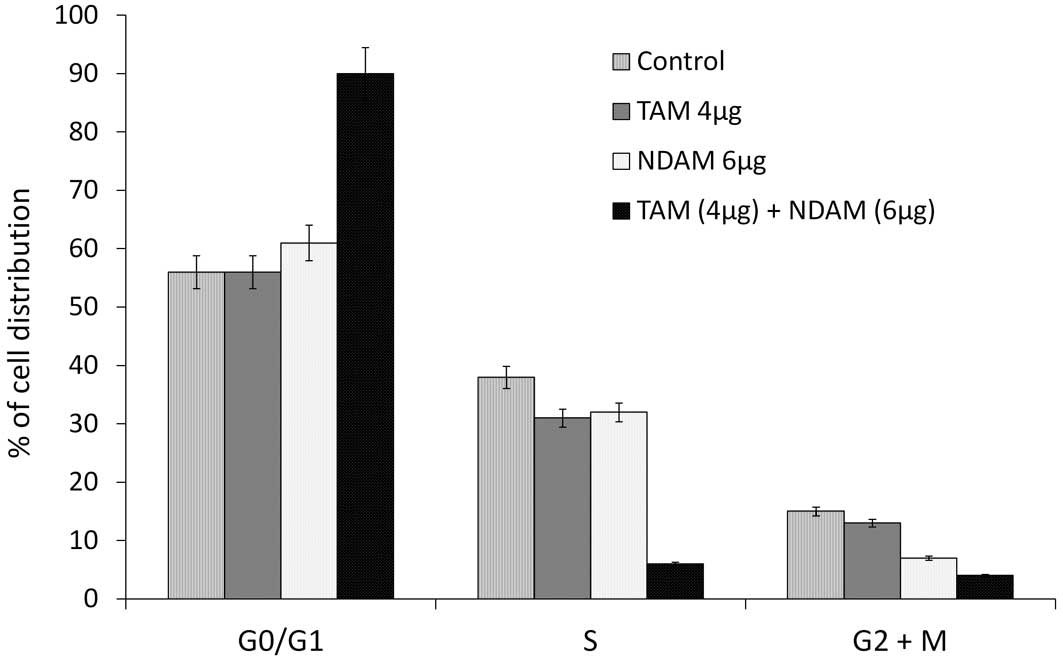
with TAM (16%) or NDAM (14%) alone (Fig. 4). Cell cycle analysis revealed an
increase in G0/G1-phase accumulation in the
TAM/NDAM-treated cells (63%; P<0.01) compared with the
DMSO-treated cells (45%), with a concomitant decrease in the
percentage of cells in the G2/M phase observed in the
TAM/NDAM-treated cells (Fig. 5).
Cell cycle arrest occurred from 24 h of treatment (data not shown)
and longer treatment durations showed that TAM/NDAM induced
G0/G1 arrest and apoptosis in the treated
cells. However, no significant increases in cell cycle arrest were
observed in the MCF-7 cells treated with NDAM (6 μg/ml) and TAM (4
μg/ml) alone.
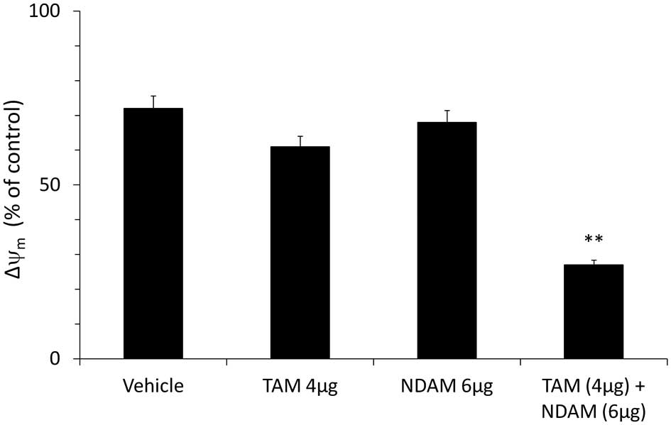
NDAM enhances the TAM-induced changes in
Δψm and lipid peroxidation
Changes in Δψm are considered to
be an indicator of mitochondrial damage. It has also been reported
that high quantities of reactive oxygen intermediates result in
lipid peroxidation (18).
Therefore, the present study analyzed the Δψm and the
lipid peroxidation end product malondialdehyde in MCF-7 cells
treated with TAM and NDAM, alone or combined. As shown in Fig. 6, changes in the Δψm were
observed in the in MCF-7 cells following TAM/NDAM exposure for 72
h. By contrast, administration of NDAM (6 μg/ml) or TAM (4 μg/ml)
alone had no significant effect on the Δψm. The
Δψm in the DMSO-treated cells was unchanged throughout
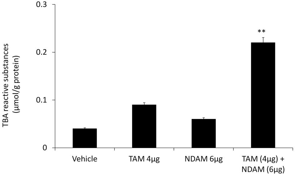
all of the incubation time-periods. Furthermore, TAM/NDAM
cotreatment was observed to have an effect on lipid peroxidation,
as demonstrated by the significant release of the malondialdehyde
equivalent TBARS from treated MCF-7 cells (Fig. 7). However, treatment with NDAM (6
μg/ml) and TAM (4 μg/ml) alone were not found to induce significant
TBARS release in the MCF-7 cells.
Discussion
TAM has been used for more than two decades for
hormone therapy in breast carcinomas expressing the estrogen
receptor. Although TAM is well tolerated and has resulted in a
5–15% absolute reduction in recurrence and mortality (4), more effective treatments for estrogen
receptor-positive breast cancer are required. In addition to the
side effects associated with TAM, it has been estimated that 90% of
patients with breast cancer acquire resistance to TAM within one
year (19). Support has increased
for the use of natural compounds that enhance the therapeutic
effect of antineoplastic agents so that lower doses may be used to
achieve the same antineoplastic effect, while simultaneously
avoiding or minimizing the side effects associated with high doses.
In the present study, the results showed that the cytotoxic effect
of TAM on breast cancer cells was enhanced through combined
treatment with NDAM, thus allowing the effective concentration of
TAM to be reduced. The growth inhibition advantage of combining
NDAM with TAM was primarily due to enhanced cell cycle arrest and
apoptosis. The cytotoxic effect was assessed through combining NDAM
(6 μg/ml) at a concentration which did not enhance apoptosis, with
the chemotherapeutic agent TAM (4 μg/ml). When NDAM was combined
with a subapoptotic dose of the chemotherapeutic agent, significant
apoptosis was induced. The primary growth inhibitory mechanism for
TAM in MCF-7 cells has been reported to be cell cycle inhibition
(20,21). In the present study, TAM/NDAM
treatment was found to induce significant
G0/G1-phase arrest in the MCF-7 cells. By
contrast, treatment with TAM or NDAM did not induce a significantly
greater G0/G1-phase arrests in the MCF-7
cells compared with TAM/NDAM cotreatment. Previous studies have
shown that TAM induces significant
G0/G1-phase arrest in breast cancer cells
(20,22). Furthermore, in agreement with the
present study, Li et al (23) showed that combined treatment of TAM
with organoselenium compounds enhanced apoptosis. These results
show that TAM/NDAM-induced cell growth inhibition is concomitant
with major changes in the cell cycle in MCF-7 cells.
In mammalian cells, the mitochondria have a
fundamental role in apoptosis. At the early stage of apoptosis,
mitochondrial damage occurs through disruption of the
Δψm which leads to the activation of caspase cascades
(24). In the present study, the
combination of TAM/NDAM was observed to induce a significant loss
of Δψm in the MCF-7 breast cancer cells. However, when
low doses of TAM and NDAM were applied individually, neither of the
drugs caused damage to the mitochondrial membrane. Furthermore, in
the present study, an increase in TBARS release was observed
following TAM/NDAM treatment for 72 h, while no significant TBARS
release was found following treatment with TAM or NDAM alone. These
findings suggest that TAM/NDAM-induced TBARS release from MCF-7
cells may have caused the loss of Δψm. In conclusion, in
the present study, NDAM was found to enhance the cytotoxic activity
of TAM, with the inhibition of cell proliferation,
G0/G1-phase arrest, the generation of
oxidative damage and the loss of Δψm cumulating in
apoptosis following TAM/NDAM cotreatment. Combining NDAM with TAM
was found to reduce the dose of TAM required to achieve the same
therapeutic effect; therefore, this combination therapy has the
potential to be a treatment regimen for breast cancer with minimal
or no side effects that are frequently associated with high doses
of TAM. However, it is important to investigate the safety and
tolerability of TAM/NDAM in vivo.
References
|
1
|
Jemal A, Siegel R, Ward E, et al: Cancer
statistics, 2009. CA Cancer J Clin. 59:225–249. 2009. View Article : Google Scholar : PubMed/NCBI
|
|
2
|
Goldhirsch A, Ingle JN, Gelber RD, et al:
Thresholds for therapies: highlights of the St Gallen International
Expert Consensus on the primary therapy of early breast cancer
2009. Ann Oncol. 20:1319–1329. 2009. View Article : Google Scholar : PubMed/NCBI
|
|
3
|
Radmacher MD and Simon R: Estimation of
tamoxifen’s efficacy for preventing the formation and growth of
breast tumors. J Natl Cancer Inst. 92:48–53. 2000. View Article : Google Scholar : PubMed/NCBI
|
|
4
|
Early Breast Cancers Trialists’
collaborative group. Tamoxifen for an early breast cancer: an
overview of the randomised trials. Lancet. 351:1451–1467. 1998.
View Article : Google Scholar
|
|
5
|
Love RR: Tamoxifen therapy in primary
breast cancer: biology, efficacy, and side effects. J Clin Oncol.
7:803–815. 1989.PubMed/NCBI
|
|
6
|
Marshall E: Tamoxifen: ‘a big deal,’ but a
complex hand to play. Science. 280:1961998. View Article : Google Scholar
|
|
7
|
Parvez S, Tabassum H, Rehman H, et al:
Catechin prevents tamoxifen-induced oxidative stress and
biochemical perturbations in mice. Toxicology. 225:109–118. 2006.
View Article : Google Scholar : PubMed/NCBI
|
|
8
|
Conklin KA: Chemotherapy-associated
oxidative stress: impact on chemotherapeutic effectiveness. Integr
Cancer Ther. 3:294–300. 2004. View Article : Google Scholar : PubMed/NCBI
|
|
9
|
Fisher B, Constantino JP, Wickerham CD, et
al: Tamoxifen for prevention of breast cancer: report of the
National Surgical Adjuvant Breast and Bowel Project P-1 Study. J
Natl Cancer Inst. 90:1371–1388. 1998. View Article : Google Scholar : PubMed/NCBI
|
|
10
|
Fornander T, Rutqvist LE, Cedermark B, et
al: Adjuvant tamoxifen in early breast cancer: occurrence of new
primary cancers. Lancet. 1:117–120. 1989. View Article : Google Scholar : PubMed/NCBI
|
|
11
|
Johnston SR, Dowsett M and Smith IE:
Towards a molecular basis for tamoxifen resistance in breast
cancer. Ann Oncol. 3:503–511. 1992.PubMed/NCBI
|
|
12
|
Osborne CK: Tamoxifen in the treatment of
breast cancer. N Engl J Med. 339:1609–1618. 1998. View Article : Google Scholar : PubMed/NCBI
|
|
13
|
Ismail NH, Ali AM, Aimi N, et al:
Anthraquinones from Morinda elliptica. Phytochemistry.
45:1723–1725. 1997. View Article : Google Scholar
|
|
14
|
Yazan LS, Ishak N and Lajis NH: BCL-2 was
downregulated in G2/M-arrest breast cancer cells MCF-7-treated with
Nordamnacanthal. J Pharm Sci & Res. 2:197–207. 2010.
|
|
15
|
Jasril, Lajis NH, Mooi LY, Abdullah MA,
Sukari MA and Ali AM: Antitumor promoting and antioxidant
activities of anthraquinones isolated from cell suspension culture
of Morinda elliptica. Asia Pac J Mol Biol Biotechnol. 11:3–7.
2003.
|
|
16
|
Kamiya K, Hamabe W, Tokuyama S, et al:
Inhibitory effect of anthraquinones isolated from the noni (Morinda
citrifolia) root on animal A-, B-, and Y-families of DNA
polymerases and human cancer cell proliferation. Food Chem.
118:725–730. 2009. View Article : Google Scholar
|
|
17
|
Frank J, Kelleher DK, Pompella A, et al:
Enhancement of oxidative cell injury and antitumor effects of
localized 44 degrees C hyperthermia upon combination with
respiratory hyperoxia and xanthine oxidase. Cancer Res.
58:2693–2698. 1998.PubMed/NCBI
|
|
18
|
Jentzsch AM, Bachmann H, Furst P and
Biesalski HK: Improved analysis of malondialdehyde in human body
fluids. Free Radic Biol Med. 20:251–256. 1996. View Article : Google Scholar : PubMed/NCBI
|
|
19
|
Johnston SR: Acquired tamoxifen resistance
in human breast cancer - potential mechanisms and clinical
implications. Anticancer Drugs. 8:911–930. 1997. View Article : Google Scholar
|
|
20
|
Danova M, Pellicciari C, Zibera C, et al:
Cell cycle kinetic effects of tamoxifen on human breast cancer
cells. Flow cytometric analyses of DNA content, BrdU labeling,
Ki-67, PCNA, and statin expression. Ann NY Acad Sci. 698:174–181.
1993. View Article : Google Scholar : PubMed/NCBI
|
|
21
|
Osborne CK, Boldt DH, Clark GM and Trent
JM: Effects of tamoxifen on human breast cancer cell cycle
kinetics: accumulation of cells in early G1 phase. Cancer Res.
43:3583–3585. 1983.PubMed/NCBI
|
|
22
|
Li S, Zhou Y, Wang R, et al: Selenium
sensitizes MCF-7 breast cancer cells to doxorubicin-induced
apoptosis through modulation of phospho-Akt and its downstream
substrates. Mol Cancer Ther. 6:1031–1038. 2007. View Article : Google Scholar : PubMed/NCBI
|
|
23
|
Li Z, Carrier L and Rowan BG:
Methylseleninic acid synergizes with tamoxifen to induce
caspase-mediated apoptosis in breast cancer cells. Mol Cancer Ther.
7:3056–3063. 2008. View Article : Google Scholar : PubMed/NCBI
|
|
24
|
Green DR and Reed JC: Mitochondria and
apoptosis. Science. 281:1309–1312. 1998. View Article : Google Scholar : PubMed/NCBI
|