Introduction
Hodgkin’s lymphoma (HL) is one of the few adult
malignancies that is usually curable (1). HL frequently presents as a
progressive, painless enlargement of the peripheral lymph nodes,
particularly around the cervical region (2). Classical HL is defined as a
well-established, proliferative neoplasm of the lymph nodes that is
composed of mononuclear Hodgkin cells and multi-nucleated
Reed-Sternberg (RS) cells in variable proportions, along with
neutrophils, eosinophils, histiocytes, fibroblasts, collagen
fibers, non-neoplastic lymphocytes and plasma cells (3,4).
Extra-nodal forms of HL are rare, accounting for <1% of all HL
cases (5,6). HL is a systemic disease, with 10–20%
of HL patients demonstrating bone involvement throughout disease
progression (7–9). However, patients presenting with
primary HL of the bone are unusual (2,10). The
histological, radiological and clinical features of HL are similar
to those of other medical conditions, including tuberculosis and
eosinophilic granuloma, and in unusual cases, may even present in
combination with multiple myeloma (1,2,7,9,10).
This makes HL difficult to diagnose and often leads to delays in
treatment.
The present study describes the case of a
22-year-old female diagnosed with primary multifocal osseous HL.
Informed consent was provided by the patient and the patient’s
family.
Case report
A 22-year-old female patient presented to the
Xiangya Hospital (Central South University, Changsha, Hunan, China)
with a five-month history of pain in the lumbar-sacral-pelvic area,
which gradually involved the left hip and subsequently involved the
left shoulder. The patient’s symptoms were accompanied by a
prolonged fever (a recurrent fever type, with a temperature of
>39°C for 1–2 days prior to returning to normal and recurring
repeatedly over the 5-month period), night sweats and weight loss.
The physical examination was mostly normal, with normal appearance
and range of motion. However, pain and tenderness were evident upon
percussion of the lumbosacral area, left hip joint and left
shoulder. The patient was previously healthy, with no exposure to
contaminated water or poisons, and no communicable diseases. The
patient’s parents were also healthy, with no history of a
hereditary or similar disease in their families.
Upon presentation, the blood cell count revealed a
persistent elevation of white blood cells (>14.0×10⁹
g/l; normal range, 4.0–10.0×10⁹ g/l), and a lowered
hemoglobin level (<90 g/l; normal range, 120–150 g/l). The
platelet count was also elevated (469.0×10⁹ g/l; normal
range, 100–300×109 g/l) and the mean corpuscular volume
was <80 fl (normal range, 80–100 fl). The mean corpuscular
hemoglobin (MCH) level was <27 pg (normal range, 27–34 pg) and
the MCH concentration was <320 g/l (normal range, 320–360 g/l),
which suggested microcytic hypochromic anemia. The liver and renal
function tests were almost normal; the biochemical evaluation
demonstrated signs of inflammation, with levels of 130 mg/l
C-reactive protein (CRP) (normal range, 0–8 mg/l) and 0.21 ng/ml
procalcitonin (normal range, <0.05 ng/ml), and a 75-mm/h
erythrocyte sedimentation rate (ESR) (normal range, 0–20 mm/h).
Further serology tests, including the human leukocyte antigen
haplotype B27, tuberculosis antibody, light chain protein and
extractable nuclear antigens tests, rheumatism immunity and lupus
tests, thyroid gland function test and an examination of 12 tumor
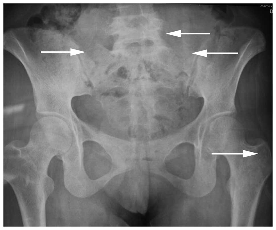
markers, were all normal. A pelvic X-ray revealed a bone lesion in
each of the sacroiliac joints five months after the onset of
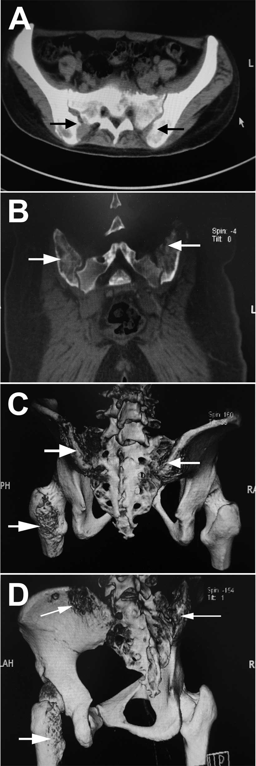
symptoms (Fig. 1). Computed
tomography (CT) identified osteolysis in each sacroiliac joint and
the left greater trochanter (Fig.
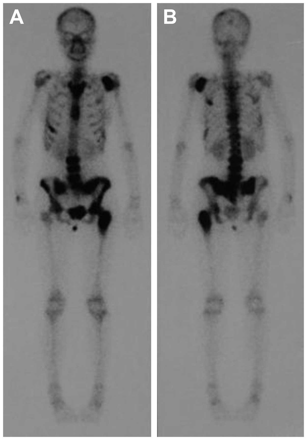
2). Single-photon emission CT revealed abnormal bone metabolism
of the lumbar, sacroiliac joint, the left greater trochanter and
the left shoulder (Fig. 3), likely
sites of bone metastases. A B-mode ultrasound scan of the cervical
and abdominal regions revealed no augmented lymph nodes, and the
organs of the abdominal cavity were normal. CT of the lungs and
mediastinum, and magnetic resonance imaging of the abdominal
region, revealed no enlargement of the lymph nodes and no
extraosseous involvement. Scans were combined with a physical
examination, which revealed no signs of lymphadenectasis. Positron
emission tomography-CT was not recommended due to its high cost and
the exposure of the patient to radioactivity. The color
ultrasonography images of gynecological features, and the color
Doppler ultrasonography of the heart, were normal. Due to the
patient’s history of multifocal bone pain and a negative bone
marrow biopsy, the clinical profile appeared to be consistent with
a diagnosis of chronic recurrent multifocal osteomyelitis (CRMO).
Furthermore, the initial biopsy from the left posterior-superior
iliac spine also revealed several giant cells against a background
of inflammation. Although combination treatment with antibiotics
[3.0 g cefperazone-sulbactam, intravenous glucose tolerance test
(i.v.g.t.t.), every 8 h; and 1.2 g clindamycin, i.v.g.t.t., every
12 h)] and non-steroidal anti-inflammatory drugs (100 mg
flurbiprofen, i.v.g.t.t., every 12 h) for three weeks greatly
improved the symptoms, CT revealed that bone destruction was still
occurring. A definitive diagnosis was not reached, as the
possibility of inflammation, a tumor or even intoxication could not
be eliminated. A second biopsy was performed, which retrieved a
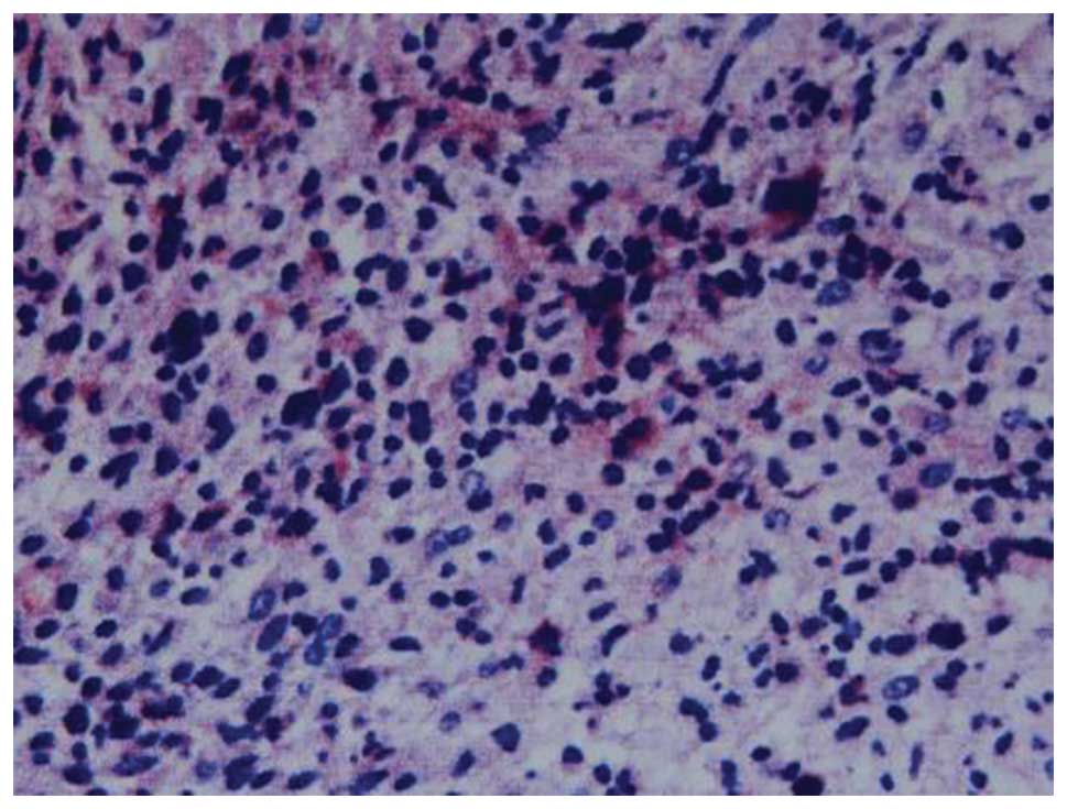
large amount of tissue for pathological examination. The results of
the pathological examination revealed that the tissue contained
cells bearing the characteristic morphology of RS cells, with
abundant cytoplasm and marked eosinophilic nucleoli, dispersed
against a background of reactive inflammation. The
immunohistochemistry staining of the RS cell population confirmed
the expression of paired box protein-5 and cluster of
differentiation (CD)30 and CD15 (Fig.
4). These results led to a diagnosis of primary multifocal
osseous lymphoma, rather than CRMO. Finally, in the hematology
ward, the patient was prescribed a course of Adriamycin, bleomycin,
vinblastine and dacarbazine (ABVD) chemotherapy, and recovered with
no pain or fever. The patient was also able to walk normally
subsequent to chemotherapy. The biochemical examination revealed
that the patient’s CRP, ESR and procalcitonin levels had returned
to normal. Furthermore, CT identified no further progression of
bone destruction of the lumbar, sacroiliac joint, the left greater
trochanter and the left shoulder. The patient returned to the
hospital six months later for a follow-up consultation, and
reported no pain or fever.
Discussion
Primary osseous HL is the diagnosis assigned to
patients who have osseous HL with no associated extraosseous
involvement. If more than one osseous site is involved, a diagnosis
of primary multifocal osseous HL is made (2). Primary osseous HL that is limited to
the bone is extremely rare, and until 2009, only 16 cases had been
identified globally (10). To the
best of our knowledge, ~20 cases of primary osseous HL, including
13 patients with primary solitary osseous HL and seven patients
with primary multifocal osseous HL without lymphatic
manifestations, have been reported (11–17).
Li et al (18) reported ~30
HL patients with extraosseous involvement. Langley et al
(2) reported that 33 cases of
primary osseous lymphoma at single or multiple sites, in a variety
of patient ages, existed in the scientific literature between 1927
and 2008. However, due to a lack of imaging equipment, it cannot be
confirmed whether certain patients presented with evidence of
lymphadenopathy within the chest or abdomen.
The case reported in the present study was the first
case of primary multifocal osseous HL, without associated
extraosseous involvement, from China. The extent of osseous
involvement included the left shoulder, the inferior segment of the
lumbar spine, the two sacroiliac joints and the left greater
trochanter. The affected region was wide and confined to the trunk
bone beside the mediastinum, however, the mediastinum appeared
normal according to a CT image of the thoracic region. The wide
range of involvement in unconnected areas had not previously been
described in other primary osseous HL cases. The ABVD chemotherapy
led to an improvement of the patient’s condition in a short period
of time, which suggested that this particular treatment regimen is
effective for classical, as well as primary multifocal osseous
HL.
According to the Ann Arbor classification system
(19), primary osseous HL is
classified as stage I, whereas systemic HL with secondary bone
involvement is classified as stage IV (18). Therefore, it is important that cases
of HL with an unusual presentation are diagnosed correctly by
clinicians, as an inaccurate diagnosis may lead to delays in
treatment. The histological, radiological and clinical features of
HL may mimic those of other medical conditions, including
tuberculosis, unusual eosinophilic granuloma, multiple myeloma and
infection with human immunodeficiency virus (HIV). There are
various types of extra-nodal manifestations of HL besides an
osseous presentation. Li et al (18) reported the case of a 38-year-old
female who presented with pain in the right upper-side of the chest
and adjacent soft-tissue swelling for three months. A surgical
biopsy, which included morphological and immunological data, was
consistent with classical HL, however, the patient had an
associated extraosseous soft-tissue mass adjacent to the ribs.
Kämmerer et al (4) presented
the case of a 73-year-old male with a suspicious ulcerating lesion
in the left retromolar region of the mandible. Prior
anti-inflammatory therapy was unsuccessful and three subsequent
biopsies identified inflammation alone. Ultimately, two biopsies
from the left retromolar region and the left inner cheek revealed
Hodgkin-Steinberg cells that were positive for the expression of
CD15 and CD30, a finding which corresponded to a diagnosis of HL.
Gandhi et al (3) described a
case of primary classical HL of the ileum. HL may also occur in the
epidural space (20) and in the
intracalvarium (21). A case of
autoimmune hemolytic anemia and immune thrombocytopenia of HL has
also been reported (22). HL not
only presents at various extra-nodal sites, but could also be
associated with HIV (23). These
patients usually have mixed cellularity, or a lymphocyte-depletion
subtype, advanced and extra-nodal disease, and systemic symptoms
(1). HL may also occur in
combination with other diseases, such as multiple myeloma (24), acute leukemia (25), peripheral T-cell lymphoma (26) and splenic marginal zone lymphoma
(27). Therefore, HL can occur at
various sites adjacent to the skeleton and may be associated with,
or occur in combination with, other conditions. Primary osseous HL
is extremely rare, and there is a possibility that clinicians may
not reach the correct diagnosis.
The present study indicates that, in the case of
complex multiple bone disease, imaging may not be sufficient to
enable a clear diagnosis. In order to improve the success rate of
diagnosis, pathological examination is necessary. During biopsies,
doctors should attempt to retrieve as much of the diseased tissue
as possible from the multiple lesions, and perform separate
pathological examinations in order to improve the success rate of
diagnosis. When a pathological diagnosis cannot be confirmed by an
initial biopsy, a second biopsy should be performed as soon as
possible.
Acknowledgements
This study was supported by the Young Teacher’s
Boosting Project of the Fundamental Research Funds for the Central
Universities in Central South University, China (no.
2012QNZT095)
References
|
1
|
Gobbi PG, Ferreri AJ, Ponzoni M and Levis
A: Hodgkin lymphoma. Crit Rev Oncol Hematol. 85:216–237. 2013.
View Article : Google Scholar
|
|
2
|
Langley CR, Garrett SJ, Urand J, Kohler J
and Clarke NM: Primary multifocal osseous Hodgkin’s lymphoma. World
J Surg Oncol. 6:342008. View Article : Google Scholar
|
|
3
|
Gandhi JS, Mehta A, Sharma A and Kamboj M:
Primary Hodgkin lymphoma of the ileum. J Cancer Res Ther.
6:342–343. 2010. View Article : Google Scholar : PubMed/NCBI
|
|
4
|
Kämmerer PW, Schiegnitz E, Hansen T, et
al: Multiple primary enoral soft tissue manifestations of a Hodgkin
lymphoma - case report and literature review. Oral Maxillofac Surg.
17:53–57. 2013. View Article : Google Scholar
|
|
5
|
Zucca E: Extranodal lymphoma: a
reappraisal. Ann Oncol. 19(Suppl 4): iv77–iv80. 2008. View Article : Google Scholar : PubMed/NCBI
|
|
6
|
Guermazi A, Brice P, de Kerviler EE, et
al: Extranodal Hodgkin disease: spectrum of disease. Radiographics.
21:161–179. 2001. View Article : Google Scholar : PubMed/NCBI
|
|
7
|
Bhagavathi S and Fu K: Primary bone
lymphoma. Arch Pathol Lab Med. 133:1868–1871. 2009.PubMed/NCBI
|
|
8
|
Franco V, Tripodo C, Rizzo A, Stella M and
Florena AM: Bone marrow biopsy in Hodgkin’s lymphoma. Eur J
Haematol. 73:149–155. 2004. View Article : Google Scholar : PubMed/NCBI
|
|
9
|
Newcomer LN, Silverstein MB, Cadman EC, et
al: Bone involvement in Hodgkin’s disease. Cancer. 49:338–342.
1982. View Article : Google Scholar : PubMed/NCBI
|
|
10
|
Oshikawa G, Arai A, Sasaki K, Ichinohasama
R and Miura O: Primary multifocal osseous Hodgkin lymphoma. Rinsho
Ketsueki. 50:92–96. 2009.(In Japanese). PubMed/NCBI
|
|
11
|
Borg MF, Chowdhury AD, Bhoopal S and
Benjamin CS: Bone involvement in Hodgkin’s disease. Australas
Radiol. 37:63–66. 1993. View Article : Google Scholar : PubMed/NCBI
|
|
12
|
Gold RH and Mirra JM: Case report 101.
Primary Hodgkin disease of humerus. Skeletal Radiol. 4:233–235.
1979. View Article : Google Scholar : PubMed/NCBI
|
|
13
|
Ostrowski ML, Inwards CY, Strickler JG, et
al: Osseous Hodgkin disease. Cancer. 85:1166–1178. 1999. View Article : Google Scholar : PubMed/NCBI
|
|
14
|
Ozdemirli M, Mankin HJ, Aisenberg AC and
Harris NL: Hodgkin’s disease presenting as a solitary bone tumor. A
report of four cases and review of the literature. Cancer.
77:79–88. 1996. View Article : Google Scholar : PubMed/NCBI
|
|
15
|
Jamshed A, Allard WF, Mourad WA and Rostom
AY: Primary Hodgkin’s disease of the mandible: a case report and
review of the literature. Oral Surg Oral Med Oral Pathol Oral
Radiol Endod. 83:680–684. 1997. View Article : Google Scholar : PubMed/NCBI
|
|
16
|
Köseoğlu RD, Senayli A, Biçakçi U, et al:
Osseous presentation of Hodgkin’s disease: a case report and review
of the literature. Turk J Pediatr. 49:218–222. 2007.
|
|
17
|
Gonzalez-Fontal GR, Rosales JD, Jaramillo
R and Henao-Martinez AF: Primary extranodal, extralymphatic hodgkin
lymphoma of the mandible. Case Rep Med. 2011:3875702011.PubMed/NCBI
|
|
18
|
Li Y, Wang XB, Tian XY, Li B and Li Z:
Unusual primary osseous Hodgkin lymphoma in rib with associated
soft tissue mass: a case report and review of literature. Diagn
Pathol. 7:642012. View Article : Google Scholar : PubMed/NCBI
|
|
19
|
Carbone PP, Kaplan HS, Musshoff K,
Smithers DW and Tubiana M: Report of the Committee on Hodgkin’s
Disease Staging Classification. Cancer Res. 31:1860–1861.
1971.PubMed/NCBI
|
|
20
|
Samadian M, Vahidi S, Khormaee F and
Ashraf H: Isolated, primary spinal epidural Hodgkin’s disease in a
child. Pediatr Neurol. 40:480–482. 2009. View Article : Google Scholar : PubMed/NCBI
|
|
21
|
Apollonsky N, Edelman M, Johnson A, Bhuiya
T and Karayalcin G: Intracerebral presentation of Hodgkin disease
mimicking meningioma in a young woman: case presentation with
literature review. J Pediatr Hematol Oncol. 30:369–372. 2008.
View Article : Google Scholar : PubMed/NCBI
|
|
22
|
Cecinati V, Brugnoletti F, D’Angiò M, et
al: Autoimmune hemolytic anemia and immune thrombocytopenia as
unusual presentations of childhood Hodgkin lymphoma: a case report
and review of the literature. J Pediatr Hematol Oncol. 34:280–282.
2012. View Article : Google Scholar : PubMed/NCBI
|
|
23
|
Shah BK, Subramaniam S, Peace D and Garcia
C: HIV-associated primary bone marrow Hodgkin’s lymphoma: a
distinct entity? J Clin Oncol. 28:e459–e460. 2010. View Article : Google Scholar : PubMed/NCBI
|
|
24
|
Huppmann AR, Liu ML and Nava VE:
Concurrent diagnoses of Hodgkin lymphoma and biclonal myeloma in
the bone marrow. Ann Diagn Pathol. 14:268–272. 2010. View Article : Google Scholar : PubMed/NCBI
|
|
25
|
Wesolowski R, Cotta CV, Khan G, Pohlman B
and Sweetenham J: Successful treatment of Hodgkin lymphoma and
acute leukemia. Leuk Lymphoma. 51:153–156. 2010. View Article : Google Scholar
|
|
26
|
Gualco G, Chioato L, Van Den Berg A, Weiss
LM and Bacchi CE: Composite lymphoma: EBV-positive classic Hodgkin
lymphoma and peripheral T-cell lymphoma: a case report. Appl
Immunohistochem Mol Morphol. 17:72–76. 2009. View Article : Google Scholar
|
|
27
|
Elmahy H, Hawley I and Beard J: Composite
splenic marginal zone lymphoma and classic Hodgkin lymphoma - an
unusual combination. Int J Lab Hematol. 29:461–463. 2007.
View Article : Google Scholar : PubMed/NCBI
|