Introduction
Bladder cancer is the most common malignant tumor of
the urinary tract, with 95% of bladder cancer patients suffering
from urothelial bladder carcinoma (1,2). The rate
of recurrence following surgical therapy is fairly high, and its
incidence and mortality rates rank first among malignant tumors of
the urinary system (3). Tumor
invasion and metastasis is the main cause of mortality in patients,
although the pathogenesis has not been fully elucidated. The
occurrence and development of bladder cancer are known to be
associated with multiple genes and multi-level cell signaling
network disorder (4). The authors of
the present study have previously conducted research into how genes
and multi-level factors are associated with bladder cancer cell
signaling network disorders (5–10);
however, the research was not thorough on the subject of cell cycle
protein with invasion and metastasis of tumors.
Dysregulation of the cell cycle is a key mechanism
for the occurrence of tumors. Cyclin D1 and cyclin E have a key
role in the regulation of the cell cycle and are a critical target
of proliferative signals in G1 phase. A high expression
of cyclin E shortens the G1 phase and induces tumor
development (11). By binding with
cyclin-dependent kinase 4 (CDK4), cyclin D1 is activated, the
compound CDK4-cyclin D1 is formed and the cell cycle is positively
regulated. Patients with tumors in which cyclin D1 is overexpressed
have a significantly lower survival rate compared with patients
exhibiting a low expression of cyclin D1 (12). Therefore, cyclin D1 is an effective
prognostic factor.
Cyclin E, as the key factor in regulating the
transformation between phases G1 and S, forms a compound
with CDK2 and enters the S phase (13). Research has revealed that the
expression of cyclin E is in direct proportion to the proliferative
activity of tumor cells, and is associated with poor prognosis of
tumors (14,15).
In the present study, a QD-based immunofluorescence
tissue chemical technique was adopted in order to detect cyclin D1
and cyclin E expression, occurrence and development in urothelial
bladder carcinoma, and evaluate the association between these
factors using clinical pathology. This method serves to provide
objective indicators for determining tumor invasion, metastasis and
prognosis of urothelial bladder carcinoma.
Materials and methods
Materials
In total, four urothelial bladder carcinoma chips
were provided by Fanpu Biotech, Inc. (Guilin, China), which had a
dot matrix of 15×10, a dot diameter of 1.1 mm and a thickness of 4
µm. The chips included 70 cases of urothelial bladder carcinoma and
5 cases of cystitis in a dual-chip matrix. The tissue samples
(Fanpu Biotech, Inc.) were surgically resected during surgery and
fixed for 24 h with 10% neutral-buffered formaldehyde. Among the
patients with urothelial bladder carcinoma, there were 42 males and
28 females, aged between 26 and 80 years (mean age, 56.8 years).
The cystitis samples were used as the control. This study was
conducted in accordance with the Declaration of Helsinki and with
the approval of the Ethics Committee of Wuhan University (Wuhan,
China). Written informed consent was obtained from all the
participants.
Patient classification
According to the 2004 World Health Organization
classification of urothelial tumor histological types (16), 42 cases were confirmed as invasive
urothelial carcinoma, 22 cases as high-grade non-invasive papillary
urothelial carcinoma and 6 cases as low-grade non-invasive
papillary urothelial carcinoma. According to the Union for
International Cancer Control (UICC) clinical stages (17), 38 cases were classified as stage T1,
22 cases as stage T2 and 10 cases as stage T3–4.
Reagents
Monoclonal rabbit anti-human cyclin D1 (cat. no.
ZA-0101) and monoclonal mouse anti-human cyclin E (cat. no.
ZM-0086) antibodies were obtained from Santa Cruz Biotechnology,
Inc., (Dallas, TX, USA) at a dilution of 1:150. Secondary
biotinylated goat anti-mouse and goat anti-rabbit immunoglobulin G
antibodies (cat. no. sfk12000) were purchased from Santa Cruz
Biotechnology, Inc., at a dilution of 1:300
Experimental methods
The QD double-staining method was employed to detect
cyclin D1 and cyclin E expression in the tissue chips of urothelial
bladder carcinoma. The experiment was conducted in strict
accordance with the instruction manual (18). The tissue chips of urothelial bladder
carcinoma (thickness, 4 µm) were dewaxed and hydrated, then
microwaved for antigen retrieval and washed in Tris-buffered saline
(TBS) (17). The chips were blocked
by incubation with blocking buffer solution (Wuhan Jiayuan Quantum
Dot Technological Development Co., Ltd.) in a wet chamber for 30
min at 37°C. Cyclin D1 and cyclin E antibodies were dripped into
the chamber and incubated for 2 h at 37°C, then washed three times
in TBS-Tween for 5 min. The chips were then incubated with blocking
buffer solution in the wet chamber for 10 min at 37°C. Next,
biotinylated goat anti-mouse or goat anti-rabbit immunoglobulin G
antibody (cat. no. sfk12000; Santa Cruz Biotechnology, Inc.) was
added to the wet chamber and incubated for 20 min at 37°C. QDs-SA
diluted with the blocking buffer solution was dripped into the wet
chamber and incubated for 30 min at 37°C, followed by washing with
TBS-Tween three times for 5 min each time and then adding 90%
glycerin buffer (19). Then, a
fluorescence microscope (DM2700 m; Leica Microsystems GmbH,
Wetzlar, Germany) was used to observe the tissue chips. Upon
observation under the microscope, the cells presenting with green
fluorescence were considered to exhibit a positive expression of
cyclin E, while cells presenting with red fluorescence were
considered to exhibit a positive expression of cyclin D1. A
positive area of ≥25% indicated that the cells exhibited a positive
expression. In the control group, the primary antibody was
substituted with TBS to form the control group, and the available
positive chip was used as the positive control.
Quantitative analysis of the cyclin D1
and cyclin E expression levels
The NuanceFX™ multi-spectral imaging system
(PerkinElmer, Inc., Waltham, MA, USA) was employed to perform
quantitative analysis of the expression of cyclin D1 and cyclin E.
Five complete and non-overlapping views were randomly selected
under a high-power lens (magnification, x400). The mean optical
density, positive area and total area of all cells with a positive
reaction under each view were measured, and the ratio of the
positive area was calculated. The mean value of the optical density
and positive area per case was used as the measured value of the
case (ratio of positive area = total area of positive reaction per
unit / total area of cells per unit × 100%).
Statistical analysis
The results of QD staining are expressed as the mean
± standard deviation. One-way analysis of variance and the
Student-Neuman-Keuls q-test were conducted using the SPSS 13.0
software (SPSS Inc., Chicago, IL, USA) to confirm the mean optical
density and ratio of the positive area in each group (α=0.05).
Prior to detection, a normality test and homogeneity test of
variances were performed. P<0.05 was considered to indicate a
statistically significant difference‥
Results
Expression of cyclin D1
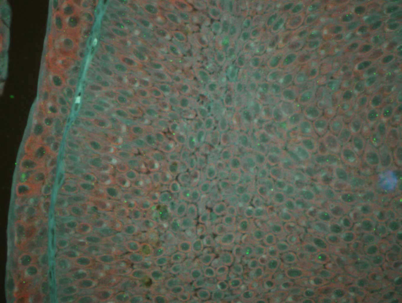
With regard to positive cyclin D1 expression, strong
red fluorescence was observed mainly in the cytoplasm of tumor
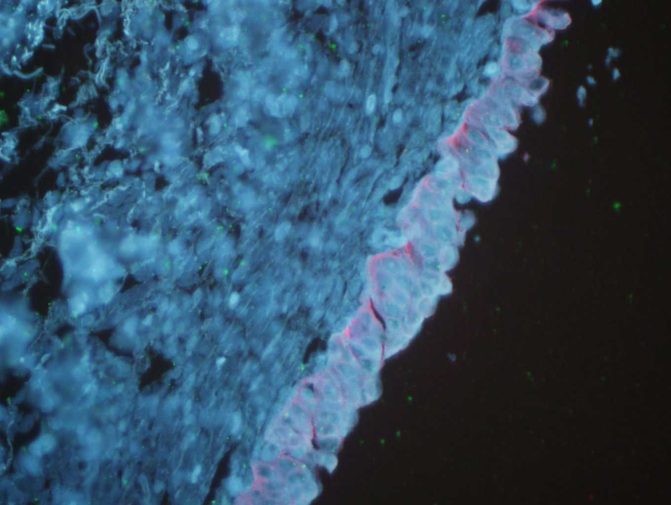
cells, indicating that cyclin D1 was highly expressed (Fig. 1). However, cyclin D1 expression was
not observed in cystitis tissues (Fig.
2). In total, 48 cases (68.6%) of urothelial bladder carcinoma
demonstrated positive cyclin D1 expression. The average optical
density and the positive area rate were found to be significantly
different between the urothelial bladder cancer and cystitis
tissues (P<0.05). The results of the image analysis are listed
in Table I. In addition, no
statistically significant difference in cyclin D1 expression
(P>0.05) in relation to patient age, gender, depth of invasion
or clinical UICC stage, as shown in Table II.
 | Table I.Quantum dot immunofluorescence
staining used to detect average optical density and rate of
positive area of cyclin D1. |
Table I.
Quantum dot immunofluorescence
staining used to detect average optical density and rate of
positive area of cyclin D1.
| Group | n | Number of slices | Average optical
density | Rate of positive
area |
|---|
| Urothelial bladder
cancer | 70 | 350 |
0.5240±0.0341a |
0.4694±0.0310a |
| Cystitis tissue | 5 | 25 |
0.1397±0.0198 |
0.1128±0.0167 |
| Table II.Correlation of cyclin D1 and cyclin E
expression with the pathological type and clinical stage of
urothelial bladder carcinoma. |
Table II.
Correlation of cyclin D1 and cyclin E
expression with the pathological type and clinical stage of
urothelial bladder carcinoma.
|
|
| Cyclin D1 |
| Cyclin E |
|
|---|
|
|
|
|
|
|
|
|---|
| Pathological
features | n | – | + | P-value | – | + | P-value |
|---|
| Gender |
|
|
| >0.05 |
|
| >0.05 |
|
Male | 42 | 14 | 28 |
| 12 | 30 |
|
|
Female | 28 | 8 | 20 |
| 9 | 19 |
|
| Age (years) |
|
|
| >0.05 |
|
| >0.05 |
|
≤60 | 37 | 10 | 27 |
| 11 | 26 |
|
|
>60 | 33 | 12 | 21 |
| 10 | 23 |
|
| Depth of tumor
invasion |
|
|
| >0.05 |
|
| <0.05 |
|
Invasive | 42 | 18 | 24 |
| 10 | 32 |
|
|
Non-invasive high-grade | 22 | 10 | 12 |
| 8 | 14 |
|
|
Non-invasive low-grade | 6 | 2 | 4 |
| 2 | 4 |
|
| UICC stage |
|
|
| >0.05 |
|
| <0.05 |
| T1 | 38 | 16 | 22 |
| 10 | 28 |
|
| T2 | 22 | 8 | 14 |
| 6 | 16 |
|
|
T3-4 | 10 | 4 | 6 |
| 2 | 8 |
|
Expression of cyclin E
With regard to positive cyclin D1 expression, strong
green fluorescence was observed mainly in the cell nuclei of tumor
cells (Fig. 1). By contrast, cyclin E
expression was not observed in cystitis (Fig. 2). In total, 49 cases (70%) of
urothelial bladder carcinoma demonstrated positive cyclin E
expression. The average optical density and the positive area rate
were found to be significantly different between the urothelial
bladder cancer and cystitis tissues (P<0.05). The results of the
image analysis are listed in Table
III. In addition, no statistically significant differences in
cyclin E expression (P>0.05) were observed in relation to
patient age and gender. However, a significant difference in the
expression of cyclin E (P<0.05) was detected in relation to the
depth of invasion and clinical UICC stage, as demonstrated in
Table II.
| Table III.Quantum dot immunofluorescence
staining used to detect average optical density and rate of
positive area of cyclin E. |
Table III.
Quantum dot immunofluorescence
staining used to detect average optical density and rate of
positive area of cyclin E.
| Group | n | Number of
slices | Average optical
density | Rate of positive
area |
|---|
| Urothelial bladder
cancer | 70 | 350 |
0.4988±0.0320a |
0.5102±0.0341a |
| Cystitis
tissue | 5 | 25 |
0.1547±0.0189 |
0.1395±0.0126 |
Correlation between cyclin D1 and
cyclin E expression
Considering the aforementioned observations, the
cyclin D1 and cyclin E expression was found to be significantly and
positively correlated in the 70 cases of urothelial bladder
carcinoma.
Discussion
Bladder cancer is the most common malignant tumor of
the urinary tract, with ~95% of bladder cancer patients suffering
from urothelial bladder carcinoma (20,21).
Approximately 80% of bladder cancer cases are superficial in the
early stage, and 70% of such patients suffer relapse following
surgery, with 30% of relapses developed towards the later stages of
the disease (1). In addition, 15–30%
of cases are invasive at the early stage, even if therapy is
received for early metastasis (22).
The prognosis of patients with urothelial bladder carcinoma is
determined by the depth of invasion, metastasis and recurrence.
Therefore, the study of factors, including tumor invasion and
metastasis of urothelial bladder carcinoma, is of great clinical
significance. Invasion and metastasis of tumors are the main causes
of mortality, although the pathogenesis has not been fully
elucidated (23,24).
The occurrence and development of bladder cancer are
known to be connected with multiple genes and multi-level cell
signaling network disorder (4).
Previous research into how genes and multi-level factors are
associated with bladder cancer cell signaling network disorders has
been conducted by the current authors (5–10);
however, the research was not thorough on the subject of cell cycle
protein with invasion and metastasis of tumors. Further
investigating the underlying mechanism of the occurrence and
development of bladder cancer, as well as searching for effective
indicators to predict its biological behavior, is crucial in order
to assist its early diagnosis and provide an experimental basis for
guiding treatment and prognosis. Studying the association between
the occurrence of tumors and the cell cycle progression is
currently a hotspot in tumor biology research (25). Cyclin D1 and cyclin E are significant
regulatory factors in the G1 and S phases of the cell
cycle; however, their correlation with bladder cancer remains
unclear.
A tissue chip (or tissue microarray) refers to a
tissue section consisting of tens of thousands of tissue blocks
that are neatly placed on a glass slide. Tissue chips have a small
volume, but contain a large quantity of information, demonstrating
substantial results in one-time experiments. Data with regard to
cyclin D1 and cyclin E expression in urothelial bladder carcinoma
may be obtained in a short period of time. In contrast to
traditional pathological techniques and methods, the results
obtained using the tissue microarray technique are consistent,
reliable and fairly comparable, which can reduce time and cost, and
provide a large quantity of data. This method has several promising
applications. Tissue chips may be used in fields including basic,
clinical and application research, as well as medicine development,
and has promising prospects for development (10).
QDs are novel fluorescence semiconductor
nanocrystals, which present a broad and continuous spectrum, high
absorptivity and intensity, a narrow and symmetrical emission peak,
and fast light bleaching, compared with the traditional organic
fluorescence labeling reagents (26–28). QDs
are able to withstand repetitive activation, have activated
pathology and may be applied in tumor diagnosis and imaging studies
(29).
In the present study, the QD immunofluorescence and
tissue chip techniques were adopted to detect cyclin D1 and cyclin
E expression, occurrence and development in human urothelial
bladder carcinoma. In addition, the association of their expression
with clinicopathological factors was investigated in order to
improve the understanding on the molecular mechanism of occurrence
and development of urothelial bladder carcinoma, as well as its
biological behavior.
The regulation of cell cycle progression mainly
relies on the formation, activation and interaction of cyclin, CDK
and CDK inhibitor. As key positive regulatory factors, cyclin D1
and cyclin E play critical roles in G1 phase regulation
and G1/S phase transformation. Cyclin D1 has been
demonstrated to be a cancer gene that is directly associated with
tumors. Cyclin D1 (11), as the most
significant positive regulatory factor of the cell cycle, plays a
key role in the occurrence and development of tumors (30). Under a physiological state (31), as the cell enters the S phase, cyclin
D1 breaks down. In cases where the cyclin D1 gene is activated,
cyclin D1 is highly and continuously expressed. G1 phase
is then shortened and cyclin D1 enters the S phase early; thus,
cell proliferation is uncontrollable and the tumor is formed.
Cyclin E is a positive regulatory factor in the
G1/S phase transformation and regulation. It is
synthesized after cyclin D, and kinase activity reaches its peak in
the late period of G1 phase. However, the chromosomal
localization of cyclin E gene remains unclear. In the body, cyclin
E combines with CDKZ to form the compound cyclin E-CDKZ, which
phosphorylates and deactivates the retinoblastoma protein (Rb)
(32). Subsequently, the
transcription factor inhibited by the phosphorylated Rb is
released, while G1/S transformation and DNA synthesis
are promoted, and cell proliferation is realized. After entering
the S phase, cyclin E breaks down quickly. Cyclin D1 and cyclin E,
as key rate-limiting factors in the G1/S transformation
of the cell cycle, play significant roles in cell proliferation
(33). In addition, cyclin E is
considered as a backup cyclin in regulating G1/S phase
transformation. As a cancer gene, cyclin E overexpression exerts
harmful effects on the cell cycle, which may cause uncontrollable
cell proliferation and promotion of tumor occurrence and
development. As previously reported in the literature, cyclin E
gene amplification and protein overexpression may be detected in
human tumors, including breast, ovarian, gastric, esophageal,
pancreatic and lung cancer (34). In
addition, previous studies have identified that cyclin E
overexpression is associated with the stage, grading, metastasis
and life cycle of certain tumors, including breast cancer, gastric
cancer, renal pelvic carcinoma and ureteral cancer (35–37).
Therefore, cyclin E expression is considered to be a significant
prognostic marker for these cancers.
In a number of tumor tissues, cyclin E generates
gene amplification and overexpression, and has already been
confirmed as a cancer gene (38). In
the majority of tumor studies, the immunolabeling technique is used
to determine cyclin E expression in the cell nucleus (39–42).
Cyclin E protein is synthesized and degraded in the cytoplasm, and
is generally quickly transferred to the cell nucleus (43). The accumulation of cyclin E in the
cytoplasm may demonstrate increased synthesis, decreased
degradation and nuclear transport disorder (32). Cyclin E is mainly engaged in cell
division within the cell nucleus; therefore, tumor cells with
negative nuclear and cytoplasm staining are considered as having
negative expression (40).
In the present study, QD staining and statistical
analysis were performed. According to the results, cyclin D1
expression in urothelial bladder carcinoma tissues was greater
compared with that in cystitis tissues, and the difference was
statistically significant (P<0.05). This indicates that cyclin
D1 protein overexpression shortens the cell cycle of G1
phase, diminishes its volume and weakens its dependence on mitogen.
Simultaneously, the overexpression results in uncontrolled cell
proliferation and leads to urothelial bladder carcinoma
development. Based on the current experimental results, cyclin D1
expression has no significant association with the
clinicopathological stages of urothelial bladder carcinoma,
indicating that cyclin D1 overexpression may be an early event in
the occurrence of urothelial bladder carcinoma, and may be
important in the initial stages where cell proliferation is a
necessary step, involving no tumor invasion or metastasis.
Using QD staining, cyclin E expression was also
detected in urothelial bladder carcinoma tissues, and statistical
analysis was conducted. Cyclin E expression was observed to be
higher in urothelial bladder carcinoma tissues compared with that
in cystitis tissues, and the difference was statistically
significant (P<0.05). In addition, cyclin E expression exhibited
a significant association with the clinicopathological stages of
urothelial bladder carcinoma (P<0.05). Therefore, cyclin E may
participate in the occurrence, development and metastasis of
urothelial bladder carcinoma and may be a significant reason for
the active proliferation of tumor cells. The present study,
reporting the overexpression of cyclin D and cyclin E in urothelial
bladder carcinoma, may assist the investigation into the
pathogenesis of this disease and the classification of the disease
into clinical stages and degrees of malignancy, thus serving as a
novel therapeutic approach.
In conclusion, cyclin D1 and cyclin E are key
regulatory factors of G1/S phase transformation. Their
overexpression shortens the duration of G1/S phase
transformation and promotes the progression of the cell cycle and
increased cell proliferation, which results in tumorigenesis. The
synergetic effect of their overexpression may affect the occurrence
and development of urothelial bladder carcinoma. Further
investigation on the correlation between cyclin and urothelial
bladder carcinoma is likely to provide an insight into the
mechanism of tumorigenesis. Previous studies have confirmed that
cyclin plays a significant role in the occurrence of tumors and is
able to provide prognostic information on a number of common tumors
(35,44). However, the underlying
mechanism through which cyclin affects tumor development remains
unclear and requires further elucidation.
References
|
1
|
Jemal A, Siegel R, Xu J and Ward E: Cancer
statistics, 2010. CA Cancer J Clin. 60:277–300. 2010. View Article : Google Scholar : PubMed/NCBI
|
|
2
|
Friedrich MG, Weisenberger DJ, Cheng JC,
et al: Detection of methylated apoptosis-associated genes in urine
sediments of bladder cancer patients. Clin Cancer Res.
10:7457–7465. 2004. View Article : Google Scholar : PubMed/NCBI
|
|
3
|
Yakasai A, Allam M and Thompson AJ:
Incidence of bladder cancer in a one-stop clinic. Ann Afr Med.
10:112–114. 2011. View Article : Google Scholar : PubMed/NCBI
|
|
4
|
Vlahou A: Back to the future in bladder
cancer research. Expert Rev Proteomics. 8:295–297. 2011. View Article : Google Scholar : PubMed/NCBI
|
|
5
|
Shan G and Xia Y: Expression and clinical
significance of RECK and MT1-MMP in bladder urothelium carcinoma
tissues. Chin J Cancer Prev Treat. 19:1722–1725. 2012.
|
|
6
|
Shan G, Shan S, Zhang X and Liu XH:
Expression and clinical sigificance of cyclin G1 and cyclin G2 in
transitional cell carcinoma of bladder. Chinese J Histochem
Cytochem. 18:268–273. 2009.
|
|
7
|
Shan G and Tang T: Expression and clinical
sigificance of ADO in transitional cell carcinoma of bladder.
Chinese J Histochem Cytochem. 20:267–271. 2011.
|
|
8
|
Shan G and Tang T: Expression and clinical
sigificance of tumor suppressor genes DPC4 and TGF-β1 in
transitional cell carcinoma of bladder. Chinese J Histochem
Cytochem. 20:491–495. 2011.
|
|
9
|
Shan G and Tang T: Expression and clinical
sigificance of CD82/KAI1 in transitional cell carcinoma of bladder.
Chinese J Histochem Cytochem. 22:185–188. 2013.
|
|
10
|
Shan G and Tang T: Expression and clinical
significance of PSCA and mesothelin in transitional cell carcinoma
of bladder. Chinese J Histochem Cytochem. 6:684–692. 2010.
|
|
11
|
Otieno S, Grace CR and Kriwacki RW: The
role of the LH subdomain in the function of the Cip/Kip
cyclin-dependent kinase regulators. Biophys J. 100:2486–2494. 2011.
View Article : Google Scholar : PubMed/NCBI
|
|
12
|
Jirawatnotai S, Hu Y, Michowski W, et al:
A function for cyclin D1 in DNA repair uncovered by protein
interactome analyses in human cancers. Nature. 474:230–234. 2011.
View Article : Google Scholar : PubMed/NCBI
|
|
13
|
Makiyama K, Masuda M, Takano Y, et al:
Cyclin E overexpression in transitional cell carcinoma of the
bladder. Cancer Lett. 151:193–198. 2000. View Article : Google Scholar : PubMed/NCBI
|
|
14
|
Farley J, Smith LM, Darcy KM, et al
Gynecologic Oncology Group: Cyclin E expression is a signifieant
predictor of survival in advanced, suboptimally debulked ovarian
epithelial cancers: a Gynecologic Oncology Group study. Cancer Res.
63:1235–1241. 2003.PubMed/NCBI
|
|
15
|
Scuderi R, Palucka KA, Pokorvskaja K, et
al: Cyclin E overexpression in relapsed adult acute lymphoblastic
leukemias of B-cell lineage. Blood. 87:3360–3367. 1996.PubMed/NCBI
|
|
16
|
Eble JN, Sauter G, Epstein JI and
Sesterhenn IA: World Health Organization Classification of
TumorsPathology and Genetics of Tumors of the Urinary System and
Male Genital Organs. IARC Press; Lyon: 2004
|
|
17
|
Sobin LH, Gospodarowicz M and Wittekind C:
Urological tumoursTNM Classification of Malignant Tumors UICC
International Union Against Cancer. 7th. Wiley-Blackwell; pp.
262–265. 2009
|
|
18
|
Chen H, Xue J, Zhang Y, Zhu X, Gao J and
Yu B: Comparison of quantum dots immunofluorescence histochemistry
and conventional immunohistochemistry for the detection of
caveolin-1 and PCNA in the lung cancer tissue microarray. J Mol
Hist. 40:261–268. 2009. View Article : Google Scholar
|
|
19
|
Tang T and Zhang DL: Study on
extracellular matrix metalloproteinase inducer and human epidermal
growth factor receptor-2 protein expression in papillary thyroid
carcinoma using a quantum dot-based immunofluorescence technique.
Exp Ther Med. 9:1331–1335. 2015.PubMed/NCBI
|
|
20
|
Parkin DM, Bray F, Ferlay J and Pisani P:
Global cancer statistics, 2002. CA Cancer J Clin. 55:74–108. 2005.
View Article : Google Scholar : PubMed/NCBI
|
|
21
|
Jemal A, Siegel R, Ward E, et al: Cancer
statistics, 2008. CA Cancer J Clin. 58:71–96. 2008. View Article : Google Scholar : PubMed/NCBI
|
|
22
|
Dhawan D, Ramos-Vara JA, Naughton JF, et
al: Targeting folate receptors to treat invasive urinary bladder
cancer. Cancer Res. 73:875–884. 2013. View Article : Google Scholar : PubMed/NCBI
|
|
23
|
Sylvester RJ, van der Meijden AP,
Oosterlinck W, et al: Predicting recurrence and progression in
individual patients with stage Ta T1 bladder cancer using EORTC
risk tables: A combined analysis of 2596 patients from seven EORTC
trials. Eur Urol. 49:466–475; discussion. 475–477. 2006. View Article : Google Scholar : PubMed/NCBI
|
|
24
|
Amit D, Tamir S, Birman T, et al:
Development of targeted therapy for bladder cancer mediated by a
double promoter plasmid expressing diphtheria toxin under the
control of IGF2-P3 and IGF2-P4 regulatory sequences. Int J Clin Exp
Med. 4:91–102. 2011.PubMed/NCBI
|
|
25
|
Das SN, Khare P, Singh MK and Sharma SC:
Correlation of cyclin D1 expression with aggressive DNA pattern in
patients with tobacco-related intraoral squamous cell carcinoma.
Indian J Med Res. 133:381–386. 2011.PubMed/NCBI
|
|
26
|
Remacle F and Levine RD: Quantum dots as
chemical building blocks: elementary theoretical considerations.
Chemphyschem. 2:20–36. 2001. View Article : Google Scholar : PubMed/NCBI
|
|
27
|
Yu WW, Chang E, Drezek R and Colvin VL:
Water-soluble quantum dots for biomedical applications. Biochem
Biophys Res Commun. 348:781–786. 2006. View Article : Google Scholar : PubMed/NCBI
|
|
28
|
Koole R, Mulder WJ, van Schooneveld MM,
Strijkers GJ, Meijerink A and Nicolay K: Magnetic quantum dots for
multimodal imaging. Wiley Interdiscip Rev Nanomed Nanobiotechnol.
1:475–491. 2009. View
Article : Google Scholar : PubMed/NCBI
|
|
29
|
Li J, Huang X, Xie X, Wang J and Duan M:
Human telomerase reverse transcriptase regulates cyclin D1 and
G1/S phase transition in laryngeal squamous carcinoma.
Acta Otolaryngol. 131:546–551. 2011. View Article : Google Scholar : PubMed/NCBI
|
|
30
|
Hwang CF, Cho CL, Huang CC, et al: Loss of
cyclin D1 and p16 expression correlates with local recurrence in
nasopharyngeal carcinoma following radiotherapy. Ann Oncol.
13:1246–1251. 2002. View Article : Google Scholar : PubMed/NCBI
|
|
31
|
Umekita Y, Ohi Y, Sagara Y and Yoshida H:
Over expression of cyclin D1 predicts for poor prognosis in
estrogen receptor-negative breast cancer patients. Int J Cancer.
98:415–418. 2002. View Article : Google Scholar : PubMed/NCBI
|
|
32
|
Stamatakos M, Palla V, Karaiskos I, et al:
Cell cyclins: triggering elements of cancer or not? World J Surg
Oncol. 8:1112010. View Article : Google Scholar : PubMed/NCBI
|
|
33
|
Freemantle SJ and Dmitrovsky E: Cyclin E
transgenic mice: discovery tools for lung cancer biology, therapy
and prevention. Cancer Prev Res (Phila). 3:1513–1518. 2010.
View Article : Google Scholar : PubMed/NCBI
|
|
34
|
Musat M, Morris DG, Korbonits M and
Grossman AB: Cyclins and their related proteins in pituitary
tumourigenesis. Mol Cell Endocrinol. 326:25–29. 2010. View Article : Google Scholar : PubMed/NCBI
|
|
35
|
Jung YJ, Lee KH, Choi DW, et al:
Reciprocal expressions of cyclin E and cyclin D1 in hepatocellular
carcinoma. Cancer Lett. 168:57–63. 2001. View Article : Google Scholar : PubMed/NCBI
|
|
36
|
Pajalunga D and Crescenzi M: Regulation of
cyclin E protein levels through E2F-mediated inhibition of
degradation. Cell Cycle. 3:1572–1578. 2004. View Article : Google Scholar : PubMed/NCBI
|
|
37
|
Richter J, Wagner U, Kononen J, et al:
High-throughput tissue microarray analysis of cyclin E gene
amplification and overexpression in urinary bladder cancer. Am J
Pathol. 157:787–794. 2000. View Article : Google Scholar : PubMed/NCBI
|
|
38
|
Li JQ, Miki H, Ohmori M, et al: Expression
of cyclin E and cyclin-dependent kinase 2 correlates with
metastasis and prognosis in colorectal carcinoma. Hum Pathol.
32:945–953. 2001. View Article : Google Scholar : PubMed/NCBI
|
|
39
|
Donnellan R, Kleinschmidt I and Chetty R:
Cyclin E immunoexpression in breast ductal cancinoma: pathologic
correlations and prognostic implications. Hum Pathol. 32:89–94.
2001. View Article : Google Scholar : PubMed/NCBI
|
|
40
|
Zhu HX: Cyclin E and tumor. Chinese
Medical Journal of Zhong Guo Yejin Gongye Yixue Zazhi. 20:86–88.
2003.(In Chinese).
|
|
41
|
Bales ES, Dietrich C, Bandyopadhyay D, et
al: High levels of expression of p27KIPl and cyclin E in invasive
primary malignant melanomas. J Invest Dermatol. 113:1039–1046.
1999. View Article : Google Scholar : PubMed/NCBI
|
|
42
|
Harwell RM, Porter DC, Danes C and
Keyomarsi K: Processing of cyclin E differs between normal and
tumor breast cells. Cancer Res. 60:481–489. 2000.PubMed/NCBI
|
|
43
|
Siu KT, Rosner MR and Minella AC: An
integrated view of cyclin E function and regulation. Cell Cycle.
11:57–64. 2012. View Article : Google Scholar : PubMed/NCBI
|