Introduction
Metanephric adenomas of the kidney are a rare and
predominantly benign type of neoplasm that are typically removed by
surgical resection (1). The clinical
presentation is similar to that of malignant renal masses, and
includes polycythemia, hematuria and abdominal pain. Furthermore,
metanephric adenomas are occasionally detected incidentally upon
imaging (2). Tumors are typically
composed of solid, rare cystic components or calcifications with a
poorly-defined border (3). To the best
of knowledge, thus far, <200 total cases of metanephric adenoma
and only one case of metastasis to the lymph nodes has been
described in the literature (4).
Despite the benign nature of this neoplasm, an improved
understanding of its pathology may lead to the development of less
invasive treatment strategies in the future. The current study
presents the case of a male patient diagnosed with atypical
metanephric adenoma with diffuse calcifications who subsequently
underwent radical nephrectomy.
Case report
A 23-year-old male patient was admitted to Subei
People's Hospital (Yangzhou, China) with a history of flank pain on
October 23, 2013, and appeared in good general condition. The
patient had no history of occupational exposure to carcinogens. At
>1 month after the onset of symptoms, a medical examination
identified a left renal mass with diffuse calcifications. The mass,
including the calcifications, was subsequently resected on October
28, 2013. The patient recovered well following the surgical
procedure.
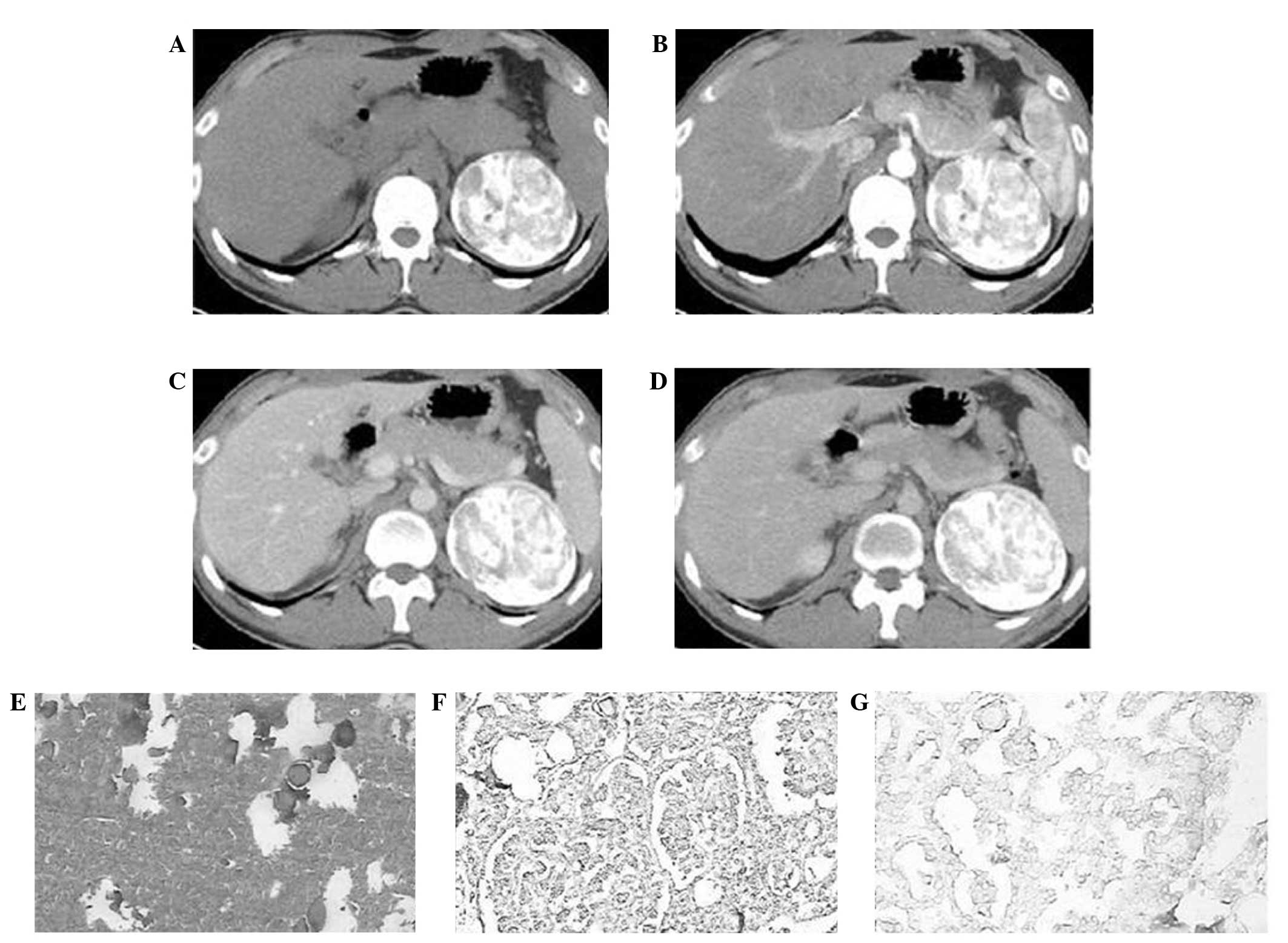
Unenhanced computed tomography (CT) scans were
performed using 64-Light speed Ultra (GE Healthcare, Milwaukee, WI,
USA), and identified a well-defined mass (diameter, 6.9 cm) with
diffuse calcifications in the medulla region of the kidney. The
attenuation of the solid component of the mass was 22 HU and
diffuse calcifications were noted (Fig.
1A). During the cortical phase contrast-enhanced CT scan,
attenuation of the solid component demonstrated mild enhancement at
a value of less than that in the cortex and medulla regions (36 HU;
Fig. 1B). The attenuation of the
solid tumor (45 HU) marginally increased during the medullary phase
(Fig. 1C). However, during the
delayed phase, the attenuation of the tumor decreased to 37 HU
(Fig. 1D). Pathological analysis of
sections revealed heterogeneous round or ovoid tumor cells arranged
in tubules and tubular glands. The tumor cells exhibited little
eosinophilic cytoplasm and few cylindrical nuclei, and no
karyokinesis was observed by hematoxylin and eosin staining
(Fig. 1E). Immunohistochemical
analysis (EnVision™ kit; Dako, Glostrup, Denmark) identified high
expression levels of vimentin (Fig.
1F) and pan-cytokeratin (Fig. 1G)
(4).
Written informed consent for the present case report
was obtained from the patient.
Discussion
Metanephric adenoma may occur in children and
adults, although it is most common in the fifth and sixth decades
of life. Furthermore, the disease has a 2:1 female preponderance
(5). To the best of our knowledge,
only one case of high grade sarcoma arising in association with
metanephric adenoma, termed metanephric adenosarcoma, has been
reported in the literature thus far (6). Approximately 50% of cases of metanephric
adenoma are incidental findings, however, symptoms include
polycythemia, abdominal or flank pain, a mass and hematuria
(7). Similarly, presenting symptoms
of metanephric adenofibroma include, polycythemia or hematuria
while certain cases have been diagnosed from incidental findings
(8). Haemorrhagic foci and necrosis
are also commonly detected in metanephric adenoma, with
calcifications and small cysts present in ~20 and 10% of cases,
respectively (9).
Previously published studies of metanephric adenoma
have described the pathological, clinical and CT imaging features
of this rare form of renal carcinoma (10). Tumors typically exhibited solid, rare
cystic components or calcifications, a poorly-defined border, and
were without retroperitoneal lymph node metastasis (11). Tumors were observed to be centred in
the medulla region, with decreased attenuation on unenhanced CT
scans and reduced enhancement compared with the adjacent cortex or
medulla (11,12). Furthermore, there is typically no
evidence of invasion into the renal pelvis or calyx, renal vein,
inferior vena cava or ureter (12).
However, the current case demonstrated a clearly defined margin
with diffuse calcifications, dissimilar to that observed in
previous cases. This formation of diffuse calcifications may
correlate with intratumoral hemorrhage (12,13).
Patients with metanephric adenoma typically have a good prognosis
after undergoing total nephrectomy or local resection with kidney
preservation (14). Therefore, an
accurate diagnosis is important for selecting the most appropriate
clinical treatment strategy.
In the present study, the degree of enhancement of
the solid component of the metanephric adenoma was lower than that
of the normal renal cortex during all enhanced phases. This
enhancement pattern is atypical of tumors with a rich blood supply,
such as clear cell renal cell carcinoma, renal medullary carcinoma,
renal angiomyolipoma and renal angioma (15,16). For
example, the degree of enhancement of clear cell renal cell
carcinoma is commonly higher than that of the renal cortex
(17). These findings indicate that
it may be relatively simple to distinguish between metanephric
adenoma and tumors with a rich blood supply on the basis of
differences in enhancement (18). For
example, metanephric adenoma, collecting duct carcinaoma,
chromophobe renal cell carcinoma and renal lymphoma are
hypovascular on enhanced CT imaging compared with renal parenchyma.
In addition, medullary involvement and an infiltrative appearance
are common findings on CT imaging and may indicate a diagnosis of
collecting duct carcinoma (19). The
majority of cases of papillary renal cell carcinoma demonstrate
marginal enhancement during all enhanced phases; however, papillary
renal cell carcinoma may be multifocal and bilateral. In such
cases, the tumor typically measures <2 cm in diameter at the
time of diagnosis and exhibits reduced enhancement.
In conclusion, metanephric adenoma is a rare subtype
of renal carcinoma with specific pathological features. Precise
diagnosis based on CT imaging findings alone remains difficult,
particularly for atypical tumors. Therefore, additional
investigation is required in larger patient populations to assist
with diagnosis in the future.
References
|
1
|
Küpeli S, Baydar DE, Canakl F, et al:
Metanephric adenoma in a 6-year-old child with hemihypertrophy. J
Pediatr Hematol Oncol. 31:453–455. 2009. View Article : Google Scholar : PubMed/NCBI
|
|
2
|
Fan H, Li HZ, Zhang YS, Zhang XB and Shi
BB: Clinical character of metanephric adenoma of the kidney: A case
report. Beijing Da Xue Xue Bao. 45:654–656. 2013.PubMed/NCBI
|
|
3
|
Zhu P, Yan F, Yang Z, Meng L and Ao Q:
Composite tumor of metanephric adenoma and Wilms' tumor of the
kidney: A case report and review of the literature. Oncol Lett.
5:1311–1314. 2013.PubMed/NCBI
|
|
4
|
Mantoan Padilha M, Billis A, Allende D,
Zhou M and Magi-Galluzzi C: Metanephric adenoma and solid variant
of papillary renal cell carcinoma: Common and distinctive features.
Histopathology. 62:941–953. 2013. View Article : Google Scholar : PubMed/NCBI
|
|
5
|
Blanco LZ Jr, Schein CO, Patel T, et al:
Fine-needle aspiration of metanephric adenoma of the kidney with
clinical, radiographic and histopathologic correlation: A review.
Diagn Cytopathol. 41:742–751. 2013. View
Article : Google Scholar : PubMed/NCBI
|
|
6
|
Masuda A, Kamai T, Mizuno T, et al: Renal
metanephric adenoma mimicking papillary renal cell carcinoma on
computed tomography: A case report. Urol Int. 90:369–372. 2013.
View Article : Google Scholar : PubMed/NCBI
|
|
7
|
Bastide C, Rambeaud JJ, Bach AM and Russo
P: Metanephric adenoma of the kidney: Clinical and radiological
study of nine cases. BJU Int. 103:1544–1548. 2009. View Article : Google Scholar : PubMed/NCBI
|
|
8
|
Moskvina LV, Andreeva IuIu, Frank GA,
Alekseev BIu and Petrov AN: Metanephric adenoma of the kidney. Arkh
Patol. 75:55–57. 2013.PubMed/NCBI
|
|
9
|
Zambrano N, Vivaldi B and Espinoza R:
Metanephric adenoma of the kidney associated with polycythemia and
erythromelalgia: Report of one case. Rev Med Chil. 140:629–632.
2012. View Article : Google Scholar : PubMed/NCBI
|
|
10
|
Kohashi K, Oda Y, Nakamori M, et al:
Multifocal metanephric adenoma in childhood. Pathol Int. 59:49–52.
2009. View Article : Google Scholar : PubMed/NCBI
|
|
11
|
McNeil JC, Corbett ST, Kuruvilla S and
Jones EA: Metanephric adenoma in a five-year-old boy presenting with
chyluria: Case report and review of literature. Urology. 72:545–7.
2008. View Article : Google Scholar : PubMed/NCBI
|
|
12
|
Zhu Q, Zhu W, Wu J, Chen W and Wang S: The
clinical and CT imaging features of metanephric adenoma. Acta
Radiologica. 55:231–238. 2014. View Article : Google Scholar : PubMed/NCBI
|
|
13
|
Jain M, Rastogi A and Gupta RK: Atypical
metanephric adenoma - a case report and review of literature. Int
Urol Nephrol. 39:123–127. 2007. View Article : Google Scholar : PubMed/NCBI
|
|
14
|
Renshaw AA, Freyer DR and Hammers YA:
Metastatic metanephric adenoma in a child. Am J Surg Pathol.
24:570–574. 2000. View Article : Google Scholar : PubMed/NCBI
|
|
15
|
Comerci SC, Levin TL, Ruzal-Shapiro C, et
al: Benign adenomatous kidney neoplasms in children with
polycythemia: Imaging findings. Radiology. 198:265–268. 1996.
View Article : Google Scholar : PubMed/NCBI
|
|
16
|
Lopez-Beltran A, Scarpelli M, Montironi R
and Kirkali Z: 2004 WHO classification of the renal tumors of the
adults. Eur Urol. 49:798–805. 2006. View Article : Google Scholar : PubMed/NCBI
|
|
17
|
Drut R, Drut RM and Ortolani C: Metastatic
metanephric adenoma with foci of papillary carcinoma in a child: A
combined histologic, immunohistochemical, and FISH study. Int J
Surg Pathol. 9:241–247. 2001. View Article : Google Scholar : PubMed/NCBI
|
|
18
|
Amodio JB, Shapiro E, Pinkney L, et al:
Metanephric adenoma in an 8-year-old child: Case report and review
of the literature. J Pediatr Surg. 40:e25–e28. 2005. View Article : Google Scholar : PubMed/NCBI
|
|
19
|
Yoon SK, Nam KJ, Rha SH, et al: Collecting
duct carcinoma of the kidney: CT and pathologic correlation. Eur J
Radiol. 57:453–460. 2006. View Article : Google Scholar : PubMed/NCBI
|