Introduction
Gastric cancer is one of the most common malignant
tumors worldwide. In particular, China is one of the countries with
the highest incidence of gastric cancer. It has been reported that
the incidence of gastric cancer in China accounts for >40% of
all novel gastric cancer cases worldwide (1). The majority of patients presents with an
advanced stage of disease, which results in frequent relapse and
metastasis, even subsequent to surgical resection. By contrast,
gastric cancer in the early stages has a better prognosis. Although
certain tumor markers, such as carcinoembryonic antigen,
carbohydrate antigen (CA)19-9, CA724 and CA125 (2–4),
contribute to the diagnosis of gastrointestinal cancer, none of the
markers demonstrate a high specificity for gastric cancer.
Therefore, the focus of the present study was to identify methods
to improve the early diagnosis rate of gastric cancer and to aid in
the prediction of the prognosis of patients subsequent to
surgery.
It is known that a large number of developmentally
regulated genes contribute to tumor genesis and metastasis,
including the members of the superfamily of the homeobox gene
(5–7).
In total, 39 HOX genes have been found in humans and they are
organized in four clusters, consisting of HOXA, HOXB, HOXC and
HOXD. The homeobox genes encode transcription factors with
important roles in the embryonic development (7,8). In
addition, abnormal expression of the HOX genes is also involved in
certain cancers, including leukemia and colorectal, breast, oral
cavity, pancreatic and prostate cancers (9–14).
However, the association between the deregulation of the HOX gene
and gastric cancer is not well understood.
In a previous study, the expression of the HOX gene
family was investigated using a cDNA microarray. The results
revealed that several HOX genes were differentially expressed in
gastric cancers, including the high-expression genes HOXA1, HOXA4,
HOXA10, HOXA13, HOXB7 and HOXC10, and the low-expression genes
HOXC5 and HOXC8 (15). Among these
genes, HOXB7 was highly expressed more frequently in gastric cancer
tissues (9/12 tissues) compared with the corresponding normal
tissues. HOXB7, an important member of the HOX family, has been
found to be involved in the process of certain cancers, including
oral squamous cell carcinoma, breast cancer and colorectal cancer
(10–12). Aberrant expression of HOXB7 was
observed in these cancers. In addition, overexpression of HOXB7 in
these cancer patients is associated with a poor prognosis (10,12).
Additional studies revealed that enforced HOXB7 expression promotes
cell proliferation (16),
angiogenesis (17), invasion and
metastasis (18). However, little is
known about the role of HOXB7 in gastric cancer. In the present
study, the expression of HOXB7 and its prognostic significance in
gastric cancer was investigated.
Materials and methods
Patients and samples
For reverse transcription-quantitative polymerase
chain reaction (RT-qPCR) and western blot analysis, 30 pairs of
fresh samples, with each pair composed of gastric cancer and
adjacent normal mucosa tissues, were snap-frozen immediately
subsequent to surgical resection in liquid nitrogen between 2012
and 2013. An additional 96 paired paraffin-embedded samples were
obtained from gastric cancer patients that had undergone surgical
resection at Shanghai First People's Hospital (Shanghai Jiao Tong
University, Shanghai, China) between 2007 and 2010. The gastric
cancer patients, 57/96 of which were male, ranged in age between 27
and 89 years, with a mean age of 66.9 years. All patients were
followed up regularly subsequent to surgery. The present study was
performed with the approval of the Institutional Review Board of
Shanghai First People's Hospital. Informed consent was also
obtained from all patients.
RNA extraction and RT-qPCR
Total RNA from fresh tissues was isolated using
TRIzol reagent (Invitrogen, Carlsbad, CA, USA) according to the
manufacturer's instructions. Subsequently, 500 ng of total RNA was
used to synthesize cDNA (Takara Bio, Inc., Otsu, Shiga, Japan). The
resulting cDNA was used for qPCR in a 20 µl reaction system. The
primers used for RT-qPCR were as follows: HOXB7 forward,
5′-ACCGACACTAAAACGTCCCTG-3′ and reverse, 5′-TTT
GTTCTGGGAAGGCTCCG-3′; and GAPDH forward, 5′-TCT
ATAAATTGAGCCCGCAGC-3′ and reverse, 5′-CCATGG TGTCTGAGCGATGT-3′.
qPCR reactions were performed on a Mastercycler ep realplex
(Eppendorf, Hamburg, Germany) with SYBR green master mix kit
(Takara Bio, Inc.). The reaction parameters were 95°C for 5 min,
followed by 40 cycles of 95°C for 10 sec, 60°C for 30 sec, and 72°C
for 30 sec, with a final extension at 72°C for 5 min. GAPDH was
used as the internal control.
Western blot analysis
Four pairs of fresh samples, which were randomly
chosen from the aforementioned 30 pairs of fresh samples, were used
for western blot analysis. The protein samples were extracted from
frozen tissues with RIPA buffer consisting of 50 mM Tris (pH 7.4),
150 mM NaCl, 1% NP-40, 0.5% sodium deoxycholate and 0.1% sodium
dodecyl sulfate. The protein concentrations were then measured
using a bicinchoninic acid assay kit (Beyotime Institute of
Biotechnology, Haimen, Jiangsu, China). In total, 60 µg of protein
per sample was resolved in 15% SDS-PAGE and transferred to
polyvinylidene difluoride membranes (EMD Millipore, Billerica,
USA). The membranes were then blocked for 1 h with 5% non-fat milk
solution, and incubated with a 1:1,000 dilution of anti-HOXB7 mouse
monoclonal antibody (ab111018; Abcam, Cambridge, UK) or a 1:1,000
dilution of rabbit monoclonal anti-GAPDH antibody (EPR16891;
Epitomics, Burlingame, CA, USA) at 4°C overnight. Subsequent to
washing, the membranes were incubated with an immunoglobulin G
horseradish peroxidase-conjugated secondary antibody (dilution,
1:5,000) for 2 h. Enhanced chemiluminescence (EMD Millipore,
Billerica, MA, USA) was then used to detect the expression of HOXB7
and GAPDH.
Immunohistochemistry
The paraffin sections were dewaxed in xylene and
hydrated in graded ethanol solutions (100-95-90-80-70%), followed
by antigen retrieval with citrate buffer (PH 6.0) in a pressure
cooker. The sections were then treated with 3%
H2O2. Subsequent to washing with
phosphate-buffered saline, the tissue sections were incubated with
mouse anti-HOXB7 monoclonal antibody (ab111018; dilution, 1:200;
Abcam, Cambridge, UK) at 4°C overnight. The Envision Detection
system peroxidase/DAB, rabbit/mouse kit (GeneTech, Co., Ltd.,
Shanghai, China), was used to visualize the immunohistochemical
staining. The evaluation of the HOXB7 expression level was based on
staining intensity and extent of staining. The staining intensity
scores ranged between 0 and 3, as follows: 0, no staining; 1, weak
staining; 2, moderate staining; and 3, strong staining. The scores
for the extent of staining ranged between 0 and 4, as follows: 0,
0% of cells stained; 1, 1–25% of cells stained; 2, 26–50% of cells
stained; 3, 51–75% of cells stained; and 4, 76–100% of cells
stained. The final staining score, calculated from the sum of the
intensity and extent scores, was classified as follows: <2,
HOXB7-negative; and ≥2, HOXB7-positive (19).
Statistical analysis
SPSS 19.0 statistical software (IBM, Armonk, NY,
USA) was used for statistical analysis of the data. To analyze the
difference between the HOXB7 mRNA expression levels in the groups,
a Student's t-test was used. The association between
immunohistochemical HOXB7 expression and the clinicopathological
parameters of gastric cancer was tested using the χ2
test or Fisher's exact test. The Kaplan-Meier method was used to
estimate the survival rates of the patients with and without HOXB7
expression, and the differences between the survival curves were
compared by a log-rank test. To investigate multivariate analysis
and independent prognostic factors, the Cox proportional hazards
model was used. P<0.05 was considered to indicate a
statistically significant difference.
Results
HOXB7 expression was upregulated in
gastric cancer
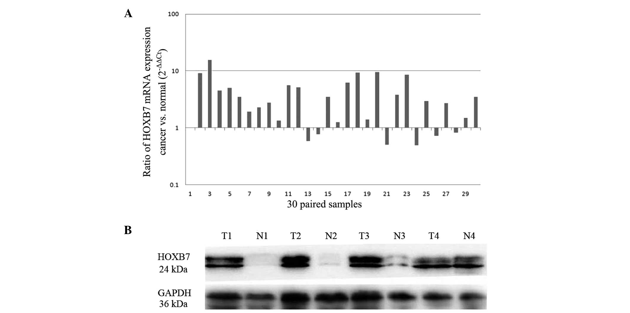
The expression of HOXB7 was detected through RT-qPCR
and western blot analysis. In total, 18 of the 30 paired cases
demonstrated a >2-fold increase in the expression of HOXB7 mRNA
in the gastric cancer tissue compared with the adjacent normal
mucosa, and the highest increase in HOXB7 mRNA expression was
15.6-fold (Fig. 1A). For the
subsequent western blot analysis, four pairs of samples were
randomly chosen from the 30 paired samples. At the protein level,
western blot analysis confirmed the upregulation of HOXB7 within
cancerous tissues compared with the adjacent non-cancerous tissues
from the same patients (Fig. 1B).
Overexpression of HOXB7 in gastric
cancer tissues was determined by immunohistochemistry
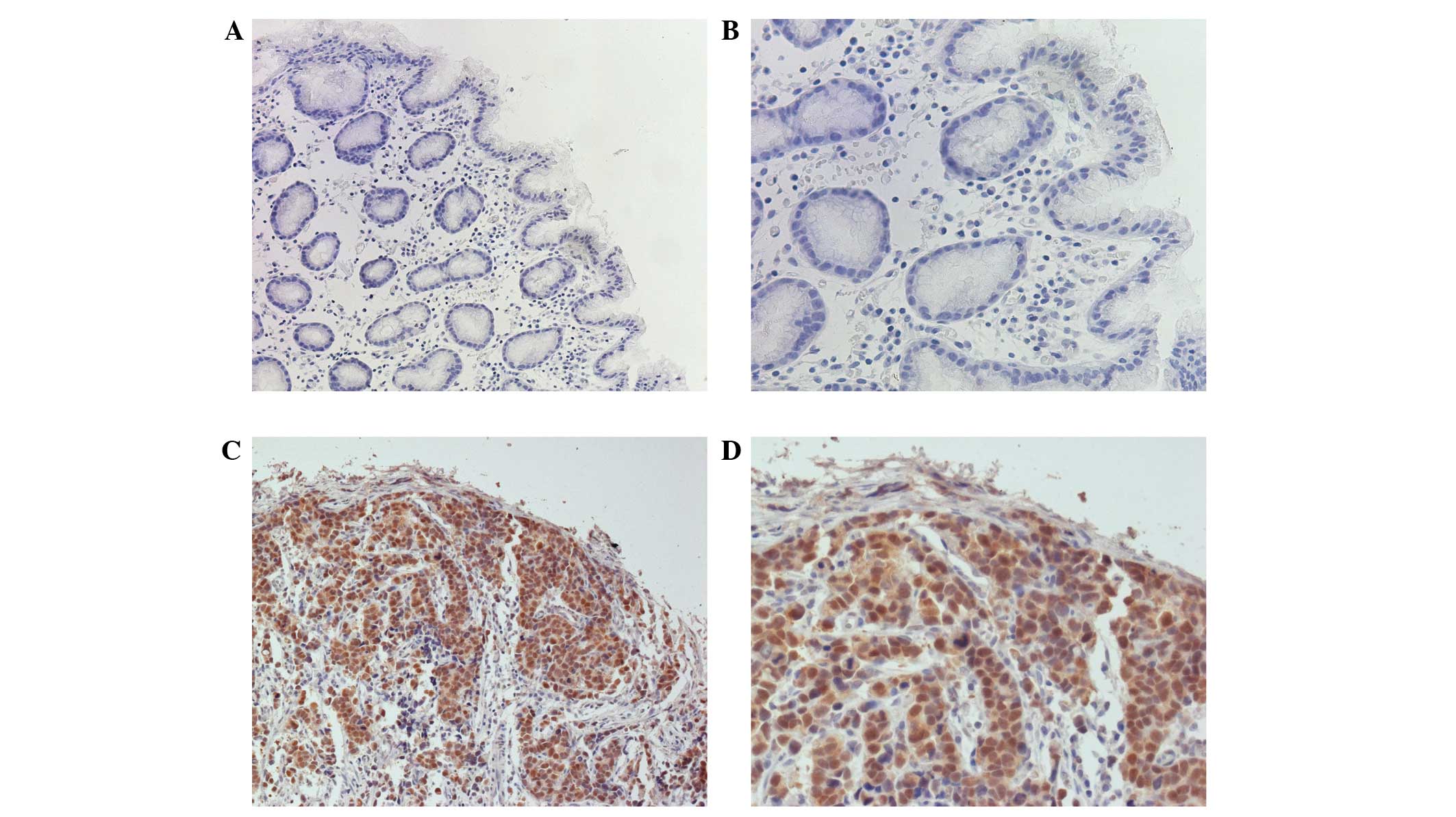
Immunohistochemistry was used to confirm the
expression of the HOXB7 protein in 96 paired paraffin-embedded
sections. Representative micrographs of HOXB7 staining in gastric
cancer samples revealed prominent nuclear expression and weaker
cytoplasmic expression (Fig. 2). Of
the 96 gastric cancer samples, 66 (68.8%) were positive for the
expression of HOXB7, whereas the HOXB7 protein was only detected in
15 out of 96 (15.6%) adjacent normal mucosa tissue samples
(Table I). The present results
indicated that HOXB7 expression was significantly upregulated in
tumor samples in comparison with the adjacent non-cancerous tissues
(P<0.001).
 | Table I.Expression of HOXB7 in normal gastric
mucosa and cancer tissues. |
Table I.
Expression of HOXB7 in normal gastric
mucosa and cancer tissues.
|
|
| HOXB7 expression |
|
|---|
|
|
|
|
|
|---|
| Tissue | Total, n | Absent, n (%) | Present, n (%) | P-value |
|---|
| Normal mucosa | 96 | 81 (84.4) | 15 (15.6) | <0.001 |
| Gastric cancer | 96 | 30 (31.2) | 66 (68.8) |
|
HOXB7 expression was associated with
the clinicopathological parameters of gastric cancer
As shown in Table II,
upregulated expression of HOXB7 was significantly associated with
the tumor size (P=0.01), T stage (P<0.001) and advanced Union
for International Cancer Control (UICC) stage (P=0.003). In
addition, out of the 66 samples that expressed HOXB7, 41 (62.1%)
tumors were classified as UICC stage III–IV and 51 (77.3%) tumors
were poorly differentiated. Thus, the results indicated that HOXB7
is associated with the progression of gastric cancer.
| Table II.Association between HOXB7 expression
and clinicopathological features in patients with gastric cancer
(n=96). |
Table II.
Association between HOXB7 expression
and clinicopathological features in patients with gastric cancer
(n=96).
|
|
| HOXB7 expression |
|
|---|
|
|
|
|
|
|---|
| Characteristic | Total, n | Absent, n (%) | Present, n (%) | P-value |
| Age, years |
|
|
| >0.05 |
|
<65 | 42 | 15 | 27 |
|
| ≥65 | 54 | 15 | 39 |
|
| Gender |
|
|
| >0.05 |
| Male | 57 | 19 | 38 |
|
|
Female | 39 | 11 | 28 |
|
| Tumor size |
|
|
| 0.01 |
| ≤2
cm | 20 | 11 | 9 |
|
| ﹥2
cm | 76 | 19 | 57 |
|
| T stage |
|
|
| <0.001 |
| T1 | 25 | 13 | 12 |
|
| T2 | 16 | 10 | 6 |
|
| T3 | 42 | 4 | 38 |
|
| T4 | 13 | 3 | 10 |
|
| N stage |
|
|
| >0.05 |
| N0 | 33 | 14 | 19 |
|
| N1 | 36 | 12 | 24 |
|
| N2 | 24 | 4 | 20 |
|
| N3 | 3 | 0 | 3 |
|
| M stage |
|
|
| >0.05 |
| M0 | 91 | 29 | 62 |
|
| M1 | 5 | 1 | 4 |
|
|
Differentiation |
|
|
| >0.05 |
|
Well-differentiated | 10 | 6 | 4 |
|
|
Moderately differentiated | 13 | 2 | 11 |
|
| Poorly
differentiated | 73 | 22 | 51 |
|
| UICC stage |
|
|
|
0.003 |
| I | 30 | 17 | 13 |
|
| II | 15 | 3 | 12 |
|
|
III | 40 | 9 | 31 |
|
| IV | 11 | 1 | 10 |
|
Survival analysis and prognostic
significance of HOXB7 expression in gastric cancer
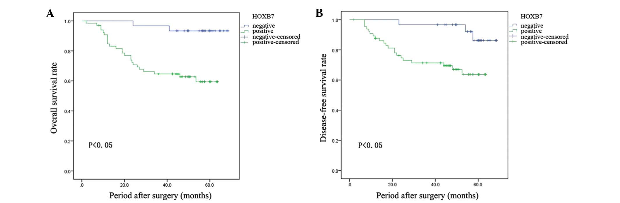
The Kaplan-Meier survival analysis indicated that
patients with HOXB7 expression demonstrated evidently decreased
overall survival (OS) and disease-free survival (DFS) rates
compared with patients with lesions that did not express HOXB7
(Fig. 3). Subsequently, the
univariate analysis revealed that the OS and DFS rates were
associated with tumor size (P=0.006 and P=0.042, respectively), T
stage (P=0.001 and P=0.011, respectively), N stage (P<0.001, for
both rates), advanced UICC stage (P<0.001 and P=0.001,
respectively) and HOXB7 expression (P=0.007 and P=0.015,
respectively). Furthermore, the multivariate survival analysis
revealed that the presence of HOXB7 expression was a significant
independent prognostic factor for the OS and DFS rates in patients
with gastric cancer (Table
III).
| Table III.Univariate and multivariate analysis
of variables affecting overall and disease-free survival after
surgery. |
Table III.
Univariate and multivariate analysis
of variables affecting overall and disease-free survival after
surgery.
| A, Overall
survival |
|
|
|
|
|
|
|---|
|
|---|
|
| Univariate
analysis | Multivariate
analysis |
|---|
|
|
|
|
|---|
| Variable | HR | 95% CI | P-value | HR | 95% CI | P-value |
|---|
| Age | 1.685 | 0.757–3.751 |
0.201 |
| NR |
|
| Gender | 0.987 | 0.458–2.127 |
0.973 |
| NR |
|
| Tumor size | 2.322 | 1.277–4.223 |
0.006a |
| NR |
|
| T stage | 2.115 | 1.377–3.250 |
0.001a |
| NR |
|
| N stage | 3.237 | 1.958–5.350 |
<0.001a | 1.972 | 1.070–3.634 | 0.029 |
| M stage | 3.319 |
0.990–11.125 |
0.052 |
| NR |
|
|
Differentiation | 1.488 | 0.744–2.976 |
0.262 |
| NR |
|
| UICC stage | 2.914 | 1.809–4.695 |
<0.001a | 1.866 | 1.033–3.372 | 0.039 |
| HOXB7 | 7.384 |
1.745–31.242 |
0.007a | 4.737 |
1.097–20.447 | 0.037 |
|
| B, Disease-free
survival |
|
|
|
|
|
|
|
|
| Univariate
analysis | Multivariate
analysis |
|
|
|
|
| Variable | HR | 95% CI | P-value | HR | 95% CI | P-value |
|
| Age | 1.712 | 0.732–4.005 |
0.215 |
| NR |
|
| Gender | 0.691 | 0.295–1.616 |
0.393 |
| NR |
|
| Tumor size | 1.853 | 1.023–3.354 |
0.042a |
| NR |
|
| T stage | 1.720 | 1.134–2.610 |
0.011a |
| NR |
|
| N stage | 3.036 | 1.778–5.184 |
<0.001a | 2.823 | 1.644–4.848 | <0.001 |
| M stage | 1.179 | 0.158–8.778 |
0.872 |
| NR |
|
|
Differentiation | 1.594 | 0.746–3.405 |
0.228 |
| NR |
|
| UICC stage | 2.519 | 1.382–3.374 |
0.001a |
| NR |
|
| HOXB7 | 4.489 |
1.331–15.141 |
0.015a | 3.742 |
1.077–13.003 |
0.038 |
Discussion
HOX genes play an important role during embryonic
development by encoding transcription factors that regulate the
expression of numerous downstream target genes (7). However, dysregulation of HOX gene
expression is frequently found in certain cancers, including oral
squamous cell carcinoma, breast cancer, colorectal cancer, and
pancreatic ductal adenocarcinoma (PDAC) (10–13).
However, little is known about the association between HOX gene
expression and gastric cancer.
It has previously been found that HOXB7 is
upregulated in gastric cancers, which was determined using a cDNA
microarray (15). The present study
identified that HOXB7 was upregulated in gastric cancer at the mRNA
and protein levels by RT-qPCR and western blot analysis. The
present findings indicated that HOXB7 was likely to be associated
with gastric cancer. Subsequently, immunohistochemistry revealed
that 66 of the 96 tumor samples (68.8%) were positive for the
expression of HOXB7, while the HOXB7 protein was only detected in
15 of the 96 adjacent normal mucosa samples (15.6%). The results
presented in the present study demonstrated that HOXB7 expression
was significantly upregulated in gastric cancer samples in
comparison with adjacent normal tissue (P<0.001). In addition,
upregulation of HOXB7 expression has also been observed in certain
cancers, including oral squamous cell carcinoma, PDAC, and
colorectal cancer (10,12,13). De
Souza Setubal Destro et al (12) found that expression of HOXB7 was
significantly increased in oral squamous cell carcinomas compared
to normal oral mucosas. In addition, Nguyen Kovochich et al
(13) revealed that the levels of the
HOXB7 mRNA and protein were significantly elevated in PDAC cell
lines and patient tumor samples relative to normal pancreas cells.
Liao et al (10) also
identified that HOXB7 was significantly upregulated in eight
examined colorectal cancer samples paired with adjacent
non-cancerous tissues. These studies indicated that HOXB7 was
highly expressed in numerous cancers and may be involved in the
process of cancer development.
The present results also revealed that the
expression of the HOXB7 protein was associated with the tumor size,
and T and advanced UICC stages of the lesion. In addition, HOXB7
overexpression was found to be associated with poor survival rates
in patients with gastric cancer. Multivariate survival analysis
revealed that the presence of HOXB7 expression was a significant
independent prognostic factor for OS and DFS rates in gastric
cancer patients. Therefore, it was hypothesized that HOXB7 may act
as a valuable prognostic biomarker for gastric cancer. Similarly to
the present results, HOXB7 overexpression was also reported to be
associated with clinical progression and a poor prognosis of
patients with colorectal cancer and PDAC (10,13). Liao
et al (10) revealed that the
expression of the HOXB7 protein was associated with an advanced
Dukes' stage, the T stage, the presence of distant metastasis, a
higher proliferation index and a poor survival of patients. In
addition, Nguyen Kovochich et al (13) found that HOXB7 protein expression was
associated with lymph node metastasis and acted as an independent
predictor of worse overall survival, which was determined by
multivariate analysis. It was therefore hypothesized that HOXB7 may
be used as a biomarker to identify the prognosis of patients with
various cancers.
Multiple studies have revealed that aberrant
expression of HOXB7 may promote cell proliferation, influence
tumorigenesis, invasion and metastasis. For instance, De Souza
Setubal Destro et al (12)
identified that overexpression of HOXB7 in the HaCAT human
epithelial cell line promoted cell proliferation, whereas the
downregulation of the endogenous levels of HOXB7 in human oral
carcinoma SCC9 cells led to decreased proliferation. In addition,
Wu et al (11) demonstrated
that HOXB7 overexpression promoted tumor invasion by upregulating
basic fibroblast growth factor (bFGF), which is known to be a
transcriptional target of HOXB7. Chen et al (18) generated mouse mammary tumor
virus-HOXB7 transgenic mice to study the function of HOXB7. It was
observed that HOXB7 promoted tumor progression and metastasis.
Overall, HOXB7 has been revealed to be involved in several cancers
through its multiple effects.
Although HOXB7 has been found to be associated with
tumorigenesis, migration and invasion, the precise mechanisms are
not well elucidated. Wu et al (11) reported that HOXB7 induced
epithelial-mesenchymal transition (EMT) through the
Ras-mitogen-activated protein kinase (MAPK) pathway in breast
cancer cell lines. It is well known that EMT is currently
considered to be a mechanism in the process of tumorigenesis
(20,21). HOXB7 was found to be able to activate
the Ras/Rho pathway by upregulating bFGF and then promoting tumor
invasion (11). In addition, Liao
et al (10) identified that
HOXB7 facilitated the G1 to S-phase cell cycle
transition in colorectal cancer cells. Enforced expression of HOXB7
was found to regulate the cell cycle factors cyclin D1 and
p27Kip1 through the activation of the MAPK and
phosphoinositide 3-kinase (PI3K)-Akt signaling pathways. This study
indicated that HOXB7 may modulate the tumor proliferation through
MAPK and PI3K-Akt pathways. However, the exact mechanisms of HOXB7
deregulation in cancer require further investigation.
In conclusion, the present study reported the
clinical significance of HOXB7 expression in gastric cancer. The
current study demonstrated that overexpression of HOXB7 may be
associated with a highly aggressive phenotype of gastric cancer.
Furthermore, HOXB7 may be a significant prognostic biomarker for
gastric cancer. These preliminary results require verification in a
larger controlled prospective clinical study.
Acknowledgements
This study was supported by the National Natural
Science Foundation of China (grant nos., 81172330 and 30700813),
New Medical Younger Talent of Shanghai Health Bureau (grant no.,
XYQ 2011035) and ‘Climbing’ Program of Shanghai Songjiang District
(grant no., 2011PD02).
References
|
1
|
Wu AW, Ji JF, Yang H, et al: Long-term
outcome of a large series of gastric cancer patients in China. Chin
J Cancer Res. 22:167–175. 2011. View Article : Google Scholar
|
|
2
|
Haglund C, Kuusela P, Roberts P and
Jalanko H: Tumour marker CA125 in patients with digestive tract
malignancies. Scand J Clin Lab Invest. 51:265–270. 1991. View Article : Google Scholar : PubMed/NCBI
|
|
3
|
Ychou M, Duffour J, Kramar A, et al:
Clinical significance and prognostic Value of CA724 compared with
CEA and CA199 in patients with gastric cancer. Dis Markers.
16:105–110. 2000. View Article : Google Scholar : PubMed/NCBI
|
|
4
|
Li Y, Yang Y, Lu M and Shen L: Predictive
value of serum CEA, CA199 and CA724 in early diagnosis of
recurrence after radical resection of gastric cancer.
Hepatogastroenterology. 58:2166–2170. 2011.PubMed/NCBI
|
|
5
|
Taipale J and Beachy PA: The Hedgehog and
Wnt signalling pathways in cancer. Nature. 411:349–354. 2001.
View Article : Google Scholar : PubMed/NCBI
|
|
6
|
Reya T and Clevers H: Wnt signaling in
stem cells and cancer. Nature. 434:843–850. 2005. View Article : Google Scholar : PubMed/NCBI
|
|
7
|
Samuel S and Naora H: Homeobox gene
expression in cancer: insights from developmental regulation and
deregulation. Eur J Cancer. 41:2428–2437. 2005. View Article : Google Scholar : PubMed/NCBI
|
|
8
|
Cillo C, Faiella A, Cantile M and
Boncinelli E: Homeobox genes and cancer. Exp Cell Res. 248:1–9.
1999. View Article : Google Scholar : PubMed/NCBI
|
|
9
|
Raval A, Tanner SM, Byral JC, et al:
Downregulation of death-associated protein kinase 1 (DAPK 1) in
chronic lymphocytic leukemia. Cell. 129:879–890. 2007. View Article : Google Scholar : PubMed/NCBI
|
|
10
|
Liao WT, Jiang D, Yuan J, et al: HOXB7 as
a prognostic factor and mediator of colorectal cancer progression.
Clin Cancer Res. 17:3569–3578. 2011. View Article : Google Scholar : PubMed/NCBI
|
|
11
|
Wu X, Chen H, Parker B, et al: HOXB7, a
homeodomain protein, is overexpressed in breast cancer and confers
epithelial-mesenchymal transition. Cancer Res. 66:9527–9534. 2006.
View Article : Google Scholar : PubMed/NCBI
|
|
12
|
De Souza Setubal Destro MF, Bitu CC,
Zecchin KG, et al: Overexpression of HOXB7 homeobox gene in oral
cancer induces cellular proliferation and is associated with poor
prognosis. Int J Oncol. 36:141–149. 2010.PubMed/NCBI
|
|
13
|
Kovochich A Nguyen, Arensman M, Lay AR, et
al: HOXB7 promotes invasion and predicts survival in pancreatic
adenocarcinoma. Cancer. 119:529–539. 2013. View Article : Google Scholar : PubMed/NCBI
|
|
14
|
Waltregny D, Alami Y, Clause N, De Leval J
and Castronovo V: Overexpression of the homeobox gene HOXC8 in
human prostate cancer correlates with loss of tumor
differentiation. Prostate. 50:162–169. 2002. View Article : Google Scholar : PubMed/NCBI
|
|
15
|
Han Y, Tu WW, Wen YG, et al:
Identification and validation that up-expression of HOXA13 is a
novel independent prognostic marker of a worse outcome in gastric
cancer based on immunohistochemistry. Med Oncol. 30:5642013.
View Article : Google Scholar : PubMed/NCBI
|
|
16
|
Caré A, Silvani A, Meccia E, Mattia G,
Peschle C and Colombo MP: Transduction of the SkBr3 breast
carcinoma cell line with the HOXB7 gene induces bFGF expression,
increase cell proliferarion and reduces growth factor dependence.
Oncogene. 16:3285–3289. 1998. View Article : Google Scholar : PubMed/NCBI
|
|
17
|
Carè A, Felicetti F, Meccia E, et al:
HOXB7: a key factor for tumor-associated angiogenic switch. Cancer
Res. 61:6523–6529. 2001.
|
|
18
|
Chen H, Lee JS, Liang X, et al: Hoxb7
inhibits transgenic HER-2/neu-induced mouse mammary tumor onset but
promotes progression and lung metastasis. Cancer Res. 68:3637–3644.
2008. View Article : Google Scholar : PubMed/NCBI
|
|
19
|
Li D, Yan D, Tang H, et al: IMP3 is a
novel prognostic marker that correlates with colon cancer
progression and pathogenesis. Ann Surg Oncol. 16:3499–3506. 2009.
View Article : Google Scholar : PubMed/NCBI
|
|
20
|
Kang Y and Massagué J:
Epithelial-mesenchymal transitions: twist in development and
metastasis. Cell. 118:227–229. 2004. View Article : Google Scholar
|
|
21
|
Petersen OW, Nielsen HL, Gudjonsson T, et
al: Epithelial to mesenchymal transition in human breast cancer can
provide a nonmalignant stroma. Am J Pathol. 162:391–402. 2003.
View Article : Google Scholar : PubMed/NCBI
|