Introduction
The major features of malignant cancer are
uncontrolled cell growth and proliferation, while cell cycle
disorder is an important pathogenesis mechanism. The primary
function of S-phase kinase-associated protein 2 (Skp2) is its role
as the substrate recognition subunit of ubiquitin ligase complex by
specific recognition of the phosphorylated substrate, and its
ability to mediate ubiquitin ligase complex polyubiquitination and
degradation (1,2). Cyclin-dependent kinase (CDK) inhibitor
(CKI), p27kinase-interacting protein 1
(p27kip1), is an important negative regulator of the
cell cycle (1). As a well-recognized
tumor suppressor gene, it is able to control cell cycle transition
from G1 to S phase. Thus far, various cell cycle
regulators have been identified, including the tumor suppressor
p27kip1, p21wafl, cyclin A and p53, all of
which may be degraded via the Skp2-mediated ubiquitin-proteasome
pathway (2,3). Thus, Skp2 is key in the
p27kip1 ubiquitin degradation pathway and may inhibit
the proliferation of a variety of cell types through the
ubiquitin-proteasome pathway (4–7). Skp2 and
p27kip1 have been to demonstrated to exhibit varying
degrees of abnormal expression in a number of solid tumors,
including lung cancer (8),
oesophageal squamous cell carcinoma and oral squamous cell
carcinoma (9,10), laryngeal squamous cell carcinoma
(11) and breast cancer (12). They are considered to be closely
associated with the occurrence, development and prognosis of
malignant tumors (8–12). The expression levels of Skp2 and
p27kip1 proteins have rarely been investigated in
hypopharyngeal squamous cell carcinoma. Therefore, the present
study used immunohistochemistry to detect the expression of Skp2
and p27kip1 proteins in normal hypopharyngeal mucosa and
hypopharyngeal squamous cell carcinoma tissues. The correlation
between their expression, and the occurrence and development of
hypopharyngeal squamous cell carcinoma, as well as clinical and
pathological features, was then analyzed.
Materials and methods
Specimen collection
Hypopharyngeal squamous cell carcinoma specimens
(n=42) were obtained from patients treated at the Second Affiliated
Hospital of Fujian Medical University (Quanzhou, China) between
January 1996 and February 2009. A diagnosis of hypopharyngeal
squamous cell carcinoma was confirmed by pathological examination.
No patients underwent radiotherapy or chemotherapy prior to the
surgery. The 42 cases included 39 males and 3 females, and the
median age was 54.8 years. Approval for the present study was
obtained from the Ethics Committee of Fujian Medical University.
Written informed consent was obtained from all the participants.
According to different primary focus of tumors, 21 cases occurred
in the piriform sinus, 15 occurred in the posterior wall and 6 in
the postcricoid area. According to the differentiation degrees,
there were 15 well-differentiated, 17 moderately-differentiated and
10 poorly-differentiated cancer cases. In addition, 37 cases
exhibited cervical lymph node metastasis, while 5 cases did not
present metastasis. According to the 2002 classification criteria
of the Union for International Cancer Control TNM staging system
(13), there were 15 T1/T2 stage and
27 T3/T4 stage cases, including T1N0M0 (2 cases), T1N1M0 (1 case),
T2N0M0 (2 cases), T2N1M0 (3 cases), T2N2M0 (7 cases),; T3N0M0 (2
cases), T3N1M0 (8 cases), T3N2M0 (9 cases), T4N0M0 (1 case), T4N1M0
(2 cases), T4N2M0 (3 cases) and T4N3M0 (2 cases). Normal
hypopharyngeal mucous membranes without cancer cells were
identified by observation from the piriform sinus or postcricoid
area of specimens obtained during total laryngectomy and used as
the control group. Specimens were obtained from 15 patients with
laryngeal carcinoma who underwent total laryngectomy at the Second
Affiliated Hospital of Fujian Medical University. The specimens
were located 3–4 cm from the tumor margin. The patients included 14
males and 1 female, and had an age range of 47–71 years (mean age,
59.1 years). There was no significant difference in age or gender
between the control and experimental groups. The normal tissue
samples (n=15) were confirmed to be healthy hypopharyngeal mucous
membranes by performing a hematoxylin and eosin staining assay.
Immunohistochemical assay
The immunohistochemical assay used a PV-9000 (SP)
two-step staining kit (Zhongshan Jinqiao Biotechnology Co., Ltd.,
Beijing, China), according to the manufacturer's instructions.
p27kip1 and Skp2 rat anti-human monoclonal antibodies
(1:40 dilutions; catalog nos. ZM-0340 and ZM-0454, respectively),
polymer helper and polyperoxidase anti-rat IgG (1:100 dilution)
were all purchased from Zhongshan Jinqiao Biotechnology Co., Ltd.
Furthermore, breast cancer tissue specimens (ZSGB-BIO Co., Ltd.,
Beijing, China) were used as the positive control and
phosphate-buffered saline (PBS) was used as the negative
control.
Analysis of experimental results
To analyze the results of the current study, Skp2
positive cell counts were performed in a minimum of 10 fields per
sample under an optical microscope (magnification, ×400; BX51;
Olympus Corporation, Tokyo, Japan). In the positive cells counts,
≥100 tumor cells were present in each field. According to a
previous study (8), Skp2-positive
cells were visualized as brown-yellow particles in the nucleus and
negative control cells were normal hypopharyngeal mucous membranes
in close proximity to the tumor tissues. Skp2 protein expression
was classified as follows: <5% positive cells, (−); 6–25%
positive cells, (+); 26–50% positive cells, (++); and >50%
positive cells, (+++). Furthermore, >25% positive cells
represented a high expression of Skp2. For the expression of
p27kip1, positive cells were visualized as brown-yellow
particles in the nucleus. p27kip1 protein expression was
classified as follows: 0% positive cells, (−); <5% positive
cells, (+); 5–50% positive cells, (++); and >50% positive cells,
(+++). Furthermore, >50% positive cells represented a high
expression of p27kip1. All the results were evaluated by
two experienced pathologists using a double-blinded method.
Statistical analysis
Statistical analyses were performed using the SPSS
software (version 13.0; SPSS, Inc., Chicago, IL, USA) and data are
presented as the mean ± standard deviation. χ2 and
Fisher's tests were used to compare data between the groups, while
Spearman's rank correlation analysis was used for data correlation.
P<0.05 was considered to indicate a statistically significant
difference.
Results
Skp2 and p27kip1 protein
expression levels
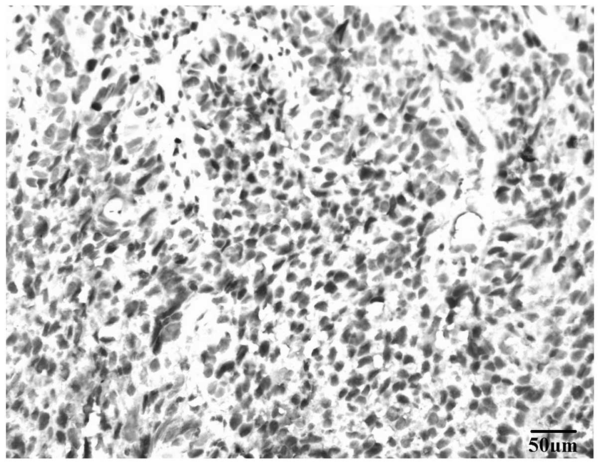
Immunohistochemical staining revealed that Skp2
protein, which was visualized as brown-yellow particles, was
predominantly located in the nucleus and rarely located in the
cytoplasm (Fig. 1). High Skp2 protein
expression was observed in 61.90% (26/42 samples) and 26.67% (4/15
samples) of hypopharyngeal squamous cell carcinoma tissues and
normal hypopharyngeal mucous membranes, respectively. Thus, the
expression of Skp2 protein was significantly higher in
hypopharyngeal squamous cell carcinoma compared with that in normal
hypopharyngeal mucous membranes (P<0.05). The expression levels
of Skp2 and p27kip1 proteins in normal hypopharyngeal mucosa of the
control group were not significantly correlated
(χ2=1.26, Pearson coefficient of continency, C=0.2756).
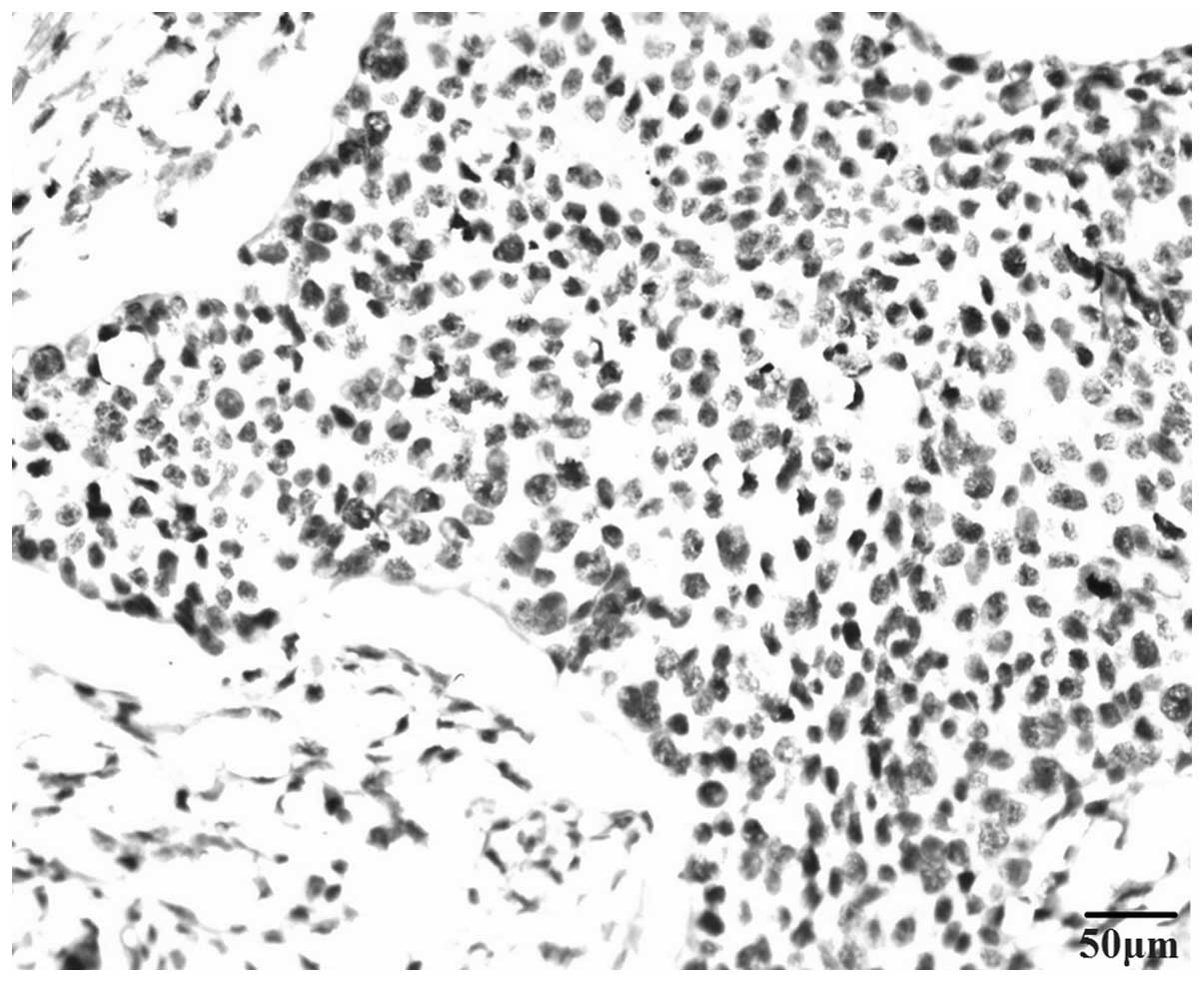
p27kip1 protein presented as brown-yellow particles
located in the nucleus (Fig. 2). Weak
p27kip1 expression was observed in hypopharyngeal
squamous cell carcinoma tissues, with only 5 cases exhibiting high
expression. In addition, high p27kip1 protein expression
was observed in 11.90% (5/42) and 53.33% (8/15) of hypopharyngeal
squamous cell carcinoma tissues and normal hypopharyngeal mucous
membranes, respectively. This difference was statistically
significant (P<0.05; Table I).
 | Table I.Comparison of Skp2 and
p27kip1 protein expression levels in hypopharyngeal
squamous cell carcinoma tissue and normal hypopharyngeal mucous
membrane samples. |
Table I.
Comparison of Skp2 and
p27kip1 protein expression levels in hypopharyngeal
squamous cell carcinoma tissue and normal hypopharyngeal mucous
membrane samples.
| Index | Hypopharyngeal
squamous cell carcinoma | Normal hypopharyngeal
mucous membrane | P-value |
|---|
| Skp2 | 26 | 4 | 0.019 |
|
p27kip1 | 5 | 8 | 0.002 |
Correlation of Skp2 and
p27kip1 protein expression levels with
clinicopathological characteristics
No statistically significant differences were
detected between the age or differentiation degree of patients and
the expression of Skp2 protein (P>0.05). However, a significant
difference was identified between gender and the expression of Skp2
protein (P<0.05). Despite this significant association, the
study population had a 1:13 female: male ratio and the P-value was
close to 0.05. Thus, the present authors consider that there may be
no significant difference between gender and the expression of Skp2
protein. Notably, there was an significant difference between high
expression of Skp2 protein and cervical lymph node metastasis
(P<0.01), as well as between T stage and the high expression of
Skp2 protein (P<0.05). Spearman's rank correlation analysis
indicated that the expression of Skp2 protein was positively
correlated with cervical lymph node metastasis [Spearman's
correlation coefficient (rs)=0.402, P<0.01] and T
staging (rs=0.033, P<0.05) in hypopharyngeal squamous
cell carcinoma.
The present study also identified no significant
correlation between the gender, age and differentiation degree of
patients, and the expression of p27kip1 protein
(P>0.05). However, low expression of p27kip1 protein
was significantly associated with cervical lymph node metastasis
(P<0.01) and T stage (P<0.05). Furthermore, Spearman's rank
correlation analysis indicated that the expression of
p27kip1 protein was negatively correlated with cervical
lymph node metastasis (rs=-0.016, P<0.05) and T stage
(rs=-0.351, P<0.05) in hypopharyngeal squamous cell
carcinoma (Table II).
| Table II.Association of high Skp2 and
p27kip1 protein expression with clinicopathological
characteristics in patients with hypopharyngeal squamous cell
carcinoma. |
Table II.
Association of high Skp2 and
p27kip1 protein expression with clinicopathological
characteristics in patients with hypopharyngeal squamous cell
carcinoma.
|
|
| Skp2 |
p27kip1 |
|---|
|
|
|
|
|
|---|
| Pathological
characteristic | Case, n | High expression,
n | P-value | High expression,
n | P-value |
|---|
| Gender |
|
|
|
|
|
|
Male | 39 | 26 | 0.049 | 4 | 0.323 |
|
Female | 3 | 0 |
| 1 |
|
| Age, years |
|
|
|
|
|
|
≥60 | 11 | 6 | 0.720 | 1 | 1.000 |
|
<60 | 31 | 20 |
| 4 |
|
| Cervical lymph node
metastasis |
|
|
|
|
|
| + | 37 | 26 | 0.005 | 2 | 0.008 |
| – | 5 | 0 |
| 3 |
|
| Primary location of
the tumora |
|
|
|
|
|
|
Piriform sinus | 21 | 12 | >0.0125 | 3 | >0.0125 |
|
Postcricoid area | 6 | 4 |
| 1 |
|
|
Posterior wall | 15 | 10 |
| 1 |
|
| T stage |
|
|
|
|
|
| T1 +
T2 | 15 | 6 | 0.029 | 4 | 0.047 |
| T3 +
T4 | 27 | 20 |
| 1 |
|
| Differentiation
degree |
|
|
|
|
|
| High to
moderate | 32 | 18 | 0.270 | 3 | 0.577 |
|
Low | 10 | 8 |
| 2 |
|
Correlation analysis between Skp2 and
p27kip1 protein expression levels
Spearman's rank correlation analysis indicated that
there was a negative correlation between the high expression of
Skp2 protein and low expression of p27kip1 protein in
hypopharyngeal squamous cell carcinoma (rs=-0.317,
P=0.041; Table III).
| Table III.Association between Skp2 and
p27kip1 protein expression in hypopharyngeal squamous
cell carcinoma. |
Table III.
Association between Skp2 and
p27kip1 protein expression in hypopharyngeal squamous
cell carcinoma.
|
| p27kip1
expression, n |
|
|
|---|
|
|
|
|
|
|---|
| Skp2
expression | High | Low | P-value | rs |
|---|
| High | 1 | 25 |
|
|
| Low | 4 | 12 | 0.041 | −0.317 |
Discussion
Skp2 was initially cloned from human fibroblasts in
1995 (13). Skp2 is predominantly
located in the S phase of malignant cells and reacts with the
cyclin A-CDK complex; thus, it is termed S-phase kinase-associated
protein 2. Skp2 behaves as an oncogene in cell systems and is an
established protooncogene causally involved in the pathogenesis of
lymphomas. Skp2 expression, encoded by the Skp2 gene,
appears to be closely correlated with the occurrence and
development of carcinomas through binding to various targeting
ubiquitinated proteins (2,14). Furthermore, it has been demonstrated
that numerous cell cycle proteins exert their effects via a
Skp2-dependent ubiquitin proteasome signaling pathway, including
cyclin D1, cyclin E, p27kip1 and p53 (2,3). Thus,
Skp2 specifically may be involved in the regulation of cell
proliferation and apoptosis (15). In
addition, Skp2 acts as oncogene through the degradation of
p27kip1, with the expression of Skp2 and
p27kip1 differing in each stage of the cell cycle.
Typically, Skp2 expression initially occurs during the
G1-S phase, increases at the S-G2 phase and
then quickly decreased at the M phase. A number of previous studies
have indicated that p27kip1 is specifically recognized
and targeted for ubiquitination by Skp2 (15,16). For
instance, Skp2 may recognize the cyclin E-CDK2 and cyclin D-CDK2
complexes, causing phosphorylation of threonine-187 in p27
(15,16). Skp2 is particularly important for
regulating the degradation of p27kip1 in the
G1-S phase. Disorder of cell cycle regulation is a
critical molecular biological event in tumorigenesis; thus, cell
cycle regulating factors may be relevant to tumorigenesis. For
example, CKIs exhibit negative effects on cell cycle regulation. In
particular, p27kip1, which a member of the CKI family
functioning as a negative regulating factor of the cell cycle, may
play a key role in cell proliferation. Currently,
p27kip1 is known as a tumor-suppressing gene (17). A previous study proposed that
mutations of the p27 gene are rare in tumors (18); however, abnormal p27 protein
expression was observed in the tumors investigated in the present
study. This abnormal expression may be the result of low gene
transcription or high protein activity. Various tumors, including
colon cancer, rectum cancer, cervical carcinoma, oral squamous cell
carcinoma, gastric cancer, lung cancer and prostatic cancer, have
been found to exhibit a high expression of Skp2 protein but low
expression of p27kip1 protein (19,20), and a
negative correlation was identified between Skp2 and
p27kip1 protein expression (8–12).
Furthermore, tumor malignancy and prognosis were correlated with
Skp2 expression (21,22).
In the present study, 26/42 samples (61.9%)
exhibited Skp2 protein expression in hypopharyngeal squamous cell
carcinoma tissues; however, Skp2 protein expression was low in
11/15 and high in 4/15 of normal hypopharyngeal mucous membranes.
In addition, p27kip1 protein expression was observed in
5/42 (11.9%) samples of hypopharyngeal squamous cell carcinoma and
high p27kip1 protein expression was observed in 8/15
(53.33%) samples of normal hypopharyngeal mucous membranes. This
difference was statistically significance (P<0.05). Spearman's
rank correlation analysis identified that overexpression of Skp2
protein was negatively correlated with low expression of
p27kip1 protein in hypopharyngeal squamous cell
carcinoma tissues (rs=-0.317, P=0.041). The current
results were similar to those of previous studies conducted
(5,10), and indicate that Skp2 downregulates
the expression of p27kip1 through ubiquitination in
hypopharyngeal squamous cell carcinoma. Thus, high expression of
Skp2 protein and low expression of p27kip1 protein may
promote tumor cell progression from G1 to S phase.
p27kip1 is unable to effectively inhibit aberrant cell
proliferation, but can accelerate cell malignancy (18,21).
Therefore, it is proposed that Skp2 promotes the development of
hypopharyngeal squamous cell carcinoma through degradation of
p27kip1 protein.
In numerous malignant tumors, including gastric
(8), breast (12), prostate (18) and colon cancer (8), increased Skp2 protein expression is
observed concurrently with reduced p27kip1 protein
expression. Increased Skp2 protein expression indicates a poorer
prognosis and more highly-differentiated tumors. Furthermore,
p27kip1 appears to be involved in regulating cell
differentiation and intercellular adhesion, which are processes
associated with tumor infiltration and metastasis, respectively.
This may be due to lack of p27kip1 transcription or
decreased p27kip1 protein expression causing inhibition
of the cell proliferation, disorder of the cell cycle and low cell
differentiation. Furthermore, p27kip1 regulates
intercellular adhesion by aggregating N-cadherin to prevent normal
cells from binding to stroma or receiving extracellular signals
(8,9,12,17,18). In a
previous study, p27kip1 protein expression caused a
reduction in tumor cell adhesion and tumor metastasis (17). The current results demonstrated that
high expression of Skp2 protein and low expression of
p27kip1 protein were significantly associated with
cervical lymph node metastasis and T-stage in 42 samples of
hypopharyngeal squamous cell carcinomas. Furthermore, correlation
analysis identified a positive correlation between high expression
of Skp2 protein, and cervical lymph node metastasis and T-stage,
and a negative correlation between low expression of
p27kip1 protein, and cervical lymph node metastasis and
T-stage. By contrast, the expression levels of Skp2 and
p27kip1 protein were not significantly associated with
age, gender or differentiation degree (P>0.05). The
aforementioned results were similar to those obtained in the
present study, which indicated that high expression of Skp2 protein
and low expression of p27kip1 protein may aid in
predicting the degree of tumor infiltration and cervical lymph node
metastasis in hypopharyngeal squamous cell carcinoma. Furthermore,
these two proteins may be useful for assessing the biological
characteristics of hypopharyngeal squamous cell carcinoma.
However, Dowen et al (23) opposed the aforementioned theory that
increased Skp2 protein expression decreases the expression of
p27kip1 protein. Instead, the authors proposed that the
significance of high Skp2 protein expression in tumors was
associated with the degradation of p27kip1 protein, as
well as other mechanisms, which may require further
investigation.
In conclusion, Skp2 may degrade p27kip1
protein through the ubiquitin-proteasome pathway, thus,
participating in the occurrence and development of hypopharyngeal
squamous cell carcinoma. Furthermore, the abnormal protein
expressions Skp2 and p27kip1 appear to be associated
with the poor prognosis of hypopharyngeal squamous cell carcinoma.
Therefore, combined detection of Skp2 and p27kip1 may
provide significant guidance for comprehensively determining the
malignant degree and prognosis of patients with hypopharyngeal
squamous cell carcinoma.
References
|
1
|
Slingerland J and Pagano M: Regulation of
the cdk inhibitor p27 and its deregulation in cancer. J Cell
Physiol. 183:10–17. 2000. View Article : Google Scholar : PubMed/NCBI
|
|
2
|
Inuzuka H, Gao D, Finley LW, et al:
Acetylation-dependent regulation of Skp2 function. Cell.
150:179–193. 2012. View Article : Google Scholar : PubMed/NCBI
|
|
3
|
Ezoe S, Matsumura I, Nakata S, et al:
GATA-2/estrogen receptor chimera regulates cytokine-dependent
growth of hematopoietic cells through accumulation of p21(WAF1) and
p27(Kip1) proteins. Blood. 100:3512–3520. 2002. View Article : Google Scholar : PubMed/NCBI
|
|
4
|
Sicari BM, Troxell R, Salim F, Tanwir M,
Takane KK and Fiaschi-Taesch N: c-myc and skp2 coordinate p27
degradation, vascular smooth muscle proliferation, and neointima
formation induced by the parathyroid hormone-related protein.
Endocrinology. 153:861–872. 2012. View Article : Google Scholar : PubMed/NCBI
|
|
5
|
Dai DM and Zhu H: Expression of P27kip1
and Skp2 in basal cell carcinoma and its clinical pathological
characters. Int Oncol. 38:158–160. 2011.
|
|
6
|
Suzuki S, Fukasawa H, Misaki T, et al: The
amelioration of renal damage in Skp2-deficient mice canceled by
p27kipl deficiency in Skp2-/-p27/-mice. PLoS One. 7:e362492012.
View Article : Google Scholar : PubMed/NCBI
|
|
7
|
Nakayama K, Nagahama H, Minamishima YA, et
al: Targeted disruption of Skp2 results in accumulation of cyclin E
and p27(Kip1) polyploidy and centrosome overduplication. EMBO J.
19:2069–2081. 2000. View Article : Google Scholar : PubMed/NCBI
|
|
8
|
Gstaiger M, Jordan R, Lim M, Catzavelos C,
Mestan J, Slingerland J and Krek W: Skp2 is oncogenic and
overexpressed in human cancers. Proc Natl Acad Sci USA.
98:5043–5048. 2001. View Article : Google Scholar : PubMed/NCBI
|
|
9
|
Kudo Y, Kitajima S, Sato S, Miyauchi M,
Ogawa I and Takata T: High expression of S-phase kinase-interacting
protein 2, human F-box protein, correlates with poor prognosis in
oral squamous cell carcinomas. Cancer Res. 61:7044–7047.
2001.PubMed/NCBI
|
|
10
|
Shintani S, Li C, Mihara M, Hino S,
Nakashiro K and Hamakawa H: Skp2 and Jab1 expression are associated
with inverse expression of p27(KIP1) and poor prognosis in oral
squamous cell carcinomas. Oncology. 65:355–362. 2003. View Article : Google Scholar : PubMed/NCBI
|
|
11
|
Dong Y, Sui L, Watanabe Y, Sugimoto K and
Tokuda M: S-phase kinase-associated protein 2 expression in
laryngeal squamous cell carcinomas and its prognostic implications.
Oncol Rep. 10:321–325. 2003.PubMed/NCBI
|
|
12
|
Signoretti S, Di Marcotullio L, Richardson
A, et al: Oncogenic role of the ubiquitin ligase subunit Skp2 in
human breast cancer. J Clin Invest. 110:633–641. 2002. View Article : Google Scholar : PubMed/NCBI
|
|
13
|
Patel SG and Shah JP: TNM staging of
cancers of the head and neck: striving for uniformity among
diversity. CA Cancer J Clin. 55:242–258; quiz 261–262, 264. 2005.
View Article : Google Scholar : PubMed/NCBI
|
|
14
|
Zhang H, Kobayashi R, Galaktionov K and
Beach D: p19Skp1 and p45Skp2 are essential elements of the cyclin
A-CDK2 S phase kinase. Cell. 82:915–925. 1995. View Article : Google Scholar : PubMed/NCBI
|
|
15
|
Carrano AC, Eytan E, Hershko A and Pagano
M: SKP2 is required for ubiquitin-mediated degradation of the CDK
inhibitor p27. Nat Cell Biol. 1:193–199. 1999. View Article : Google Scholar : PubMed/NCBI
|
|
16
|
Yokoi S, Yasui K, Saito-Ohara F, et al: A
novel target gene, SKP2, within the 5p13 amplicon that is
frequently detected in small cell lung cancers. Am J Pathol.
161:207–216. 2002. View Article : Google Scholar : PubMed/NCBI
|
|
17
|
Coats S, Flanagan WM, Nourse J and Roberts
JM: Requirement of p27Kip1 for restriction point control of the
fibroblast cell cycle. Science. 272:877–880. 1996. View Article : Google Scholar : PubMed/NCBI
|
|
18
|
Nakatsuka S, Liu A, Yao M, et al:
Methylation of promoter region in p27 gene plays a role in the
development of lymphoid malignancies. Int J Oncol. 22:561–568.
2003.PubMed/NCBI
|
|
19
|
Ewald JA and Jarrard DF: Decreased skp2
expression is necessary but not sufficient for therapy-induced
senescence in prostate cancer. Transl Oncol. 5:278–287. 2012.
View Article : Google Scholar : PubMed/NCBI
|
|
20
|
Rose AE, Wang G, Hanniford D, et al:
Clinical relevance of SKP2 alterations in metastatic melanoma.
Pigment Cell Melanoma Res. 24:197–206. 2011. View Article : Google Scholar : PubMed/NCBI
|
|
21
|
Baresova P, Pitha PM and Lubyova B: Kaposi
sarcoma-associated herpesvirus vIRF-3 protein binds to F-box of
Skp2 protein and acts as a regulator of c-Myc protein function and
stability. J Biol Chem. 287:16199–16208. 2012. View Article : Google Scholar : PubMed/NCBI
|
|
22
|
Tschen SI, Georgia S, Dhawan S and Bhushan
A: Skp2 is required for incretin hormone-mediated β-cell
proliferation. Mol Endocrinol. 25:2134–2143. 2011. View Article : Google Scholar : PubMed/NCBI
|
|
23
|
Dowen SE, Scott A, Mukherjee G and Stanley
MA: Overexpression of Skp2 in carcinoma of the cervix does not
correlate inversely with p27 expression. Int J Cancer. 105:326–330.
2003. View Article : Google Scholar : PubMed/NCBI
|